High-Speed Time-Lapse Atomic Force Microscopy to Capture Nucleosome Dynamics
Abstract
Source: Stumme-Diers, M. P., et al., Probing The Structure And Dynamics Of Nucleosomes Using Atomic Force Microscopy Imaging. J. Vis. Exp. (2019)
In this video, we demonstrate high-speed time-lapse atomic force microscopy, AFM, imaging techniques to study the dynamics of nucleosome complexes. As the AFM tip scans across the surface of the nucleosome, it detects alterations in the nucleosome height and records the DNA unwrapping over time.
Protocol
1. Continuous Dilution Assembly of Mono-nucleosomes
- Generate and purify an approximately 400 bp DNA substrate that contains an off-centered Widom 601 nucleosome positioning sequence.
NOTE: To limit the unwanted formation of di-nucleosomes, each ‘arm' flanking the positioning sequence should not exceed ~150 bp.- Use plasmid pGEM3Z-601 along with the designed primers and amplify the substrate DNA using PCR. For the 423 bp substrate with 122 and 154 bp arm lengths used here, use forward (5'-CAGTGAATTGTAATACGACTC-3') and reverse (5'-ACAGCTATGACCATGATTAC-3') primers.
- Add tubes containing the reaction mixture to a thermal cycler preheated to 95 °C. Run the following program for 33 cycles after an initial denaturation for 5 min at 95 °C: 30 s denaturation at 95 °C, 30 s annealing at 49 °C, and 35 s extension at 72 °C. Set a final extension at 72 °C for 10 min following the 33 cycles.
- Purify the DNA from the PCR mixture using a commercially available PCR Purification Kit. When eluting the DNA from the PCR cleanup column, use 10 mM Tris buffer (pH 7.5) in place of the kit-provided elution buffer.
NOTE: Take extra care not to transfer buffers between the purification steps. A contaminated eluent can cause issues downstream when measuring DNA concentration and/or can alter the starting salt concentration of the nucleosome assembly mixture.
- Determine the DNA concentration by measuring the absorbance of purified DNA at 260 nm.
- Collect a blank on the UV VIS Spectrophotometer using only the 10 mM Tris pH 7.5 elution buffer. Collect a measurement of the purified DNA.
- With the concentration determined, aliquot 25 pmol of the purified DNA into a 0.6 mL microfuge tube and place it in a vacuum centrifuge until the solution is barely visible; this is typically 30 min to 1 h.
NOTE: The DNA substrate is now ready for nucleosome assembly. Otherwise, the protocol can be paused here and the DNA stored at -20 °C until use. - Place the microfuge tube containing the 25 pmol DNA on the ice and add the nucleosome assembly components in Table 1, in the order listed. When all components have been added, remove the mixture from the ice and incubate at room temperature (RT) for 30 min.
NOTE: It is critical that the salt content of the stock histone buffer is considered when calculating the NaCl needed to achieve the 2 M final concentration. - Assemble the nucleosomes by reducing the 2 M salt concentration of the mixture to 200 mM using a continuous rate dilution.
- Fill a syringe with 100 µL of dilution buffer containing 10 mM Tris pH 7.5 and place it on a syringe pump.
- Direct the syringe's needle through a pre-punctured hole in the cap of the microfuge tube, ensuring contact is made with the assembly mixture (Figure 1).
- Run the syringe pump at a rate of 0.75 µL/min for 120 min.
NOTE: The resulting 100 µL solution contains 250 nM nucleosomes and 200 mM NaCl. - Transfer the mixture to a 10 K MWCO dialysis button and dialyze against 200 mL of a pre-chilled (4 °C) low salt buffer containing 10 mM Tris pH 7.5, 0.25 mM EDTA and 2.5 mM NaCl, for 1 hr at 4 °C.
- To assess the histone content of the nucleosome assembly, prepare a discontinuous SDS-PAGE gel with a 15% separating and 6% stacking as previously described.
- Aliquot 10 – 20 µL of the nucleosome stock to a microfuge tube and add 4x Laemmli Sample Buffer to a working concentration of 1 – 2x.
- As a control, repeat this preparation in a separate microfuge tube for 1 – 2 µg of the histone stock. Heat the samples at 95 °C for ~ 5 min.
- Load the samples in adjacent lanes to one another on the gel. Add sample buffer to the unused lanes to promote even band migration.
- Run the gel at 65 V until the dye front moves through the stacking gel. When the separating gel is reached, increase it to 150 V and run until the dye front has migrated completely out of the gel.
- Dismantle the electrophoresis unit and gently transfer the gel to a staining container filled with dd H2O. Let the gel sit for 5 min with gentle agitation. Repeat this process twice more with fresh dd H2O used each time.
NOTE: The Coomassie stain used in this protocol does not require the typical fixing steps needed for Coomassie stains (see Table of Materials). If another Coomassie preparation is being used, adjust the fixing steps as needed. - Remove the water from the final rinse and add just enough stain to cover the gel. Let the gel sit with gentle agitation for at least 1 h.
NOTE: For the agitation, the staining container can be placed on any apparatus that promotes movement of the stain over the gel while also keeping the gel covered in liquid. - Remove the stain from the container and rinse the gel with dd H2O. Replace the dd H2O and soak the gel for 30 min with gentle agitation. (The gel should appear like that shown in Figure 2, with clear separation of the histone bands.)
- Store the nucleosomes at 4 °C until use.
NOTE: When stored in these conditions, nucleosomes remain stable for several months. The protocol can be paused here.
2. High-Speed Time-Lapse AFM Imaging of Nucleosome Dynamics
NOTE: The protocol below is provided for the HS-AFM instrument developed by the Ando group (Kanazawa University, Kanazawa, Japan).
- Prepare APS-mica for liquid imaging.
- Attach the glass rod to the AFM scanner stage using the glass rod-scanner glue (see Table of Materials). Let this dry for a minimum of 10 min.
- Make ~0.1 mm thick circular pieces of mica with a 1.5 mm diameter by punching them from a larger mica sheet. Use the HS-AFM mica-glass rod glue (see Table of Materials) to attach this mica piece to the glass rod on the HS-AFM and dry, untouched for a minimum of 10 minutes. Cleave layers using a pressure-sensitive tape until a well-cleaved layer is seen on the tape.
- Dilute 1 µL of 50 mM APS stock in 99 µL of dd H2O to make a 500 µM APS solution. Deposit 2.5 µL of this solution on the freshly cleaved mica surface and let functionalize for 30 min.
NOTE: To prevent drying of the surface while functionalizing, the cap of a 50 mL conical centrifuge tube can be fit with a damp piece of filter paper and placed over the scanner. The APS stock is diluted 3 times less for liquid imaging than for static imaging to control the dynamics to a rate that can be observed with AFM. - Rinse the mica with 20 µL of dd H2O by applying several ~3 µL rinses. Remove water completely following each rinse by placing a non-woven wipe at the edge of the mica. After the final rinse, place ~3 µL of dd H2O on the surface and let it sit for a minimum of 5 min to remove any non-specifically bound APS.
- Place the probe in the HS-AFM holder and position the holder on the AFM stage with the tip facing up. Rinse the holder using ~100 µL of dd H2O followed by two ~100 µL rinses of 0.22 µm filtered nucleosomes imaging buffer which contains 10 mM HEPES pH 7.5 and 4 mM MgCl2.
- With the rinses done, fill the chamber with ~100 µL of nucleosome imaging buffer, submerging the tip. Adjust the cantilever position until it is hit with the laser. Rinse the APS-mica with 20 µL of filtered nucleosome imaging buffer, using ~4 µL per rinse.
- Dilute 1 µL of the nucleosome assembly stock into 250 µL of filtered nucleosome imaging buffer for a final nucleosome concentration of 1 nM. Deposit 2.5 µL of this dilution on the surface and let it sit for 2 min. Rinse the surface with ~ 4 µL of nucleosome imaging buffer two times. After the final rinse, leave the surface covered in the imaging buffer.
NOTE: If the surface is not rinsed after depositing the nucleosome sample, the surface will rapidly become overcrowded. - Set the scanner and sample on top of the tip holder so that the sample is face down. To begin the approach, use the auto-approach function with a set point amplitude, As close to the free oscillation amplitude A0.
NOTE: Ideally, As = 0.95 A0, however, operating at 82% of A0 will work as well if careful. - Adjust the set point until the surface is being well-tracked.
NOTE: To minimize the transfer of energy from the AFM tip to the nucleosome sample, the amplitude of the cantilever should be kept small, with amplitudes as low as 1 nm optimal. - Set the image area around 150 x 150 nm to 200 x 200 nm with a data acquisition rate of ~300 ms per imaging frame.
NOTE: This image size is typically sufficient to capture the dynamics of several nucleosomes simultaneously. A less populated surface may call for changes to these parameters. The suggested frame rate is sufficient to capture nucleosome dynamics such as looping, sliding, and unwrapping, among others (see the Representative Results section below).
Table 1: Reagents needed for continuous salt gradient nucleosome assembly. Each of the components listed is added to the microfuge tube containing the purified DNA. This should be done in the order in which the reagents are listed in the table, with water and NaCl added first, followed by the H2A/H2B dimer and the histone tetramer added last. If pre-folded histone octamers are to be used, add them at the same ratio as for the tetramer above. *Take note of the NaCl content in each of the histone stocks and adjust the 5M NaCl to add accordingly, the final [NaCl] should equal 2M. (See Table of Materials).
Representative Results
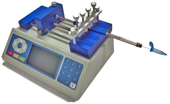
Figure 1: Schematic of the syringe pump used for microscale nucleosome assembly. The assembly mixture is positioned to be in contact with the end of the syringe needle. As the dilution buffer is delivered by the syringe pump to the assembly mixture the concentration of NaCl is decreased, promoting nucleosome assembly. This figure is adapted from Stumme-Diers et al.

Figure 2: SDS-PAGE of assembled nucleosomes. Lanes 1 and 2 contain the H3 octamer and the CENP-A assembly of histones, respectively. Lanes 3 and 4 contain the assembled H3 nucleosomes and the assembled CENP-A nucleosomes, respectively. The comparison of the assembled nucleosomes to the histone-only controls in lanes confirms that nucleosomes were properly assembled. The cartoon schematic above each lane indicates which histone components are present. This figure is adapted from Stumme-Diers et al.
Disclosures
The authors have nothing to disclose.
Materials
| Plasmid pGEM3Z-601 | Addgene, Cambridge, MA | 26656 | |
| PCR Primers | IDT, Coralville, IA | Custom Order | (FP) 5'- CAGTGAATTGTAATACGACTC-3' (RP) 5'-ACAGCTATGACCATGATTAC-3' |
| DreamTaq polymerase | ThermoFischer Scientific, Waltham, MA | EP0701 | Catalog number for 200 units |
| PCR purification kit | Qiagen, Hilden, Germany | 28104 | Catalog number for 50 units |
| Tris base | Sigma-Aldrich, St. Louis, MO | 10708976001 | Catalog number for 250 g |
| EDTA | ThermoFischer Scientific, Waltham, MA | 15576028 | Catalog number for 500 g |
| (CENP-A/H4)2, recombinant human | EpiCypher, Durham, NC | 16-0010 | Catalog number for 50 ug |
| H2A/H2B, recombinant human | EpiCypher, Durham, NC | 15-0311 | Catalog number for 50 ug |
| H3 Octamer, recombinant human | EpiCypher, Durham, NC | 16-0001 | Catalog number for 50 ug |
| Slide-A-Lyzer MINI Dialysis Device Kit, 10K MWCO, 0.1 mL | ThermoFischer Scientific, Waltham, MA | 69574 | Catalog number for 10 devices |
| Sodium Chloride | Sigma-Aldrich, St. Louis, MO | S9888-500G | Catalog number for 500 mg |
| Amicon Ultra-0.5 mL Centrifugal Filters | Millipore-sigma, Burlington, MO | UFC501008 | Catalog number for 8 devices |
| HCl | Sigma-Aldrich, St. Louis, MO | 258148-25ML | Catalog number for 25 mL |
| Tricine | Sigma-Aldrich, St. Louis, MO | T0377-25G | Catalog number for 25 g |
| SDS | Sigma-Aldrich, St. Louis, MO | 11667289001 | Catalog number for 1 kg |
| Ammonium Persulfate (AmmPS) | Bio-Rad, Hercules, CA | 1610700 | Catalog number for 10 g |
| 30% Acrylamide/Bis Solution, 37.5:1 | Bio-Rad, Hercules, CA | 1610158 | Catalog number for 500 mL |
| TEMED | Bio-Rad, Hercules, CA | 1610800 | Catalog number for 5 mL |
| 4x Laemmli protein sample buffer for SDS-PAGE | Bio-Rad, Hercules, CA | 1610747 | Catalog number for 10 mL |
| 2-ME | Sigma-Aldrich, St. Louis, MO | M6250-10ML | Catalog number for 10 mL |
| PageRuler Prestained Protein Ladder | ThermoFischer Scientific, Waltham, MA | 26616 | Catalog number for 500 uL |
| Bio-Safe™ Coomassie Stain | Bio-Rad, Hercules, CA | 1610786 | Catalog number for 1 L |
| Nonwoven cleanroom wipes: TX604 TechniCloth | TexWipe, Kernersvile, NC | TX604 | |
| Muscovite Block Mica | AshevilleMica, Newport News, VA | Grade-1 | |
| Aminopropyl silatrane (APS) | Synthesized as described in 22 | ||
| HEPES | Sigma-Aldrich, St. Louis, MO | H4034-25G | Catalog number for 25 g |
| Scotch Tape | Scotch-3M, St. Paul, MN | ||
| Aron Alpha Industrial Krazy Glue | Toagosei America, West Jefferson, OH | AA480 | Catalog number for 2 g tube |
| MgCl2 | Sigma-Aldrich, St. Louis, MO | M8266-100G | Catalog number for 100 g |
| Millex-GP Filter, 0.22 µm | Sigma-Aldrich, St. Louis, MO | SLGP05010 | Catalog number for 10 devices |
| BL-AC10DS-A2 afm probe (for HS-AFM) | Olympus, Japan | ||
| Compound FG-3020C-20 | FluoroTechnology Co., Ltd., Kagiya, Kasugai, Aichi, Japan | ||
| Compound FS-1010S135-0.5 | FluoroTechnology Co., Ltd., Kagiya, Kasugai, Aichi, Japan | ||
| High-Speed Time-Lapse Atomic Force Microsocopy | Toshio Ando, Nano-Life Science Institute, Kanazawa University, Kakuma-machi, Kanazawa, Japan |
