This study was conducted in compliance with the Animal Welfare Act and the Implementing Animal Welfare Regulations in accordance with the principles of the Guide for the Care and Use of Laboratory Animals. All animal procedures were approved by the Institutional Animal Care and Use Committee at the University of Texas Health Science Center at San Antonio. For an overview of the protocol, see Figure 1; see the Table of Materials for details on all materials, reagents, and instruments used in this protocol.
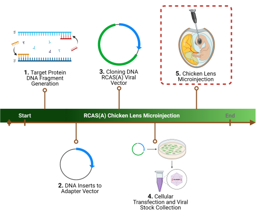
Figure 1: Experimental outline. 1. The first step of the protocol is the determination of a specific target protein(s), the identification of the associated gene sequence(s), and DNA fragment generation. 2. Cloning of the gene sequence(s) into a retroviral vector by initial cloning into an adaptor vector, 3. followed by a viral vector. 4. Preparation of high-titer viral particles using packaging cells to harvest and concentrate. 5. The final step, and the focus of this protocol, is the chicken lens microinjection of the RCAS(A) viral particles into the lens lumen. Please click here to view a larger version of this figure.
1. Preparation of high-titer recombinant retroviruses
- Polymerase chain reaction (PCR) to make target protein DNA fragments
NOTE: This section aims to amplify the DNA sequence corresponding to the target lens protein. For more detail, see7,8.- Design primers for PCR, corresponding to the protein of interest, to make DNA fragments with their carboxyl termini in-frame with or without a FLAG epitope sequence (5'-GACTACAAGGACGACGATGACAAG-3'), a stop codon, and a restriction enzyme site (i.e., EcoRI) present inside the polylinker region of CLa12NCO)) according to the specifications noted in previously published protocols, with sequences for adequate restriction enzymes1,7,8,10,11.
- Perform the PCR reaction10. Combine 100 pmol of sense and antisense of the designed primers with 0.5 µg of connexin cDNA (such as Cx50, Cx43, or chimeric Cx50*43L combinations), to the PCR reaction buffer of choice. Perform 30 cycles each consisting of 94 °C for 1 min, 58 °C for 1 min, and 72 °C for 1 min, followed by a final extension for 10 min at 72 °C.
- Isolate and purify PCR products with a gel purification kit.
- Digest the purified PCR products using restriction enzymes of choice according to the manufacturer's instructions.
- Cloning of DNA inserts into adaptor plasmid
NOTE: Insertion of DNA fragments into an adaptor vector eliminates the need for helper cells/viruses to increase RCAS(A) vector stability14. For more detail, see7,8.- Subclone DNA fragments into an adapter plasmid, Cla12NCO using a sticky-end ligation reaction. Mix the PCR fragments with restriction enzymes and Cla12NCO at a 2-5:1 ratio. Perform ligation and DNA transformation using competent cells.
- Isolate DNA using a DNA isolation kit.
- Sequence the DNA to ensure accuracy.
- Cloning into the RCAS(A) viral vector
NOTE: DNA fragments are inserted into a vehicle (RCAS(A) retrovirus) for the stable transduction of a gene into cells in the developing chick embryo14,15. For more detail, see7,8,15.- Digest the Cla12NCO-DNA fragment of interest-containing vector with ClaI (1 unit per 1 µg of DNA) and gel-isolate the fragments.
- Subclone the DNA fragments into a ClaI-linearized RCAS(A) plasmid. Use the restriction enzyme SalI to distinguish the ClaI site on the RCAS(A) plasmid.
- Confirm the correct orientation of the inserted fragments on the constructs by digestion with ClaI and SalI restriction enzymes.
- Isolate DNA using a DNA isolation kit.
- Determine the DNA concentration.
- Transfection of chick embryonic fibroblast (CEF) cells and RCAS(A) harvest
NOTE: Virus packaging cells, CEFs, are used to obtain/harvest cell culture media containing viral particles15,16. For more detail, see7,8,15.- Transfect CEF cells with recombinant retroviral DNA constructs using the transfection agent according to the manufacturer's instructions.
- Perform western blotting of the transfected cells to examine their expression of the protein of interest.
- When the transfected cells reach confluency, begin collecting the supernatant medium, processing/filtering it appropriately (to remove any cellular debris using a 0.22µm filter), and store at -80 °C until ready for concentration.
- Concentration and titering of viral stocks
NOTE: The pellet and large volumes of media derived from the cells containing the viral particles must be filtered. Additionally, a viral titer assay must be performed to establish the strength of the virus against the host cells. For more detail, see6,7,8,15.- Thaw the culture media containing the virions.
- Spin the culture media at 72,000 × g for 2 h at 4 °C.
- Decant the supernatant and resuspend the viral pellet with the residual (~50 µL) medium and store at -80 °C until use, in ~10 µL viral stocks.
- For titering, using either QT-6 or CEF cells, transfect the cells with a serial dilution of the viral stocks.
- Post confluency, fix the cells using 4% formaldehyde.
- Immunostain for viral gag proteins or process for alkaline phosphatase histochemically to obtain the titer, defined as (colony-forming units) CFU per milliliter (CFU/mL).
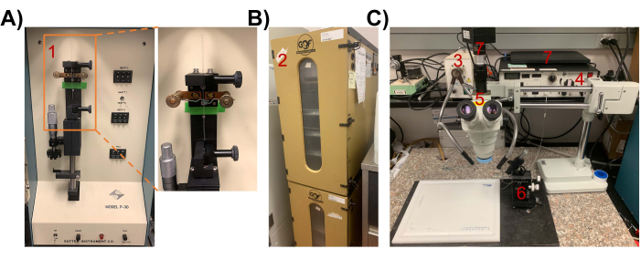
Figure 2: Instruments and setup for chick lens microinjection. (A) P-30 manual vertical microelectrode micropipette puller. (1) inset showing a glass micropipette being pulled. (B) (2) Egg Incubator. Incubate chicken egg for ~65-68 h in a 37 °C incubator to reach stage 18 (with a sealed, empty central lumen) for injection of retrovirus. (C) Microinjection setup. (3) lighting equipment, (4) Pico-Injector, (5) dissecting microscope, (6) Drummond micromanipulator, (7) computer/camera for visualization. Please click here to view a larger version of this figure.
2. Chick lens microinjection
- Preparation of supplies
- Preparation of glass micropipettes for microinjection17
NOTE: Glass capillaries are used to make glass micropipettes with a tip point and an outer diameter (OD) of ~11 µm for microinjection. The setup is shown in Figure 2A.- Attach the borosilicate glass capillary to the rubber cushioned clips in the manual vertical micropipette puller.
- Set heat temperature (HEAT 1) to 950 °C and perform prepulling to obtain a thinner and softer glass capillary.
- Set heat temperature (HEAT 2) to 790 °C and perform a secondary pulling to produce final micropipettes.
- With a micropipette grinder, sharpen the tip opening to an OD of ~11 µm.
- For future experiments, keep the glass micropipettes in a sponge clamping pad inside a glass jar.
- Incubation of fertilized eggs until development stage 18
NOTE: During lens development, at ~65-68 h of embryonic development, the lens separates from the ectoderm and forms a sealed vesicle with a central lumen; injection into this empty lens lumen primes for restricted expression to proliferative lens cells7,8,18.- Incubate fertilized chicken eggs for ~65-68 h in a 37 °C humidified rocking incubator (Figure 2B).
- Preparation of glass micropipettes for microinjection17
- Opening of the chicken egg
NOTE: This step describes the identification and exposure of the embryo for microinjection. For more detail, see7,8.- Wipe down the work area thoroughly with 70% ethanol.
- Remove the eggs from the incubator, spray them down heavily with 70% ethanol, and let them air-dry.
- Place the egg, with the larger end of the egg facing upwards, on an egg holder.
- Using a pair of sharp-toothed forceps, produce a hole of ~2 cm diameter on the larger end of the egg by carefully tapping on the eggshell and removing fragments of the eggshell (Figure 3A).
- Using a dissecting microscope, locate the embryo.
- Once the embryo and chick lens are located, use the dissecting microscope and fine dissecting forceps and scissors to cut off the amnionic membrane immediately covering the top of the embryo (Figure 3B).
NOTE: Do not cut too widely (only enough to expose the embryo ~0.5 cm x 0.5 cm), or else the embryos might sink into the yolk mass; also avoid touching blood vessels, if possible. - Cover the opening of the egg with a 60 mm Petri dish in preparation for the next steps.
- Microinjection of concentrated viral stock
NOTE: Viral particles containing target protein DNA fragments for transduction are inserted into cells inside the lens lumen. The setup is shown in Figure 2C. For more detail, see6,7,8.- Thaw the viral stock stocks on ice.
- For visualization of the viral stock during injection, dilute 1 µL of 10x Fast green into one vial of viral stock containing 10 µL.
- To ensure there are no large chunks of undissolved material that could clog the glass micropipette, centrifuge the solutions for 10 s at 10,000 × g at 4 °C to pellet "large chunks" and transfer the supernatants into new tubes, all while keeping the solutions on ice.
- On a 35 mm x 10 mm culture dish, place a stretched laboratory parafilm and add 1 µL of sterile saline (PBS) on top of the film (non-sterilized, with the covered side considered "clean").
- Connect a prepared glass micropipette to an automatic pico-injector.
- Press the fill mode to fill the sterile saline (PBS) into a glass micropipette, and then use the inject mode to test the allocated 1 µL of liquid.
- Place a second stretched laboratory parafilm on the surface of a new 35 mm x 10 mm cell culture dish and add 1 µL of Fast green-stained viral stocks on top of the film.
- Lower the tip of the micropipette into the viral stock and press the fill-model to fill the glass micropipette with ~1 µL of viral stock.
NOTE: To avoid drying out, put the tip of the filled micropipette in sterile saline (PBS) whenever there is a pause in the procedure. - Lower the filled glass micropipette, connected to an automatic injector, into the target region of the embryo lens lumen with the assistance of a dissecting microscope.
- Adjust the light source to ensure clear visualization of the outline of the lens vesicle.
NOTE: The lumen of the right lens (facing up) is typically used for injection and the left (facing down) is kept intact as a contralateral control. - Once sure that the placement of the micropipette is within the correct location inside the lens lumen, inject 5-40 nL of the viral stock (Figure 3C).
NOTE: Practice is very necessary for optimal injection placement. - Wait for ~45 s and then gently remove the glass micropipette.
- Check for the successful microinjection into the lens lumen using the dissecting microscope by examining Fast Green dye that should stay in the empty lumen of the lens without any leakage.
- After the injection procedure, seal the eggshell opening with scotch tape.
- Return the embryo to the 37 °C humidified incubator, without any rotating motion, until the desired embryonic age has been reached for lens dissection.
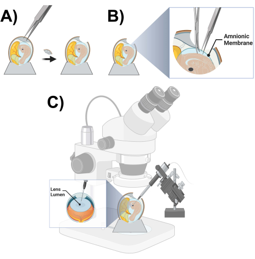
Figure 3: Microinjection chick prep and schematic. (A) Opening of a chicken egg. (B) Cutting of the amniotic membrane. (C) Chicken lens lumen microinjection schematic. Please click here to view a larger version of this figure.
After the determination of a specific target protein(s) and the identification of the associated gene sequence(s), the overall experimental approach involves the cloning of the gene sequence(s) into a retroviral RCAS(A) vector by the initial cloning into an adaptor vector, followed by a viral vector. Second, high-titer viral particles are prepared using packaging cells to harvest and concentrate the virions. These first two major steps have been largely described and representative results presented elsewhere6,7,8,14,16.
For this protocol, the main area of focus is the step of microinjection. It is imperative to determine the success of the in situ microinjection of RCAS(A) viral particles containing DNA fragments of the protein target of interest, both at the time of injection and after lens isolation. Figure 4 shows an image of the lens lumen pre- and post-injection of the viral vectors dyed with Fast Green for visualization and confirming the proper localization into the lens lumen. Fast Green is a dye with a high degree of safety and is approved by the U.S. Food and Drug Administration as a color additive used to color food, drugs, and cosmetics19.
Figure 5 shows the histological evaluation through immunofluorescence of the target protein, the chimeric connexin Cx50*43L, which was introduced into high-titer recombinant retroviruses, microinjected into the lens lumen and evaluated. Since the C-termini of the chimeric connexins were epitope-tagged with FLAG sequences, anti-flag labeling was used to identify the exogenous connexins from the endogenous ones, using standard staining methodology, with sagittal and coronal sections (sagittal shown here), as described in Liu et al.10 Although Cx50*43L is localized on the plasma membrane, because of the orientation of the tissue sections prepared, it appears to localize inside the lens in certain regions. In this study, the interaction between the intercellular loop domain of Cx50 and AQP0 was evaluated and stained accordingly10.
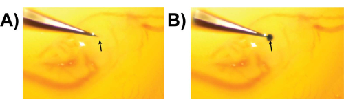
Figure 4: Example of microinjection into the chicken lens lumen. (A) Preinjection into lens lumen. (B) Post injection into lens lumen. Arrow = injection site. Please click here to view a larger version of this figure.
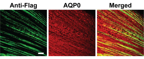
Figure 5: Microinjection and histological evaluation. At stage 18 of embryonic development, which is ~65-68 h of embryonic development, a microinjection of recombinant retroviruses containing chimeric Cx50*43L mutant was done into the empty lumen of a chick lens. We examined the cryosections of chick lenses dissected out on embryonic day 18, which were immunolabeled with FLAG (green) and AQP0 (red) antibodies. Fluorescein-conjugated anti-mouse IgG was used to detect primary antibodies against anti-FLAG, while rhodamine-conjugated anti-rabbit IgG was used to detect primary antibodies against anti-AQP0. The visualization of immunostaining was done using confocal fluorescence microscopy. The corresponding merged images, labeled as "Merged", can be seen on the right. Scale bar = 50 µm. Please click here to view a larger version of this figure.
