The significance of petite colonies in Candida spp. drug resistance has not been fully explored. Antimicrobial photodynamic therapy (aPDT) offers a promising strategy against drug-resistant fungal infections. This study demonstrates that rose bengal-mediated aPDT effectively deactivates Candida glabrata and induces petite colonies, presenting a unique procedure.
Facing a 40% mortality rate in candidemia patients, drug-resistant Candida and their petite mutants remain a major treatment challenge. Antimicrobial photodynamic therapy (aPDT) targets multiple fungal structures, unlike antibiotics/antifungals, potentially thwarting resistance. Traditional methods for inducing petite colonies rely on ethidium bromide or fluconazole, which can influence drug susceptibility and stress responses. This study investigated the application of green light (peak 520 nm) and rose bengal (RB) photosensitizer to combat a drug-resistant Candida glabrata isolate. The findings revealed that aPDT treatment significantly inhibited cell growth (≥99.9% reduction) and effectively induced petite colony formation, as evidenced by reduced size and loss of mitochondrial redox indicator staining. This study provides initial evidence that aPDT can induce petite colonies in a multidrug-resistant C. glabrata strain in vitro, offering a potentially transformative approach for combating resistant fungal infections.
Fungal infections, particularly those caused by Candida albicans and increasingly drug-resistant Candida glabrata, pose a serious global threat1. These infections can be deadly, especially for hospitalized patients and those with weakened immune systems. Rising antifungal resistance threatens the control of invasive candidiasis, a severe fungal infection with high mortality, especially from Candida albicans2. Resistant strains hinder effective treatment, potentially increasing both complexity and death rates. In Alameda County, California, USA, C. glabrata has become the most prevalent invasive species3. This shift in the prevalence and distribution of Candida species may be influenced by local healthcare practices, patient demographics, the utilization of antifungal agents, and the prevalence of risk factors for Candida infections.
Petite mutants in Candida, lacking functional mitochondria, reveal how this organelle affects drug response, virulence, and stress resistance4,5. C. glabrata readily forms these colonies, gaining sensitivity to polyenes while losing it to azoles6. Azole sensitivity and respiratory function are intricately linked, with diminished respiration leading to resistance via mitochondrial DNA loss7. Petite colonies of C. glabrata with azole resistance have been isolated from human stool samples from a bone marrow transplant recipient undergoing fluconazole treatment8 and from blood culture bottles of patients with bloodstream infections9. Their potential implications in drug resistance, virulence, and stress response highlight their clinical significance. Additionally, their distinct properties make them valuable tools for investigating fundamental questions in mitochondrial biology5. As research into petite mutants continues, their applications in both clinical and basic research are likely to expand.
This study discovered that photodynamic therapy (PDT) can induce petite colonies in C. glabrata, expanding the range of methods beyond the traditional techniques of exposing C. glabrata to ethidium bromide or fluconazole.
1. Culturing of C. glabrata
NOTE: A multidrug-resistant C. glabrata (C2-1000907) that is resistant to most antifungal agents, including fluconazole, is used for the experiments. The experimental conditions may need to be adapted to the specific strain, as variations may exist among different strains. All experiments used log-phase Candida grown at 25 °C (mimicking natural infection) for consistency. C. glabrata's lack of hyphae simplifies quantification compared to C. albicans, which forms hyphae at 37 °C10.
2. Induction of petite colonies by ethidium bromide, fluconazole and photodynamic therapy
3. Mitochondrial function analysis (TCC staining test)
NOTE: TTC is a redox indicator and an electron acceptor. It turns red when white compounds are broken by electrons11. Note that not all cells necessarily form visible colonies within 24 h after culture on an agar plate. Colonies with functional mitochondria turn red, while those with non-functional mitochondria remain white. This allows differentiation between colonies with different mitochondrial functionality.
4. Growth kinetics of normal and petite C. glabrata
NOTE: Three strains of yeast were compared: C. glabrata C2-1000907 T0 (clinical isolate without PDT treatment), T3n (C. glabrata C2-1000907 after 3 consecutive RB-PDT exhibiting colonies with an average diameter of 1.5 ± 0.8 mm similar to the parental cells), and T3p (petite colonies of C. glabrata C2-1000907 after 3 consecutive RB-PDT).
The data is presented as the mean with ± standard error and was obtained from three independent experiments, with at least triplicates in each group. Experimental data, including colony counts, OD600 measurements, and TTC staining results, were graphed and statistically analyzed using graphing and statistical software (see Table of Materials). One-way ANOVA or t-test was used to analyze the data, and a p-value <0.05 was considered significant. Scanning was performed with a 48-bit full-color optical scanner at a resolution of 1200 dpi, and subsequent measurements of the colony diameters were conducted using ImageJ software.
As depicted in the growth curves in Figure 2, petite mutants of C. glabrata C2-1000907 (T3p) exhibited slower growth compared to the untreated parental fungi of regular size (T0). When C. glabrata was mixed with 0.2% RB for 15 min and exposed to 4.38 J/cm2 green light (PDT treatment), the titer of the fungi decreased from 107.5 CFU/mL to 104.5 CFU/mL (at least 3 logs, Figure 3) compared with the control group (RB only), which had a cell density of around 107 CFU/mL. Additionally, after aPDTs, petite colonies were observed. These petite colonies had an average size of 0.4 mm ± 0.25 mm instead of the usual size of 1.5 mm ± 0.8 mm (Figure 4A). Small-size colonies with mitochondrial dysfunction can be identified by their size and color after staining with 20% TTC. The petite colonies that maintained a white color (Figure 3B, white arrows) exhibited mitochondrial dysfunction, while some smaller red-colored colonies and normal-sized colonies retained normal mitochondrial function (Figure 4B). The traditional DNA intercalating agent ethidium bromide (Figure 4C) and fluconazole (Figure 4D) can effectively induce petite mutants of Candida. Disk diffusion is a simple method to define drug susceptibility in fungi. In Figure 4D, a clear circular zone of no growth surrounded the fluconazole disk containing 25 µg fluconazole in the T0 culture (without PDT), indicating less drug resistance in this parental fungus. Nonetheless, the C. glabrata C2-1000907 is still defined as a resistant strain by minimal inhibition concentration toward fluconazole (data not shown) and the diameter of the clear zone. In the T3 culture, many petite colonies formed near the disk (arrow, lower panel), suggesting drug resistance after three repeated PDT treatments.
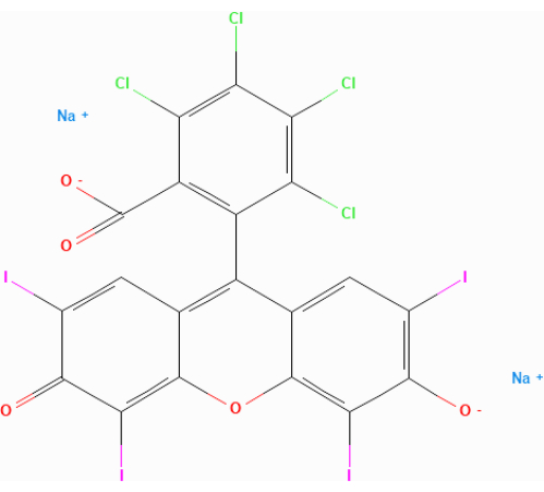
Figure 1: Molecular structure of rose Bengal (RB). Please click here to view a larger version of this figure.
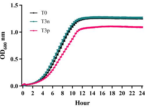
Figure 2: Growth Curves of C. glabrata C2-1000907 24 h following repetitive RB-PDT. Growth curves were compared for three strains: C. glabrata C2-1000907 T0 (naïve parental cells), T3n (colonies with normal size after 3 repeated treatments), and T3p (petite colonies after 3 repeated treatments). Compared to T0 and T3n, T3p colonies exhibited a significantly slower growth rate (n = 3, p < 0.05). All cells are grown in YPD medium. Please click here to view a larger version of this figure.
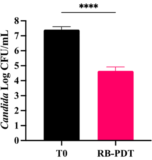
Figure 3: Growth Inhibition of C. glabrata C2-1000907 following aPDT. A single aPDT treatment with a 4.38 J/cm² light dose and 0.2% RB significantly inhibited C. glabrata C2-1000907 growth by 3 logs compared to the naïve control (p < 0.0001, unpaired t-test). Error bars represent mean ± SEM, data is pooled from 3 independent experiments. Please click here to view a larger version of this figure.
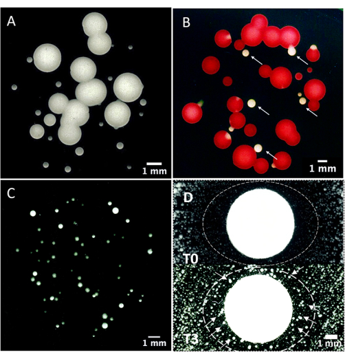
Figure 4: Mitochondrial dysfunction analysis using triphenyltetrazolium chloride (TCC) staining. (A) C. glabrata C2-1000907, 24 h following aPDT treatment (0.2% RB, 4.38 J/cm² green light), displayed a bimodal colony size distribution, with large (1.5 mm ± 0.8 mm) and small (petite) colonies (0.4 mm ± 0.25 mm). (B) TCC staining revealed that the large colonies were functional, as they turned red, while the petite colonies were non-functional, as they remained white. (C) Petite colonies were also induced by ethidium bromide (100 µg/mL for 30-40 min) and (D) fluconazole (25 µg/mL) treatment. A clear circular zone of no growth surrounded the fluconazole disk containing 25 µg fluconazole in the T0 culture (without PDT), indicating susceptibility. In the T3 culture, many petite colonies formed near the disk (arrow, lower panel), suggesting drug resistance after three repeated PDT treatments. Scale bars: 1 mm. Please click here to view a larger version of this figure.
This study unveils PDT as the first reported method to induce petite colony formation in Candida, surpassing the established effects of ethidium bromide and fluconazole. This novel observation necessitates further exploration to unravel its implications for both fungal eradication by decreasing virulence and the emergence of resistance mechanisms.
RB-mediated PDT effectively inhibits the growth of C. glabrata, suggesting a potential alternative treatment approach for Candida infections. As a light-activated photosensitizer, RB produces singlet oxygen and reactive oxygen species (ROS) when exposed to a specific wavelength. This oxidative stress causes damage to essential cellular components such as lipids, proteins, and nucleic acids13, leading to mitochondrial dysfunction, as shown in the present study. This makes it difficult for Candida to develop resistance through conventional mechanisms observed with standard antifungal drugs.
The three yeast strains used in this study, T0, T3n, and T3p, exhibit distinct growth patterns. T0 remains untreated, whereas T3n and T3p undergo RB-PDT three times. T3n displays normal-sized colonies, whereas T3p exhibits petite colonies. The slower growth of the petite colonies may indicate alterations in cellular energy and metabolism. The petite phenotype, often associated with mitochondrial dysfunction4, could lead to impaired aerobic respiration, consequently influencing the overall growth kinetics. The impaired aerobic respiration in petite colonies might affect their virulence, drug susceptibility, or ability to persist in different environments. This connection holds potential clinical implications and provides avenues for future research directions.
Ethidium bromide, a DNA intercalating agent, inhibits mitochondrial DNA synthesis and degrades existing mitochondrial DNA, transforming Candida into petite colonies with mitochondrial dysfunction14. Fluconazole, commonly used to treat Candida infections, can induce drug resistance and petite colony development in C. glabrata6. Petite mutants may exhibit azole drug resistance through the activation of the transcription factor PDR1 and its regulation of genes CDR1 and CDR215. Moreover, due to the partial or complete loss of mitochondrial DNA, mitochondrial function is compromised16.
PDT has demonstrated efficacy in reducing bacterial virulence17, inducing susceptibility to antibiotics18, and reducing biofilm formation19 in bacteria. Studies on PDT against fungal infections are limited20. This discovery raises intriguing questions about how RB-PDT affects mitochondria in C. glabrata. To fully comprehend how RB-PDT impacts yeast cells, understanding the molecular mechanisms causing growth variations is crucial. However, petite mutants induced by PDT are currently understudied, and their detailed mechanistic underpinnings remain unclear. Further investigation is needed to determine if there are any differences between small colonies created by various methods. Understanding how petites form in Candida species is important for studying the relationship between mitochondrial function and pathogenicity. Furthermore, petite mutants can serve as a model for exploring antifungal drug resistance mechanisms.
The current study, while providing valuable insights into the effects of RB-mediated PDT on C. glabrata and the emergence of petite colonies, has certain limitations that should be considered. Firstly, the study primarily focused on in vitro experiments, and the translation of these findings to clinical settings warrants further investigation. Additionally, the specific molecular mechanisms underlying the formation of petite colonies following RB-PDT and their potential implications for antifungal drug resistance require more in-depth exploration. Furthermore, while the study compared the growth patterns of different yeast strains, additional molecular and genetic analyses could provide a deeper understanding of the metabolic and genetic changes associated with petite colony formation.
Moreover, the potential variability in response to RB-PDT among different clinical isolates of C. glabrata was not addressed in this study, highlighting the need for broader strain-specific investigations. Future studies addressing these limitations will be crucial for a more comprehensive understanding of the implications of RB-mediated PDT and the emergence of petite colonies in the context of antifungal treatment strategies.
In summary, this investigation reveals that aPDT induces C. glabrata (C2-1000907) petite mutants with impaired mitochondrial function. This unique mechanism might offer a new method for antifungal studies. The precise mechanism of action underlying PDT-induced petite colonies remains to be elucidated, and exploring potential differences from other methods will be crucial for optimizing therapeutic strategies.
The authors have nothing to disclose.
This work has received funding from the Ministry of Science and Technology, Taiwan [MOST 110-2314-B-006-086-MY3], National Cheng Kung University [K111-B094], [K111-B095], National Cheng Kung University Hospital, Taiwan [NCKUH-11204031], [NCKUMCS2022057].
0.22 μm filter | Merck, Taipei, Taiwan | Millex, SLGVR33RS | |
1.5 mL microfuge tube | Neptune, San Diego, USA | #3745 | |
20% Triphenyltetrazolium chloride (TTC) | Sigma-Aldrich, MO, USA | T8877 | |
5 mL polypropylene round bottom tube | Corning, AZ, USA | 352059 | |
5 mL round-bottom tube with cell strainer cap | Corning, AZ, USA | Falcon, #352235 | |
96-well plate | Alpha plus, Taoyuan Hsien, Taiwan | #16196 | |
Agar | BRS, Tainan, Taiwan | AG012 | |
Blank disk | Advantec, Tokyo, Japan | 49005040 | |
Centrifuge | Eppendorf, UK | 5415R | |
Ethidium bromide solution | Sigma-Aldrich, MO, USA | E1510 | |
Fluconazole, 2 mg/mL | Pfizer, NY, USA | BC18790248 | |
GraphPad Prism | GraphPad Software | Version 7.0 | |
Green light emitting diode (LED) strip | Nanyi electronics Co.,Ltd, Tainan, Taiwan | 5050 | Excitation wave: 500~550 nm |
Low Temperature. shake Incubators | Yihder, Taipei, Taiwan | LM-570D (R) | |
Mouth care cotton swabs | Good Verita Enterprise, Taipei, Taiwan | 161357 | |
Muller Hinton II agar | BD biosciences, California, USA | 211438 | |
Multimode microplate reader | Molecular Devices | SpectraMax i3x | |
OD600 spectrophotometer | Biochrom, London, UK | Ultrospec 10 | |
Rose Bengal | Sigma-Aldrich, USA | 330000 | stock concentration 40 mg/mL = 4%, prepare in PBS, stored at 4 °C |
Sterilized glass tube | Sunmei, Tainan, Taiwan | AK45048-16100 | |
Yeast Extract Peptone Dextrose Medium | HIMEDIA, India | M1363 |