Isolation of Cytotoxic Neutrophils from Tumor-Bearing Mouse Blood

Published: August 31, 2023

Abstract

Source: Sionov, R. V., et al., Isolation and Characterization of Neutrophils with Anti-Tumor Properties. J. Vis. Exp. (2015)

In this video, we demonstrate the isolation of cytotoxic high-density neutrophils and low-density neutrophils from the blood of a tumor-bearing mouse by discontinuous sucrose gradient centrifugation.

Protocol

All procedures involving animal models have been reviewed by the local institutional animal care committee and the JoVE veterinary review board.

1. Neutrophil Isolation

  1. Isolation of cytotoxic neutrophils from blood of tumor-bearing mice.
    1. Dilute 1 ml of blood drawn from a tumor-bearing mouse in PBS containing 0.5% (w/v) bovine serum albumin (BSA) to a final volume of 6 ml.
    2. Fractionation of diluted blood on a freshly prepared discontinuous sucrose gradient:
      1. Add 3 ml of sterile-filtered sucrose 1.119 g/ml to the bottom of a 15 ml conical polypropylene centrifuge tube.
      2. Slowly and carefully, layer 3 ml of sterile-filtered sucrose 1.077 g/ml on top of the 1.119 g/ml layer. Thereafter, add slowly and carefully the 6 ml of diluted blood on top of the 1.077 g/ml layer (Figure 1A). It is recommended to hold the tube tilted and add the different components by a slow, but continuous flow, keeping the mouth of the pipette towards the lower wall of the tube, such that no turbulence is formed.
    3. Centrifuge the tube containing the diluted blood on the sucrose gradient at 700 x g for 30 min at RT without brake.
    4. Carefully remove the tube from the centrifuge without causing any turbulence. Most of the erythrocytes will be at the bottom of the tube. High-density neutrophils (HDN) are found as a white-to-red ring at the interface between the 1.119 g/ml and the 1.077 g/ml layers (around the 3 ml mark), while the low-density leukocytes are found in a white ring at the interface between the 1.077g/ml layer and the BSA-containing PBS (around the 6 ml mark, see Figure 1B).
    5. Aspirate the PBS + 0.5% BSA until reaching 5 mm above the low-density cell layer. Pipette out the low-density cells by slow suction into a 1 ml tip through slowly swirling around the cells. Transfer the cells into 30 ml of PBS with 0.5% BSA.
    6. Aspirate the upper layer in the same gradient tube until reaching 5 mm above the high-density cell band. Pipette out the high-density cells, which are mostly high-density neutrophils, and transfer the cells into 30 ml of PBS containing 0.5% BSA.
    7. Centrifuge the cells at 400 x g for 10 min at RT.
    8. Aspirate the supernatant and lyse erythrocytes by resuspending the cells in 36 ml sterile HPLC grade water for 30 sec. Isotonicity should be restored by adding 9 ml of a 5x concentrated PBS supplemented with 2.5% (w/v) BSA.
    9. Centrifuge the cells at 400 x g for 10 min at RT.
    10. Aspirate the supernatant, and resuspend the cells in PBS-BSA. Count the number of neutrophils in a hemocytometer and use trypan blue to distinguish between live and dead cells.
    11. Centrifuge the cells at 400 x g for 10 min at RT and resuspend the neutrophils in desired incubation medium to final cell density. Use the neutrophils immediately.
    12. Then, after another centrifugation, resuspend the neutrophils in an incubation medium to final cell density. Use the neutrophils immediately.

Representative Results

Figure 1
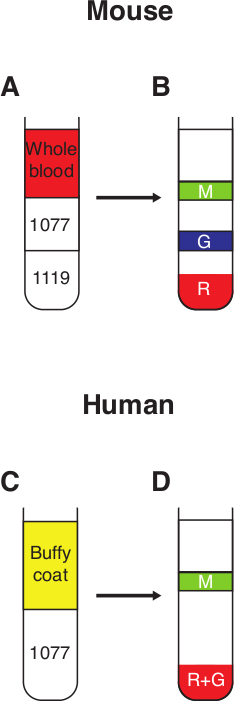
Figure 1. Neutrophil purification from whole blood. (A) 3 ml of 1.077 g/ml sucrose is carefully layered on top of 3 ml 1.119 g/ml sucrose to form a discontinuous gradient. Whole murine blood, diluted in PBS-BSA (0.5%) to a final volume of 6 ml, is then layered on top of the 1.077 g/ml sucrose. (B) Following a 30 min spin at 700 x g with no break, 3 distinct fractions can be observed; R – red blood cells in the pellet, G – the granulocytic fraction containing high-density neutrophils, M – mononuclear fraction containing mononuclear cells and low-density neutrophils. (C) Freshly drawn human blood is mixed with an equal volume of Dextran 500 (3%) and incubated at RT for 30 min. The top fraction containing the white blood cells (buffy coat) is then layered on top of 10 ml 1.077 g/ml sucrose. (D) Following a 30 min spin at 400 x g with no break, 2 distinct fractions may be observed; R+G – pellet containing red blood cells and high-density neutrophils, M – mononuclear fraction containing mononuclear cells and low-density neutrophils.

Disclosures

The authors have nothing to disclose.

Materials

Mouse 4T1 breast carcinoma cells ADCC CRL-2539 Growth medium: DMEM + 10 % heat-inactivated FBS
24-well Tissue Culture Plate Falcon 353047
100 mm Tissue Culture Plate Corning 430167
25 cm2 Tissue Culture Flask Nunc 156340
90 mm Bacterial Grade Culture Dish Miniplast, Ein Shemer, Israel 20090-01-017
15 ml Sterile Conical Centrifuge Tube Miniplast, Ein Shemer, Israel 835015-40-111
50 ml Sterile Conical Centrifuge Tube 835050-21-111
Falcon 12×75 mm Round-Bottom Polystyrene Tube Becton Dickinson 352058
HPLC grade water J.T. Baker 4218-03 Autoclave
Histopaque-1119 Sigma 11191 Sterile filter through a 0.2 mm filter.
Histopaque-1077 Sigma 10771 Sterile filter through a 0.2 mm filter.
PBS+0.5% BSA Dissolve 2.5g BSA in 500 ml PBS, and sterile filter through a 0.2 mm filter.
PBS+1% BSA Dissolve 1g BSA in 100 ml PBS, and sterile filter through a 0.2 mm filter.
Bovine serum albumin (BSA), fraction V Sigma A7906

Tags

check_url/kr/21564?article_type=t

Play Video

Cite This Article
Isolation of Cytotoxic Neutrophils from Tumor-Bearing Mouse Blood. J. Vis. Exp. (Pending Publication), e21564, doi: (2023).

View Video