Detection of Vitronectin Binding to Bacterial Surfaces using Flow Cytometry

Published: November 30, 2023

Abstract

Source: Singh, B. et al., Assays for Studying the Role of Vitronectin in Bacterial Adhesion and Serum Resistance. J. Vis. Exp. (2018)

This video demonstrates the detection of vitronectin's interaction with bacterial surface proteins. The flow-cytometry analysis measures the fluorescence signal of fluorescently labeled antibodies bound to vitronectin, confirming its presence at the bacterial surface. This technique provides insights into host-pathogen interactions and potential bacterial infection therapies.

Protocol

1. Analysis of Vitronectin, Vn as a Bacterial Surface Protein-Ligand

  1. Detection of Vn-binding at the bacterial surface using flow cytometry
    NOTE: In flow cytometry, we used side scatter and forward scatter to gate positive events. To examine the interactions with Vn, Haemophilus influenzae type f, Hif clinical isolates (n=10) were selected together with E. coli BL21 (DE3) as a negative control (Figure 1A).
    1. Culture Hif clinical isolates in brain-heart infusion (BHI) medium supplemented with 10 µg/mL nicotinamide adenine dinucleotide (NAD) and hemin at 37 °C with shaking at 200 rpm. Use Luria-Bertani medium to culture E. coli. Harvest Hif at mid-log phase (Optical density, OD600 = 0.3), and resuspend the bacteria in phosphate-buffered saline (PBS), pH 7.2, containing 1% (w/v) bovine serum albumin (BSA) (blocking buffer; PBS-BSA). Adjust the suspension to 109 colony-forming units, CFU/mL.
    2. Transfer the aliquots containing 5 x 10⁶ CFU to 5 mL polystyrene round-bottom tubes (12 x 75 mm2) and add 1 mL of blocking buffer. Centrifuge the suspension at 3,500 × g at room temperature (RT) for 5 min to pellet the bacteria, then carefully aspirate to remove the supernatant without disturbing the pellet.
    3. Resuspend the bacterial pellets with 50 μL of blocking buffer containing 250 nM Vn and incubate the samples for 1 h at RT without shaking. After incubation, pellet the bacteria by centrifugation at 3,500 x g for 5 min, then wash the pellets three times using PBS and similar centrifugation steps.
    4. To the bacterial pellet, add 50 μL of primary sheep anti-human Vn polyclonal antibodies (pAbs) at 1:100 dilution in PBS-BSA. Incubate the suspension for 1 h at RT, then wash the bacteria three times with PBS to remove unbound antibodies (as described in step 1.1.3).
    5. Next, add 50 μL of PBS-BSA containing fluorescein isothiocyanate (FITC)-conjugated donkey anti-sheep pAbs (1:100 dilution) and incubate at RT for 1 h in the dark.
    6. Prepare a blank control by incubating bacteria with blocking buffer only and pAbs without Vn.
    7. Wash bacteria three times with 1 mL of blocking buffer and pellet the suspension by centrifugation, as described in section 1.1.2. Finally, resuspend the bacterial pellet with 300 µL of PBS and analyze it by flow cytometry.

Representative Results

Figure 1
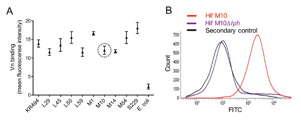
Figure 1: Haemophilus influenzae serotype f binds Vn via surface-expressed PH. (A) Flow cytometry results showing binding of Vn to cells of Hif clinical isolates. Each clinical isolate (5 x 10⁶ CFU) was incubated with 250 nM human Vn. The bound ligand was detected using sheep anti-Vn pAbs and FITC-conjugated donkey anti-sheep secondary antibodies. Escherichia coli BL21 (DE3) was included as a negative control. Data are presented as the mean fluorescence intensity from triplicate analyses in three separate experiments, and error bars represent SD. (B) Flow cytometry histograms demonstrating binding of Vn to the surface of WT Hif M10 and PH-deletion Hif M10Δlph mutant. Representative data from one of three separate experiments are shown.

Disclosures

The authors have nothing to disclose.

Materials

5 mL polystyrene round-bottom tube  BD Falcon 60819-138 12 × 75 mm style
Bovine Serum Albumins (BSA) Sigma-Aldrich A2058 Suitable for cell culture
Flow cytometer  BD Biosciences 651154 Cell analysis grade for research applications 
Vitronectin (Vn) from human plasma Sigma-Aldrich V8379-50UG Cell culture grade
E. coli host (E. Coli BL21) Novagen 69450-3 Protein expression host
FITC-conjugated donkey anti-sheep antibodies  AbD Serotec STAR88F Polyclonal
Sheep anti-human Vn antibodies AbD Serotec AHP396 Polyclonal
check_url/kr/21806?article_type=t

Play Video

Cite This Article
Detection of Vitronectin Binding to Bacterial Surfaces using Flow Cytometry. J. Vis. Exp. (Pending Publication), e21806, doi: (2023).

View Video