1. Pressure myograph set-up
- Summarized from the manufacturer instructions (Danish Myotechnology). A schematic of the complete system with optional accessories is shown in Figure 1.
- Turn on the air tank and myograph control boxes and start the MyoView software (Danish Myotechnology). In the software, load the appropriate diameter calibration file, inflation protocol file and name the file for saving data for the first protocol. Recommended inflation protocols for each age are given in Table 1.
- Choose the required cannulae and suture size for the test system depending on the mouse age. Dimensions of the custom-designed stainless steel cannulae for young and adult mice are shown in Figure 2. Place the bath under the dissecting microscope and mount the cannulae in the test system, being careful not to apply excessive force (> 1 N) to the force transducer.
- Fill the inflow bottle to 150 mL with physiological saline solution (PSS) (Table 2)8. Fill the bath and inflow tubing with PSS. Fill the outflow tubing halfway with PSS. Leaving the outflow bottle empty eliminates having to clean this side of the system. However, if PSS enters this side of the system, clean and flush after use following the manufacturers instructions.
- When the temperature reaches 37.5 °C, place the filled bath on the inverted microscope stage and zero the force transducer using the MyoView software. The force transducer drifts with temperature and location, therefore it is important to zero it in the condition it will be used for testing.
2. Carotid dissection
- Sacrifice the mouse using approved euthanasia methods (we use CO2 asphyxiation). All methods shown in this protocol have been approved by the Institutional Animal Care and Use Committee.
- Secure the mouse by taping the hands and feet to a dissecting board and place under a dissecting microscope. Open the chest to expose the right and left carotid arteries using dissecting scissors and curved forceps. Both arteries can be used for mechanical testing to increase numbers or to provide an internal control for different treatment protocols.
- Carefully clear fat and nearby tissue using curved forceps and fine tweezers until the full length of both carotids from the aorta to the common carotid bifurcation can be visualized (Figure 3). Make sure to keep the arteries moist with PSS during the dissection procedure.
- Cut a piece of 7-0 suture to the desired length using the digital calipers and dissection scissors. Suggested lengths for each age are given in Table 1. Lay the suture on the vessel and use an 18 G needle dipped in activated charcoal to mark the ends of the suture on the vessel (Figure 4A).
- Make sure that the complete length of the artery is lying in a single horizontal plane, otherwise the in vivo length measurements may be incorrect. The head of the mouse may have to be tilted up to accomplish this. Take a picture of each carotid using a camera connected to the dissection microscope. Using known calibrations, measure the in vivo length using Image J software. The length is measured from the image rather than the suture length because the charcoal markers for the cut sites often cannot be applied directly at the ends of the suture.
- Use a syringe attached to an 18 G needle to inject PSS into the left ventricle and clear all blood out of the carotid arteries. Cut the carotids at the charcoal markers using micro-scissors.
- Place the cut carotids in a petri dish with a small amount of PSS. Remove any fat from the outside of the arteries and let equilibrate for 2 – 3 minutes. Take a picture of each artery (Figure 4B) and use Image J software and known calibrations to measure the ex vivo length. Calculate the in vivo stretch ratio using the in vivo and ex vivo lengths.
- Place the carotids in a microfuge tube filled with PSS until ready for testing. They can be stored in the refrigerator for up to 3 days with no changes in passive mechanical behavior9.
3. Mounting an artery in the test system
- If the arteries have been in the refrigerator, remove and allow to come to room temperature.
- Place the heated, filled bath under the dissection microscope. Use the micrometer to bring the cannulae tips together until they are just touching and record this value. This is the “zero length” of the micrometer.
- Place a small loop of suture tied with a loose overhand knot on each end of the cannulae. Using fine tweezers, grasp the end of an artery and place it in the bath. Do not use the tweezers on any other part of the artery as the tips may puncture the wall.
- Use two tweezers to hold one end of the artery and slip it over one cannula. For mice ≥ 21 days, tighten the suture knot and repeat the process for the other side. If it is difficult to mount the second side, apply a slight pressure to open up the artery. For mice < 21 days it may be easier to slide the vessel completely onto one side, bring the cannulae together until they are touching, slide the vessel back over the second cannula, separate the cannulae and tighten the suture knot. Avoid twisting the artery during mounting.
- Calculate the necessary micrometer reading for the artery to be at the measured ex vivo length. We tie our arteries onto the cannulae with about 0.6 mm on each side, so our total unstretched artery length can be calculated from the increase in the micrometer distance from the zero length + 1.2 mm. The artery length that will be stretched in the mechanical test protocols does not include the extra 1.2 mm beyond the suture ties. The distance beyond the suture ties may need to be adjusted for different ages.
- Turn the micrometer to set the total artery length to the measured ex vivo distance and note the appearance and the axial force readings. Check the ex vivo length using the digital calipers. The artery should look unstretched (Figure 5) and the force should be close to zero. We have found that setting the artery at the measured ex vivo length is easier than trying to estimate the unloaded length based on zero force measurements or visualization alone.
- Calculate the necessary micrometer reading for the artery length between the suture ties to be at the measured in vivo stretch ratio. Turn the micrometer to set the artery at this length and begin testing protocols. This is the “in vivo testing length” of the artery.
4. Testing protocols
- Diameter tracking. Place the bath on the inverted microscope stage, position the central portion of the artery over the lens and focus on the edges of the artery wall. Start the diameter tracking in MyoView and check tracking of the outer diameter (Figure 6). The inner diameter will not track well for the carotid artery and must be calculated in the data analysis. Tracking problems can be remedied by ensuring no fat is on the artery wall, adjusting the contrast and brightness of the image and eliminating all bubbles from the fluid tubing.
- Pressure preconditioning. Biologic tissues must be preconditioned to obtain repeatable mechanical loading curves10. We have found that mouse arteries show repeatable behavior after 3 preconditioning cycles. With the artery at the in vivo testing length, manually increase and decrease the pressure 3x from zero to the maximum pressure for the specimen age (Table 1). The artery is pressurized by fluid flowing in from both cannulae. When the artery is at the in vivo stretch ratio, the axial forces should decrease slightly as the pressure is increased11.
- Axial stretch preconditioning. Manually set the pressure to 1/3 of the maximum pressure and stretch the artery axially 3x by manually turning the micrometer from the in vivo stretch ratio to the maximum axial stretch. The maximum axial stretch should be 1.2 x the in vivo testing length for mice < 21 days and 1.4 x the in vivo testing length for mice ≥ 21 days. In preliminary tests, this was the approximate maximum stretch that could be applied without any permanent creep of the artery in the axial direction.
- Pressure protocols. After preconditioning, three pressure and three axial stretch protocols are performed on each artery. With the vessel at the in vivo stretch ratio, run the appropriate inflation protocol for the specimen age (Table 1) to automatically increase pressure from zero to the maximum pressure 3x and record the data. Stretch the artery to half the maximum axial stretch and repeat, recording the data in a new file. At this length, the axial force should remain approximately constant as the pressure increases. Stretch the artery to the maximum axial stretch and repeat. At this length, the axial force should increase as the pressure increases.
- Axial stretch protocols. The Danish Myotechnology myograph system does not have automated axial stretch capabilities. Therefore, this is done manually with care taken to apply the correct stretch distance and a constant stretch rate during each protocol. Set the pressure to 1/3 the maximum pressure, manually stretch the artery to the maximum axial stretch 3x at approximately 10 μm/sec, recording the data in a new file. Set the pressure to 2/3 of the maximum pressure and repeat. Set the pressure equal to the maximum pressure and repeat.
5. Unloaded dimensions and opening angles
- Remove the artery from the bath and place in a petri dish with PSS.
- Under the dissection microscope, hold one end of the artery with fine tweezers and cut 3 – 5 rings about 0.5 mm thick using a disposable scalpel. Use the tweezers to gently open up each ring into a circle. Take pictures of the most circular rings for unloaded dimension measurements (Figure 7A). Use the micro-scissors or a scalpel to cut each ring at one location and allow it to open into an arc. After 10 minutes to equilibrate to their final shape, take a picture of the open rings for opening angle measurements12 (Figure 7B).
- If desired, fix the remainder of the carotid artery in 10% formalin for histology processing.
6. Data analysis
- We use custom Matlab (Mathworks Inc.) scripts to plot the pressure, diameter and force versus time for the 3 cycles of each protocol (Figure 8). The software records all data at 1 Hz. The third cycle is used for analysis unless there is something wrong with the data, i.e. the system lost the automatic diameter tracking. Because the axial stretch is manual, we enter the starting and final stretch values in Matlab and assume a constant stretch rate.
- We isolate the loading portion for a single cycle and save the measured pressure, diameter and force data, as well as the calculated axial stretch ratios (Figure 9). For the inflation cycles, the average pressure, diameter and force is calculated for each pressure step. The recorded data can be used to determine differences in the mechanical behavior between arteries.
- The deformed outer diameter, unloaded dimensions and axial stretch ratio are necessary to calculate the deformed inner diameter based on conservation of volume11. Additional calculations include compliance, stiffness, average stress and average strain3. The opening angle measurement is necessary for calculating stress and strain distributions through the wall thickness12. The complete set of six test protocols and the opening angle can be used to fit constitutive equations and fully characterize the mechanical behavior of each artery13-16.
7. Clean up
- Flush and clean the myograph system according to the manufacturers instructions.
- If there is a suspected clog in a cannula, clear it by inserting fine wire into the cannula tip. Do not try to force fluid through a clogged tip, as this will overload the pressure transducers.
8. Representative Results
All results are shown for C57BL6J 3-day-old mouse carotid arteries. Example closed and open rings are shown in Figure 7 for calculating unloaded dimensions and opening angles12. Example raw pressure, diameter and force data versus time are shown in Figure 8. Single loading cycles with no artifacts are isolated from this data for each protocol (Figure 9), which can then be used for further calculations, analyses and modeling to determine differences in the mechanical behavior between arteries3,11,15,16.
| Approximate age (days) | Max pressure (mmHg) | Wait time (sec) | Pressure steps (mmHg) | Deflation time (sec) | Suture length (mm) |
| 3 | 90 | 8 | 9 | 90 | 3 |
| 7 | 120 | 9 | 12 | 90 | 3.5 |
| 14 | 140 | 10 | 14 | 90 | 4.5 |
| 21 | 160 | 11 | 16 | 90 | 5 |
| 30 and above | 180 | 12 | 18 | 90 | 6 |
Table 1. Recommended automatic myograph inflation protocols and suture lengths for different age specimens. The maximum pressure avoids damage to the artery from overinflation, while capturing the nonlinear mechanical behavior, and ranges from 1.5 to 3 times the systolic pressure for each age3. The wait time provides enough time for the system to stabilize at each pressure and allows operator intervention if necessary to correct tracking problems. The pressure steps provide ten steps for each inflation cycle and an overall rate of 1 – 2 mmHg/sec from a starting pressure of 0 mmHg. This is considerably slower than the physiologic loading rate in an adult mouse artery (about 330 mmHg/sec for a 40 mmHg pulse pressure at 600 bpm), but soft biologic tissues are generally insensitive to loading rates over about three orders of magnitude10. Preliminary data showed no differences between mechanical behavior of mouse arteries when pressurized at the maximum rate of the system (approximately 7 mmHg/sec) and the rates listed here11. The deflation time allows the artery to fully return to the starting dimensions, while minimizing total cycle time. The suture length is approximated from the available carotid artery length at each age.
| Chemical | Conc (mM) | For 1 Liter (g) |
| NaCl | 130 | 7.6 |
| NaHCO3 | 15 | 1.25 |
| Dextrose | 5.5 | 1 |
| KCl | 4.7 | .35 |
| MgSO4-7H2O | 1.2 | .29 |
| KH2PO4 | 1.2 | .16 |
| EDTA | .026 | .01 |
| CaCl2 solution, pH 7.2 | 1.6 | 1.6 mL of 1M solution |
Table 2. Recipe for physiological saline solution (PSS). All chemicals from Sigma. PSS can be stored in the refrigerator for up to 3 days.
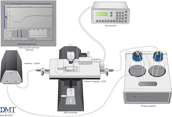
Figure 1. Schematic of the mechanical test system with optional accessories. Used with permission from Danish Myotechnology.
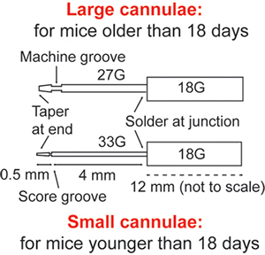
Figure 2. Dimensions of the custom-made cannulae for different aged specimens. All parts are made from 316 stainless steel hypodermic tubing (Small Parts). 7-0 suture should be used to secure the arteries to the large cannulae and 10-0 suture should be used for the small cannulae.
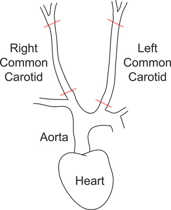
Figure 3. Diagram of the carotid artery dissection. The heart, aorta, right and left common carotid arteries and cut locations for the arteries are shown.
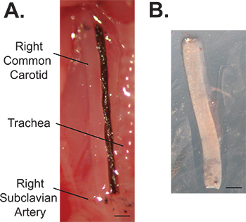
Figure 4. Images of a 3-day-old right common carotid artery in vivo (A) and ex vivo (B). The reference length is determined with a suture and the cut locations are marked with carbon particles. Note the decrease in length upon dissection. Scale bar = 0.25 mm.
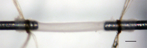
Figure 5. 3-day-old mouse carotid artery mounted on the cannulae in the myograph bath at its unstretched, ex vivo length. Scale bar = 0.2 mm.
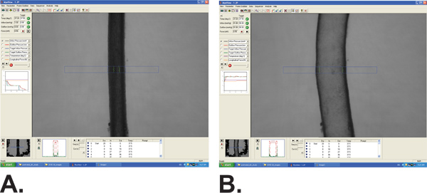
Figure 6. Screen shot of the MyoView software showing outer diameter tracking of a 3-day-old carotid artery stretched to 1.2 times the in vivo length and pressurized to 0 mmHg (A) and 90 mmHg (B).
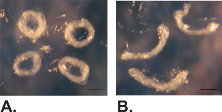
Figure 7. Example closed (A) and open (B) rings of a 3-day-old carotid artery cut to measure unloaded dimensions and opening angle. Scale bar = 0.1 mm.
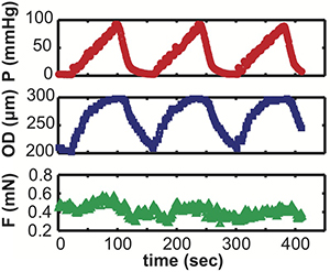
Figure 8. Example raw pressure, diameter and force data versus time for a single inflation protocol for a 3-day-old carotid artery.
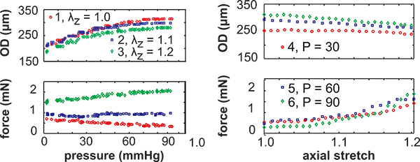
Figure 9.Example isolated loading cycles showing the recorded pressure, diameter, force and calculated axial stretch for all six mechanical test protocols for a 3-day-old carotid artery. The left panels show the inflation protocols at constant axial stretch (λz) with respect to the in vivo length and the right panels show the axial stretch protocols at constant pressure (P, mmHg). The manual axial stretch protocols are performed at a faster rate than the automated inflation protocols, so less data points are recorded.
