Kv channel dysfunction is associated with cardiac arrhythmias. In order to study the molecular mechanisms that lead to such arrhythmias we utilize a systematic protocol for isolation of atrial and ventricular cardiomyocytes from Kv channel ancillary subunit knockout mice. Isolated cardiomyocytes can then immediately be used for cellular electrophysiological studies, biochemical or immunofluorescence (IF) assays.
KCNE genes encode for a small family of Kv channel ancillary subunits that form heteromeric complexes with Kv channel alpha subunits to modify their functional properties. Mutations in KCNE genes have been found in patients with cardiac arrhythmias such as the long QT syndrome and/or atrial fibrillation. However, the precise molecular pathophysiology that leads to these diseases remains elusive. In previous studies the electrophysiological properties of the disease causing mutations in these genes have mostly been studied in heterologous expression systems and we cannot be sure if the reported effects can directly be translated into native cardiomyocytes. In our laboratory we therefore use a different approach. We directly study the effects of KCNE gene deletion in isolated cardiomyocytes from knockout mice by cellular electrophysiology – a unique technique that we describe in this issue of the Journal of Visualized Experiments. The hearts from genetically engineered KCNE mice are rapidly excised and mounted onto a Langendorff apparatus by aortic cannulation. Free Ca2+ in the myocardium is bound by EGTA, and dissociation of cardiac myocytes is then achieved by retrograde perfusion of the coronary arteries with a specialized low Ca2+ buffer containing collagenase. Atria, free right ventricular wall and the left ventricle can then be separated by microsurgical techniques. Calcium is then slowly added back to isolated cardiomyocytes in a multiple step comprising washing procedure. Atrial and ventricular cardiomyocytes of healthy appearance with no spontaneous contractions are then immediately subjected to electrophysiological analyses by patch clamp technique or other biochemical analyses within the first 6 hours following isolation.
1. Animal Anesthesia and Organ Harvesting
2. Preparation of Heart and Langendorff Perfusion
3. Microsurgical Dissociation of Cardiac Chambers
4. Further Dissociation of Cardiomyocytes
5. Preparation of Cardiomyocytes for Cellular Electrophysiology, Biochemical or IF Studies
6. Cellular Electrophysiology
Isolation of adult murine cardiomyocytes from genetically engineered mice to study the function of specific genes of interest in vitro has become a powerful tool to further understand cardiac pathophysiology. This method is currently used by only a small but increasing number of basic science laboratories worldwide. However, isolation of adult ventricular murine cardiomyocytes can be tricky and needs to be done thoroughly and repetitively by experienced hands. Figure 1 shows freshly isolated exemplar atrial and ventricular cardiomyocytes. For ventricular cardiomyocytes we recommend to only use rod- shaped, striated ventricular myocytes of healthy appearance with no spontaneous contractions. Compared with ventricular myocytes, atrial myocytes are shorter and thinner. The characteristic striation and rod-shape of ventricular cardiomyocytes is missing in adult atrial murine cardiomyocytes. The yield for ventricular cardiomyocytes isolated from an intact mouse heart is 5×10⁵ – 1×10⁶. For atrial cardiomyocytes it is significantly less. We generally expect to isolate about 5 – 25,000 atrial cells from one mouse heart. Once isolated, cardiomyocytes can be subjected to different in vitro techniques including electrophysiological recordings (Figure 1). We recommend to use isolated cardiomyocytes within the first 6 hr after isolation. Figure 1 shows exemplar Kv channel outward recordings (on the right) of atrial and ventricular cardiomyocytes evoked by different depolarization steps by whole-cell patch clamp technique. The characteristic shape of murine adult ventricular and atrial Kv channel repolarizing currents can be seen over a defined time course (here 4 sec, Figure 1). Please note that the current amplitude is significantly lower in murine adult atrial cardiomyocytes compared to ventricular cardiomycytes.
Solution | Contents (in mM, if not specified differently) |
1 | 117 NaCl, 4 KCl, 10 HEPES, 1 KH2PO4, 4 NaHCO3, 1.7 MgCl2, 10 Glucose |
2 | 117 NaCl, 4 KCl, 10 HEPES, 1 KH2PO4, 4 NaHCO3, 1.7 MgCl2, 10 Glucose + 229.5 μM EGTA |
3 | 117 NaCl, 4 KCl, 10 HEPES, 1 KH2PO4, 4 NaHCO3, 1.7 MgCl2, 10 Glucose, CaCl2 0.1 + 0.8 g/L Collagenase Type 2 (300 U/L)* |
4 | 117 NaCl, 4 KCl, 10 HEPES, 1 KH2PO4, 4 NaHCO3, 1.7 MgCl2, 10 Glucose, CaCl2 0.2 + 0.8 g/L Collagenase Type 2 (300 U/l)* + BSA (1 g/L) |
5 | 117 NaCl, 4 KCl, 10 HEPES, 1 KH2PO4, 4 NaHCO3, 1.7 MgCl2, 10 Glucose, CaCl2 0.5 + BSA (1 g/L) |
6 | 117 NaCl, 4 KCl, 10 HEPES, 1 KH2PO4, 4 NaHCO3, 1.7 MgCl2, 10 Glucose, CaCl2 1.0 + BSA (1 g/L) |
Table 1. Isolation solutions. Equilibrated for 10 min with Carbogen (95% O2, 5%CO2), at 37 °C pH=7.4 (NaOH). * Activity may depend on batch number, so previous testing of collagenase activity is recommended.
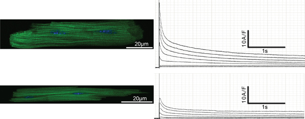
Figure 1. Upper Left: Exemplary ventricular cardiomyocyte stained with DAPI (nuclei, blue) and Alexa Flour 488 Phalloidin (α-actinin, green). Upper Right: Exemplary traces of whole cell patch clamp recording. Lower Left: Exemplary atrial cardiomyocyte stained with DAPI (nuclei, blue) and fluorochrome conjugated Phalloidin (α-actinin, green). Lower right: Exemplary traces of whole cell patch clamp recording.
With the growing development of genetically engineered mouse strains to study cardiac function and cardiac pathology related to gene deletion there is also an increasing interest in specialized methods to study effects of the specific gene deletion in vitro. In our laboratory we study the roles of a family of Kv channel ancillary subunits on cardiac repolarization. The KCNE genes comprise a family of 5 genes (KCNE1-5) that play important roles in human ventricular and atrial repolarization 11, 15. KCNEs are single transmembrane domain Kv channel ancillary subunits that cannot pass any potassium currents on their own, but they can form complexes with Kv channel alpha subunits and modulate their functional properties such as gating, conductance, pharmacology and trafficking within the cell significantly. Mutations and common polymorphisms in KCNE2 for example are associated with inherited and acquired forms of the Long QT Syndrome (LQTS) 1, 16.
Pioneer work in isolation and electrophysiological characterization of different Kv channel alpha subunits and their contribution to rat but also murine ventricular and atrial repolarization has been done by the group of Dr. Nerbonne at the Washington University of St. Louis in the 1990s 3, 5, 7. However, significantly less is known about the contribution of Kv channel ancillary subunits to atrial and ventricular repolarization within the rodent myocardium. KCNEs have mainly been studied in heterologous expression systems such as CHO cells and Xenopus laevis oocytes. Using these methods KCNE subunits have been shown to be highly promiscuous in forming heteromeric Kv channel complexes identifying a range of different Kv channel alpha subunits as potential partners for KCNEs10. However, we cannot be sure whether these specific heteromeric Kv channel complexes also occur in native cardiomyocytes or if the observed heteromeric KCNE Kv channel alpha subunit partnerships are heterologous expression artifacts 1. We therefore adopted the isolation technique and Kv channel recording protocols from the Nerbonne laboratory and modified them as indicated in the protocol section of this article to dissect the different Kv channels in murine repolarization, which are controlled by KCNE genes. Using this approach we recently found that the Kv channel ancillary subunit KCNE2 controls two distinct Kv currents in the murine ventricular myocardium (IKslow,1 and Ito,f). By application of specific inhibitors for different potassium channels we were able to show that deletion of the murine KCNE2 gene causes a significant reduction in both Kv1.5 and Kv4.2 currents 14. Regulation of Kv1.5 by KCNE2 was not previously known, whereas regulation of Kv4.2 by KCNE2 has already been demonstrated in vitro by heterologous expression studies 20.
Atrial fibrillation (AF) is the most frequent sustained arrhythmia in clinical practice and is associated with significant morbidity and mortality 4. Mutations in all different KCNEs have recently been associated with AF in humans. Recently, two non-synonymous mutations were found in KCNE1 in patients with AF that were not present in the control group (Olesen et al., 2012). Mechanistically, heterologous expression studies identified a gain-of-function effect in KCNQ1 currents as the likely underlying mechanism. Furthermore, recently it was shown that KCNE1-/- mice suffer from spontaneous episodes of paroxysmal AF 17. In a study evaluating 28 unrelated Chinese families with lone AF, Yang et al. identified a mutation in KCNE2, which resulted in an arginine-to-cysteine substitution (R27C). Functional analysis of the mutant channel in heterologous expression studies revealed a gain-of-function effect on KCNQ1 channels (IKs) 18. However, KCNE2 can also form complexes with a range of other Kv channel α-subunits including Kv1.5, which has also been implicated in AF. We have recently shown that KCNE2 can modulate Kv1.5 currents in the murine ventricle leading to a prolongation of the ventricular APD in mice 15. Therefore, disruption of atrial Kv1.5 currents by KCNE2 dysfunction could also represent the underlying arrhythmogenic mechanism for AF in our KCNE2-/- mouse model and/or in patients harboring mutation in KCNE2 suffering from AF. Mutations in KCNE3 were also recently identified in a patient with familial AF 8. Electrophysiological recordings revealed an increased activity of Kv4.3/KCNE3 and Kv11.1/KCNE3 generated currents by the mutation, thereby conferring susceptibility of mutation carriers to faster cardiac action potential repolarization and thus vulnerability to re-entrant wavelets in the atria. The KCNE3 V17M missense mutation however had no effect KCNE3/KCNQ1 currents despite the fact that KCNE3 forms functional complexes with KCNQ1 in the myocardium. Furthermore, KCNE3 was recently shown to be up regulated in a human population with valvular AF supporting the hypothesis that KCNE3 plays a role not only in familial AF but also valvular AF 6. Recently, a single nucleotide polymorphism (G/T) was identified in KCNE4 resulting in a glutamic acid (Glu, E)/aspartic acid (Asp, D) substitution at position 145 of the KCNE4 peptide 19. Subsequent functional analysis of this polymorphism revealed that the KCNE4 polymorphism exerts the effect of “gain of function” on the KCNQ1 channels 9. Again, these studies were undertaken in heterologous expression systems and experimental evidence from native cardiomyocytes is still lacking. More recently, an isolated non-familial case of AF with a missense (L65F) mutation has been identified in a Danish cohort of 158 patients with AF 12. Furthermore, a polymorphism in KCNE5 (C97T) was identified within the same study population, which was associated with a higher risk for developing AF 13. Interaction of both mutant β-subunits (KCNE2 and KCNE5) with the KCNQ1 channel produced a gain-of-function effect with increased IKs currents. The KCNQ1 α-subunit of the IKs channel can associate with any one of the five accessory β-subunits (KCNE1-5). Therefore KCNQ1 seems to be a likely candidate α-subunit partner when searching for a molecular substrate in the pathogenesis of KCNE-associated AF. However, given the pronounced promiscuity of KCNE subunits other molecular targets are possible and we therefore intend to specifically investigate these possible KCNE/Kv alpha-subunit interactions. Studies in our laboratory are therefore also currently aimed to elucidate the mechanisms behind KCNE-associated AF utilizing the technique we describe in this article – isolation of murine atrial cardiomyocytes and subsequent in vitro electrophysiological recordings with application of specific inhibitors to different Kv channel alpha subunits and biochemical analyses of native cardiomyocytes from KCNEx knockout mice.
The authors have nothing to disclose.
This work was funded by grants from Deutsche Forschungsgemeinschaft (DFG), Fritz-Thyssen-Stiftung and Charité/MDC.
Name of the Reagent | Company |
Tetrodotoxin | Alomone |
4-aminopyridine | ICN Biomedicals |
Heteropodatoxin 2 | Alomone |
Tetraethylammonium | Sigma Chemicals |
Collagenase Type 2 | Worthington |