The STA-PUT method allows for the separation of different populations of spermatogenic cells based on size and density.
Mammalian spermatogenesis is a complex differentiation process that occurs in several stages in the seminiferous tubules of the testes. Currently, there is no reliable cell culture system allowing for spermatogenic differentiation in vitro, and most biological studies of spermatogenic cells require tissue harvest from animal models like the mouse and rat. Because the testis contains numerous cell types – both non-spermatogenic (Leydig, Sertoli, myeloid, and epithelial cells) and spermatogenic (spermatogonia, spermatocytes, round spermatids, condensing spermatids and spermatozoa) – studies of the biological mechanisms involved in spermatogenesis require the isolation and enrichment of these different cell types. The STA-PUT method allows for the separation of a heterogeneous population of cells – in this case, from the testes – through a linear BSA gradient. Individual cell types sediment with different sedimentation velocity according to cell size, and fractions enriched for different cell types can be collected and utilized in further analyses. While the STA-PUT method does not result in highly pure fractions of cell types, e.g. as can be obtained with certain cell sorting methods, it does provide a much higher yield of total cells in each fraction
(~1 x 10⁸ cells/spermatogenic cell type from a starting population of 7-8 x 10⁸ cells). This high yield method requires only specialized glassware and can be performed in any cold room or large refrigerator, making it an ideal method for labs that have limited access to specialized equipment like a fluorescence activated cell sorter (FACS) or elutriator.
Mammalian spermatogenesis is a complex differentiation process that occurs in several stages in the seminiferous tubules of the testes1. Briefly, stem-like spermatogonia that reside near the epithelium of the seminiferous tubule divide and differentiate into spermatocytes, which then undergo meiotic divisions. After meiosis is complete, the resulting haploid cells, or round spermatids, undergo spermiogenesis, a differentiation process that involves the shedding of cytoplasm and compaction of the nucleus. Spermatids gradually develop a flagellum and undergo elongation and condensation of the nucleus, producing elongating and then condensing spermatids, respectively. The end products are spermatozoa, which are released into the lumen of the seminiferous tubule and ultimately into the epididymis where they mature further.
Because the process of spermatogenesis relies on special hormonal and molecular conditions in the testes, a reliable in vitro culture system for the entire process of spermatogenesis has not yet been developed2,3. Culture methods have been developed for creating “primordial germ cell-like cells” and haploid, “round spermatid-like cells” from stem cells, but these methods are not yet able to generate large numbers of these cells and fail to produce later spermatogenic cell types4,5. Fortunately, the spermatogenic cell types differ significantly in size, which allows for a single-cell suspension obtained from whole testes to be separated with a liquid gradient. The STA-PUT method, demonstrated here, uses a linear BSA gradient and simple sedimentation to separate spermatogenic cells based on size and mass6-9.
The STA-PUT method has several advantages over the other two most widely used methods to separate spermatogenic cell types: FACS and elutriation10-13. The STA-PUT apparatus requires only several pieces of specialized glassware assembled in a cold room or large refrigerator. Thus, it is less expensive than using a cell sorter or an elutriator. The STA-PUT method yields higher amounts of cells per cell type and testis than can be sorted by FACS in a comparable time frame, although the purity of each cell population is not as high as those obtained with FACS11. Cell sorting utilizing magnetic beads (magnetic activated cell sorting, MACS) has recently been successfully employed for enrichment of spermatogonia from a mixed testicular cell population, but it is currently unsuitable for separating spermatocytes or spermatids due to lack of knowledge of appropriate surface markers14. An additional advantage of the STA-PUT method over FACS or MACS is the ability to isolate viable cells suitable for subsequent culture because, in contrast to most FACS protocols, it does not require any DNA or other types of staining. For studies that require large yields of spermatogenic cells types at ~90% purity, the STA-PUT is an ideal method.
The STA-PUT protocol involves three stages: 1) Set up of the apparatus and reagents, 2) Preparation of cell suspension from whole testes, and 3) Cell loading, sedimentation, and fraction collection. When performed by a team of two researchers, the protocol takes eight hours on average.
1. Setting up the STA-PUT Apparatus (Figure 1)
***STA-PUT apparatus should be placed in a 4°C large refrigerator or a cold room that can also accommodate a fraction collector, if that method of collection is preferred.
2. Isolating Spermatogenic Cells from Whole Testes
3. Cell Loading and Sedimentation
4. Fractionation and Analysis of Fractions
***As you perform the following steps, be careful not to disturb the gradient. If the BSA gradient is disturbed, the procedure will not work!
The ideal result from the STA-PUT procedure is a fairly noticeable separation of cells from the testes based on cell size and density. While cells isolated from the testes are sedimenting through the BSA gradient, several distinct bands of cells can be observed. Any clumps of cells tend to sink to the bottom of the gradient and will not contaminate the other fractions. A little further up the gradient will be the large somatic and meiotic cells. Farther up the gradient still will be smaller round spermatids. At the top of the gradient will be condensed spermatids, sperm, and contaminating red blood cells (these appear to be small round cells without a nucleus).
Fractions can be analyzed quickly using a combination of light and fluorescent microscopy (Figure 2)15. Meiotic, spermatogonial, and somatic diploid cells are the largest cells found in the testes and will contain large nuclei that stain relatively homogeneously with DAPI. Round spermatids are smaller cells with smaller round nuclei, generally with a brightly staining chromocenter. Condensing/elongating spermatids are small cells that often look oblong, as if a small tail is forming. These cells have smaller, compact nuclei that stain brightly with DAPI and are shaped like a sickle. Once cell fractions are combined, purity can be further determined by western blot analysis of the cell lysates (Figure 3). Common markers of meiotic cells are the synaptonemal complex 1 proteins Scp1 and Sycp216. Common markers of condensing spermatids are transition proteins (e.g. TP1) or protamines17.
Although each STA-PUT run can be different, usually meiotic cells, spermatogonia and somatic diploid cells will be found in fractions ca. 25-40, round spermatids in fractions ca. 55-65, and condensing/elongating spermatids in fractions ca. 65-75 Due to a smaller difference in size between somatic/meiotic cells and round spermatids, fractions ca. 45-50 often have an even percentage of somatic/meiotic cells and round spermatids. Staining with DAPI will help to distinguish these two populations of cells. There is less overlap of the round spermatid and condensing/elongating spermatid fractions due to the larger difference in size between these two populations of cells. Usually, fractions below 15 will contain many large clumps of cells and fractions above 85 will contain few cells and many residual bodies, or membrane-bound cytoplasm that is shed by spermatids.
Generally, when cells from ~22 testes are fractionated with the STA-PUT procedure, it yields ca. 108 cells/spermatogenic cell type (meiotic/somatic diploid cells, round spermatids, and condensing/elongating spermatids). Fractions that are combined to create the final population of cells should be at least 80% pure for the type of cell in question. If you do not see this degree of purity or higher, there may be a problem with cell separation or the BSA gradient. Also, if there are few cells in the first 20 fractions and an abundance of cells in fractions 80+, or if there is an abundance of cells in the first 20 fractions and hardly any in fractions 70+, the sedimentation time needs to be further optimized. Please see the Discussion for suggestions on how to trouble shoot.
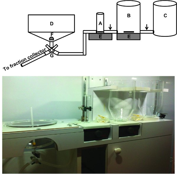
Figure 1. Setting up the STA-PUT Apparatus: A schematic and actual image of the STA-PUT apparatus are shown. All glassware is connected by plastic tubing, including the tube that connects the apparatus to the fraction collector. Arrows indicate location of clamps. A) Cell loading chamber, contains a stir bar; B) 2 L cylinder for 2% BSA, contains a stir bar; C) 2 L cylinder for 4% BSA; D) Sedimentation chamber; E) Stir plates; F) Baffle; G) Stopcock. Click here to view larger image.
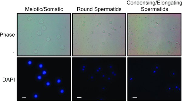
Figure 2. Cell populations obtained from the STA-PUT: Fractions were combined into three separate populations of cells: meiotic and somatic cells, round spermatids, and condensing and elongating spermatids. Each population is stained with DAPI to show differences in nuclear size and morphology. Phase contrast imaging conveys differences in cell size and shape. White bar represents 10 μm. Click here to view larger image.
Figure 3. Markers of different cell populations obtained from the STA-PUT: Whole cell extracts were made from each cell population shown in Figure 1. and western blot analysis was performed to show protein expression differences for each population. Synaptonemal complex protein 1 (Scp1) is a protein expressed exclusively during meiosis and is found enriched in the meiotic fractions, while the condensing spermatid fraction is enriched for transition protein 1 (TP1), a protein expressed late in spermiogenesis. Click here to view larger image.
Those who study spermatogenesis rely on animal models for spermatogenic cell samples, as a reliable cell culture system does not yet exist for generating all spermatogenic cell types3. Although spermatogenic cells are readily collected from whole testes, only a mixed population results. This poses a problem for those who wish to study specific subtypes of these cells, such as meiotic cells, round spermatids, and condensing spermatids. Three different methods are currently used to separate these subtypes of spermatogenic cells: STA-PUT, FACS, and elutriation6-13. The latter two methods require access to expensive pieces of equipment: a cell sorter and an elutriator, respectively. Although the FACS method yields highly pure populations of the different spermatogenic cell types, the process takes six hours and yields only 0.5-2.0 x 10⁶ cells per cell type per two to three testes11. Elutriation, like the STA-PUT method, separates cells based on size and density, but requires access to an elutriator, which is more expensive than the STA-PUT apparatus.
The STA-PUT procedure uses a simple BSA gradient to separate spermatogenic cells of different sizes with a fairly high yield (~108 cells/population per ~22 testes). Relative to FACS methods, the STA-PUT procedure yields more cells/testis and takes much less time to separate cell types. Compared to FACS and elutriation, the STA-PUT procedure is relatively simple and inexpensive. The STA-PUT requires only a cold room or large refrigerator and a set of specialized glassware, making it an ideal method for labs without access to a cell sorter or elutriator. When performed properly, the STA-PUT can provide an approximately 90% pure population of round spermatids or condensing spermatids.
The STA-PUT method is very useful, but requires optimization at several different steps, especially sedimentation. Sub-optimal separation of cell types can be caused by several different issues, most relating to the BSA gradient. To make a proper gradient, turn on the stir bars in the cell loading chamber and the 2 L cylinder holding the 2% BSA while you are creating the gradient. Also clear all tubing of bubbles and put the baffle in place in the sedimentation chamber before loading the cells. Reducing the flow rate of the BSA into or out of the sedimentation chamber may help. Most importantly, the STA-PUT apparatus should not be disturbed during the creation of the gradient, sedimentation, or fraction collection.
Sub-optimal cell yields can be the result of insufficient number/size of testes, inadequate cell separation during collagenase/trypsin treatment, and cell clumping. If one is unable to obtain the appropriate number of cells for this STA-PUT protocol due to the use of neonatal mice or genotypes that produce small numbers of spermatogenic cells, it may be necessary to use more animals per STA-PUT or to order a STA-PUT glassware kit optimized for smaller volumes and cell numbers (available from ProScience)18. To obtain an optimal number of cells (700-800 million), use at least 11 male mice of reproductive age, ideally at least 8-9 weeks old. However, no more than 800 million cells should be loaded into one STA-PUT. If cells have not dissociated into a single cell suspension after trypsin treatment, the DNAse concentration can be increased up to 1 μg/5 ml solution. DNAse is sensitive to repeat cycles of freeze/thaw, and using fresh DNAse each time will result in a more effective dispersion of tubules into a single cell suspension. One can use a wide bore pipette to help break apart cell clumps before filtering through mesh.
One aspect of the STA-PUT method that will require optimization is the amount of time the cells are allowed to sediment through the BSA gradient. One hour and 45 min usually works well, but this time may differ from lab to lab. Usually, different layers of cells can be seen visually throughout the BSA gradient. If there are few cells in the first 20 fractions collected and an abundance of cells in fractions 80+, the sedimentation time may need to be extended. If there are too many cells in the first 20 fractions collected and there is insufficient separation of cell types, the sedimentation time may need to be lowered.
The cells acquired with the STA-PUT procedure can be used for many different types of experiments. The STA-PUT provides ample material for western blot analysis, immunofluorescence, and RNA analysis, although large-scale biochemistry experiments may require combining material from several different STA-PUT runs. When separated from other cell types, haploid spermatids can be cultured and subjected to in vitro molecular manipulation for up to three days, which can be easier than in vivo treatment or creating a knockout animal19. Cells obtained with the described STA-PUT protocol have been cultured for one day without obvious signs of contamination, but if cells are to be used for longer cell culture experiments, equipment should be sterilized with ethanol, all solutions should be filter sterilized, and all steps before cell loading and after fraction collection should be performed in a tissue culture hood. In addition, culture media containing antibiotics should be used. The fact that the STA-PUT method does not require cell fixation makes it an ideal procedure for experiments that require viable cells.
The authors have nothing to disclose.
This research was supported by NIH grants GM055360 to SLB and U54HD068157 to RGM. JMB was supported by the T32 Genetics Training Grant at the University of Pennsylvania (GM008216).
BSA | Affymetrix/USB | 10857 100MG | Alternate can be used. |
0.5%, 2%, and 4% BSA solutions | Dissolve 0.25 g, 11 g, and 22 g (respectively) in total volumes of 50 ml, 550 ml, and 550 ml of 1x KREBS (respectively). | To be made day of the STA-PUT procedure. Filter sterilize. | |
KREBS (10x) | 3.26 g KH2PO4 + 139.5 g NaCl +5.89 g MgSO4/7H20 + 40 g Dextrose + 3.78 g CaCl2/2H20 + 7.12 g KCl, bring to 2 L with ddH20 | Autoclave/filter and store at 4 °C for several months. | |
KREBS (1x) | Dissolve 4.24 g NaHCO3 in 100 ml ddH20. Add 200 ml 10x KREBS. Bring to 2 L with ddH20. | To be made day of the STA-PUT procedure. Filter sterilize. | |
Collagenase | Sigma | C9891-1G | |
DNAse | Sigma | DNEP-5MG | |
Trypsin | Sigma | T9201-1G | |
DAPI | Preference of researcher | ||
Triton-X100 | Preference of researcher | ||
Formaldehyde (~37%) | Preference of researcher | ||
TABLE 2: EQUIPMENT | |||
Equipment | Company | Catalog # | Comments |
Complete STA-PUT apparatus | ProScience Glass Shop | STA-PUT (this procedure uses the standard sedimentation chamber: Cat. No. 56700-500) | Includes all glassware and equipment for the apparatus. Alternate glassware is available for performing STA-PUT with smaller cell numbers. |
10 μm mesh filter | Fisherbrand | 22363549 | |
Mouse dissection tools | Preference of researcher | ||
14 ml round bottom fraction collection tubes with caps | Preference of researcher | ||
Need approximately 100 tubes | |||
Fraction collector | GE Healthcare Life Sciences | Model: Frac-920, Product code: 18-1177-40 | Alternate can be used. |
Microscope slides, cover glass | Preference of researcher | ||
Light/Fluorescent Microscope | Preference of researcher |