Whole-organ decellularization produces natural biological scaffolds that may be used for regenerative medicine. The description of a nonhuman primate model of lung regeneration in which whole lungs are decellularized and then seeded with adult stem cells and endothelial cells in a bioreactor that facilitates vascular circulation and liquid media ventilation is presented.
There are an insufficient number of lungs available to meet current and future organ transplantation needs. Bioartificial tissue regeneration is an attractive alternative to classic organ transplantation. This technology utilizes an organ's natural biological extracellular matrix (ECM) as a scaffold onto which autologous or stem/progenitor cells may be seeded and cultured in such a way that facilitates regeneration of the original tissue. The natural ECM is isolated by a process called decellularization. Decellularization is accomplished by treating tissues with a series of detergents, salts, and enzymes to achieve effective removal of cellular material while leaving the ECM intact. Studies conducted utilizing decellularization and subsequent recellularization of rodent lungs demonstrated marginal success in generating pulmonary-like tissue which is capable of gas exchange in vivo. While offering essential proof-of-concept, rodent models are not directly translatable to human use. Nonhuman primates (NHP) offer a more suitable model in which to investigate the use of bioartificial organ production for eventual clinical use.
The protocols for achieving complete decellularization of lungs acquired from the NHP rhesus macaque are presented. The resulting acellular lungs can be seeded with a variety of cells including mesenchymal stem cells and endothelial cells. The manuscript also describes the development of a bioreactor system in which cell-seeded macaque lungs can be cultured under conditions of mechanical stretch and strain provided by negative pressure ventilation as well as pulsatile perfusion through the vasculature; these forces are known to direct differentiation along pulmonary and endothelial lineages, respectively. Representative results of decellularization and cell seeding are provided.
Bioengineering of tissues and organs is an attractive addition to the field of regenerative medicine. The creation of "lab-grown" organs that are suitable for transplant into patients to replace functionality of diseased organs is highly desirable in order to meet the current and future demand for transplantation needs. The principles of tissue engineering center around the seeding of desired cell types, or progenitors thereof, into a scaffold that supports the shape of the engineered tissue while supplying the appropriate growth factors and culture conditions necessary to mimic developmental or regenerative processes. While synthetic scaffolds have been used for tissue engineering, and the natural extracellular matrix (ECM) may be the best source of organ-specific scaffolds for this purpose. Whole-organ decellularization is a process which allows the removal of cells while leaving the chemical and structural aspects of the native ECM intact. The resulting acellular matrix scaffold can be used as a platform onto which regenerative cells can be seeded and cultured in vitro1,2.
Several rodent models of lung decellularization and subsequent recellularization have been developed to study the feasibility of this technology3-6. While offering essential proof-of-concept, rodent models are not directly translatable to human clinical needs. A recent study pointed out that genomic responses to traumatic injury (and related inflammation) do not correlate well between mice and humans; these findings raise questions of the validity of using mice as models for such complex biochemical processes in humans7. Nonhuman primate (NHP) models offer the advantage of closely resembling the biology of humans at the genomic, anatomic, and physiologic levels and allow more flexible manipulation for greater extrapolation to human use. The rhesus macaque has been used in a variety of preclinical applications and is an excellent model in which to study tissue engineering8-11. We recently described the successful decellularization of rhesus macaque (Macacca mulatta) lungs utilizing a procedure that has minimum impact on the lung ECM12. Lung decellularization is accomplished by treating the tissue consecutively with four decellularization solutions composed of detergents, salts, and enzymes with intermittent washing with deionized water (dH2O) and PBS. We have optimized this procedure by modifying a protocol originally described by Price et al.4 A variety of histological and protein analytical techniques were used to characterize the components of resulting acellular matrices relative to native macaque lungs.
In this report, we demonstrate a detailed protocol for the decellularization of nonhuman primate lungs and the recellularization of the resulting acellular lung scaffolds in a large-organ bioreactor originally demonstrated by Calle et al.13 in JoVE. By modifying their original protocol to accommodate the size, ventilation, and perfusion requirements for large-animal lungs, the technology was successfully moved from the rodent model to the rhesus macaque model. All studies presented in this report were performed in accordance with the Institutional Biosafety Committee (IBC) policies in place at the Tulane National Primate Research Center. Demonstration of this technique is essential because identification of anatomical structures and physical manipulation of larger organs is sometimes difficult without visualizing the steps of the protocol. The studies made possible by these methods provide a basis for essential preclinical studies in decellularized rhesus macaque lungs in which recellularization with rhesus mesenchymal lineage stem cells and rhesus microvascular endothelial cells can be assessed. Our version of this bioreactor simulates the developmental environment and applies forces of mechanical stretch and strain in large-animal lungs and allows the investigation of lung recellularization under conditions known to facilitate pulmonary and endothelial development13-15.
1. Whole-organ Macaque Lung Decellularization
2. Large-organ Bioreactor
3. Troubleshooting and Alternative Approaches
Results shown below represent separate experiments in which either airway or vascular compartments were seeded with rhesus macaque bone marrow-derived mesenchymal stem cells or rhesus lung-derived microvascular endothelial cells, respectively, that were isolated and characterized as previously described16-18.
Throughout the decellularization process, macaque lungs displayed a progressive whitening culminating in a translucent appearance at the end of the process; however, the lungs maintained their gross anatomical features and remain largely elastic and able to produce natural recoil after inflation with liquid (Figures 3A and 3B). At the microscopic level, the histologic ultrastructure remained intact after decellularization; that is, bronchioles, respiratory bronchioles, alveolar sacs, blood vessels, and capillaries were still distinguishable quite clearly by low-power microscopy (Figures 3C and 3D). Histological microanatomy, however, demonstrated that while the gross anatomy and ultrastructure of the lung were mildly disturbed by decellularization, the tissue completely lacked intact cells (Figures 3E and 3F).
Cellular DNA that was released as cells were lysed by the detergents was degraded by DNase treatment and efficiently removed by subsequent washing steps. DNA was extracted from native and decellularized macaque lung tissues using Qiagen's DNeasy kit. As seen in Figure 4A, only trace amounts of DNA remained in decellularized tissues; moreover, the trace DNA (concentrated by alcohol precipitation and visualized in a 0.8% agarose gel) was composed of mostly low molecular weight degraded fragments (Figure 4A, inset). The efficiency of cellular protein removal was assessed by Western blot analyses of both native and decellularized lung protein lysates using an antibody to β-actin. Briefly, equal protein amounts (25 µg) in triplicate for each native and decellularized sample were subjected to PAGE at 200 V for 1 hr. Proteins were transferred to nitrocellulose using an Invitrogen iBlotter. Blots were probed with a mouse anti-human β-actin primary antibody (IgG1) followed by a goat anti-mouse IgG-HRP secondary antibody. Equal protein loading was confirmed by staining an identical membrane with Ponceau-S and performing densitometry to determine total protein stain intensity between native and decellularized samples (not shown). The blot was developed using ECL chemiluminescent detection reagent (Invitrogen) and imaged using the ImageQuant LAS 4000 system (GE). β-actin was easily detected in native lung lysates but not in decellularized lung lysates, suggesting that decellularization dramatically depleted cells and removed cell-associated protein material (Figures 4B).
An example of the assembled bioreactor with organs installed is shown in Figure 5. A cell culture water pan easily held all three chambers and provided containment in the event that a leak occurred. The latex tubing used as sealing gaskets for the lids of the bioreactor chambers also functioned to prevent kinks in the tubing by providing added rigidity in areas where the tubing would otherwise fold on itself. The bioreactor, 6-port syringe pump, and peristaltic pump fit easily into a standard cell culture incubator. The electronic components were powered via a flat-cord electrical extension that is sandwiched between the inner incubator door and the rubber seal. The incubator was maintained at 37°C, 5% CO2 throughout experiments.
Fourteen days after airway seeding with macaque BMSC (1.0 x 10⁶ cells/ml) and bioreactor culture with ~1 inspiration/expiration cycle every 2 min, the parenchyma of decellularized macaque lungs was effectively recellularized (Figure 6A). BMSCs lined the alveolar septae while maintaining a clear and open alveolar lumen. Inoculation of cells into the trachea (or bronchioles for single lobes) was an efficient route for inoculating distal lung tissues as is evident in by the recellularization of alveoli near the pleura at the extremities of the lung. The denuded matrix of large airways was also recellularized by BMSC using the bioreactor. Figure 6B shows the luminal surface of a mainstem bronchus that was lined with a monolayer of squamous-like BMSC after 14 days of culture in the bioreactor.
Histological analysis of a decellularized rhesus lung lobe five days after seeding the vasculature with microvascular endothelial cells (8.0 x 10⁴ cells/ml) and providing constant vascular perfusion with endothelial culture medium at 5 ml/min, revealed cells lining the small vasculature in the lung parenchyma (Figures 6C and 6D). In some instances, cells appeared to be attached to the matrix via several cellular projections across the lumen (Figure 6C) while other cross sections of vessels showed cells lining the endothelial surface with clear lumen (Figure 6D).
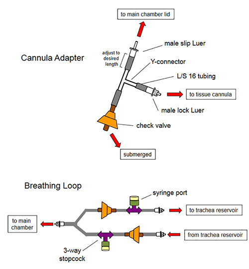
Figure 1. Assembly schematic for tracheal and vascular cannulae adapters and breathing loop. Cannulae adapters allow removal of air from the cannula tubing after the tissues are installed in the bioreactor. A one-way check valve allows medium to be aspirated from the main chamber as to not produce undue force on the tissue airway or vasculature. Liquid traveling toward the tissue will be diverted into the airway or vasculature because reverse pressure on the check valve will cause it to close. This same principle produces the main function of the breathing loop; here, air is removed by aspirating liquid through the one-way valves using a needle and syringe via the in-line injection ports.
Click here to view larger image.
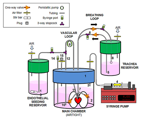
Figure 2. Assembly schematic for the large-organ bioreactor. The following components constitute the main functionality of the lung bioreactor: (1) Modified 2 L jar lid; (2) main chamber containing culture media (3) air intake/outlet for negative pressure ventilation; (4) syringe pump cycles air in and out of main chamber; (5) culture media moves from trachea reservoir into the lung airway when syringe withdraws air from main chamber; (6) one-way valves allow constant cycling of culture media into and out of lung via a breathing loop — airway cells are directly injected into the breathing loop to inflate the lung statically for 18 hr before mechanical cyclic ventilation; (7) media enters lung via the trachea and lung inflates; (8) when syringe pump returns air to the main chamber, lungs deflate and return culture media to the trachea reservoir; (9) a peristaltic pump moves culture media from the main chamber (10) into the pulmonary artery (11), through the lung vasculature, and back into the main chamber; (12) to seed the vasculature with endothelial cells, the endothelial seeding reservoir is temporarily connected to the vascular loop via a 3-way stopcock, the position of the valve is moved so that media containing endothelial cells in the endothelial seeding reservoir is drawn up (13) through the peristaltic pump into the pulmonary artery (11) and lung vasculature. When endothelial seeding is complete (volume in the endothelial seeding reservoir is completely transferred to the main chamber through the lung vasculature), the stopcock is returned to its original position to allow media to be drawn up from the main chamber (10). (14) Injection port for changing main chamber media; (15) air filter is capped during operation; the cap is removed to allow changing of air inside the main chamber.
Click here to view larger image.
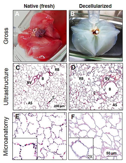
Figure 3. Decellularization of rhesus macaque lungs. A) Native lungs obtained from necropsy are perfused via the pulmonary artery to remove excess blood from the pulmonary capillary bed. B) After treatment with Triton X-100, Deoxycholic acid, hypertonic NaCl, and DNase, the lungs become white and translucent, indicative of decellularization. C) The ultrastructure of the native lung is represented in normal histology (H&E) by the appearance of bronchioles "B", blood vessels "BV", respiratory bronchioles "RB", capillaries "C", and alveolar sacs "AS". D) Lung ultrastructure is largely unaffected by decellularization, as all structural components of normal lung histology are visible after decellularization. E) H&E stain of normal native lung parenchyma shows the lacy appearance of alveoli in a 5 µm paraffin section with alveolar epithelial cells and some remaining red blood cells. F) The parenchyma of decellularized lungs contains alveoli that are intact relative to its native counterpart; however, these alveoli are completely devoid of cells as noted by the absence of dark-staining nuclei or red blood cells. Insets represent 2X magnification relative to their respective panels. Reprinted with permission from reference19.
Click here to view larger image.
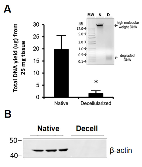
Figure 4. DNA and cellular protein is efficiently removed during decellularization. A) DNase treatment removes >90% (p<0.006) of DNA from the detergent-decellularized tissue. The small amount of remnant DNA that could be collected from the decellularized tissue was concentrated and run on a 0.8% agarose gel; the result (inset) showed that only low molecular-weight degraded fragments of DNA remained in the decellularized tissue. MW=molecular weight, N=native, D=decellularized. B) Western blot analysis showed that the representative cytoplasmic protein β-actin was not detectable in decellularized lung relative to native lung where this protein was abundantly detected. Reprinted with permission from reference19.
Click here to view larger image.
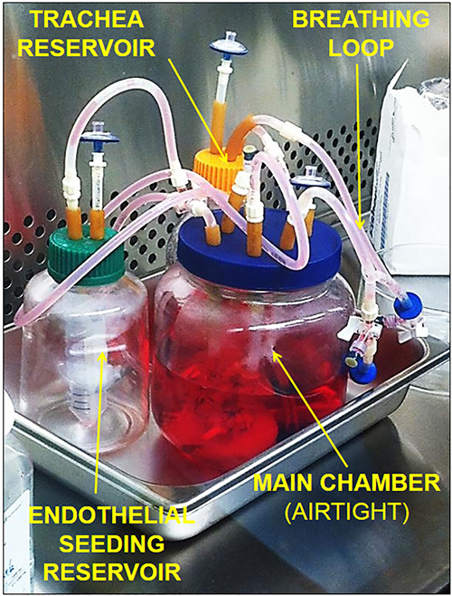
Figure 5. Fully assembled lung bioreactor. The bioreactor is assembled in a laminar flow hood to reduce the risk of contamination. A cell culture incubator water pan holds all bioreactor components and functions to collect spills if a leak occurs. The entire unit can be housed in an incubator. Levels of media in the various chambers should be determined empirically according to the researcher's needs.
Click here to view larger image.
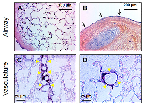
Figure 6. Bone marrow-derived mesenchymal stem cells (BMSC) and endothelial cells can be cultured within decellularized lung scaffolds. A) BMSC attach to the matrix scaffold of decellularized lung parenchyma and can be maintained with mechanical ventilation of culture media for extended periods; the panel shows a lung two weeks after seeding with BMSC. The cells line the alveolar septae creating the appearance of native lung. B) The lumenal surfaces of large airways serve as cell-attachment points as well. BMSC cultured for two weeks in the bioreactor grow along these surfaces and resemble simple squamous epithelium (black arrows). C) Seeding the vascular compartment of decellularized lungs with microvascular endothelial cells results in the recellularization of small vessels. Endothelial cells can be found attaching to the vascular basement membrane (yellow arrowheads). In some instances, the cell attachment points span the lumen (C). D) A cross-sectional view of a small vessel shows endothelial cells attached to the vessel basement membrane while maintaining an open vascular lumen.
Click here to view larger image.
Tissues can be efficiently and effectively decellularized by a number of methods employing physical, chemical, and enzymatic agents12,20. The challenges of producing 3D biological matrices from large organs include the requirement for large volumes of decellularization solutions, expensive commercial equipment (i.e. bioreactors), and a dizzying amount of methodological perturbations required to achieve the final tissue-derived product. Our method provides a straightforward approach that minimizes physical manipulation, reagent volume, and cost. Complete decellularization and sterilization of rhesus macaque lungs takes only three days; moreover, our decellularized tissues can be stored at 4 °C for several weeks without contamination or signs of scaffold degradation. We recently reported a detailed characterization of our decellularized macaque lung scaffolds in which we found that the Triton X-100/Deoxycholic acid method (modified from Price et al.4) efficiently removed cells while having minimal impact on the lung matrix4,19.
Our in-house bioreactor is comprised of parts that are routinely found in biological laboratories. The total estimated cost of consumable or replaceable components of the bioreactor is <$500. While this bioreactor is a "base-model," it can be fitted with various components to monitor or improve its function (e.g. pressure, flow, temperature, and pH sensors). From our experience with recellularizing macaque lung scaffolds, an ideal cell density for initiating in vitro culture of the cell-seeded scaffolds is 1.0 x 10⁶-1.5 x 10⁶ cells/ml for bone marrow- and adipose-derived mesenchymal stem cells in the airway compartment. The volume of cell suspensions at this density varies with the size and origin of the tissue (i.e. for single lobes, smaller volumes suffice; for half or whole lungs, larger volumes and, therefore, more cells are required). Initial vascular seeding studies with macaque lung microvascular endothelial cells (LMVEC) employed suspensions of 8.0 x 10⁴-1.0 x 10⁵ cells/ml in varying volumes perfused through single, small decellularized lung lobes. For other cell types that may be used, optimization of cell density for initiating cultures should be determined empirically. These experiments are ideal for determining cell-matrix interactions and feasibility of using decellularized scaffolds for engineering pulmonary tissue. For complete recellularization of whole macaque lungs, it is expected that a substantially large number of cells will be required, and as such, this type of culture will have immense nutrient (media) demand. Further optimization is required to reach a consensus on the appropriate means by which whole-organ recellularization can be accomplished. We were able to maintain culture of MSC- and endothelial cell-seeded lung matrices for up to two weeks without sign of contamination or tissue destruction. Histological analyses confirmed that these cells were able to attach to the decellularized lung matrix and maintain growth in culture. Additional studies are needed to determine whether the cells upregulate the expression of pulmonary- or endothelial-specific genes during culture with decellularized matrices in the bioreactor. These methods facilitate an easy and cost-effective way to optimize the recellularization of acellular, lung-derived, native-organ scaffolds in order to continue testing of laboratory-produced bioartificial lungs via clinically relevant in vivo models.
All components of the large-organ bioreactor are autoclavable with the exception of the disposable 0.22 µm filters and injection ports which are commercially available in sterile packaging. The lid of the 2 L main chamber jar is modified by drilling six 0.125-0.25 in holes through which 1/8 in silicone tubing is threaded; 1/8 in inside-diameter rubber tubing is used as an airtight gasket around the silicone tubing where they pass through the lid of the jar. Corning 500 ml laboratory jars are used for the tracheal and endothelial seeding reservoirs. The caps are similarly modified to allow tubing to be passed through.
It should be noted that since air is more compressible and expandable than liquid culture media, a larger volume of air is required to be able to produce enough pressure to move a given volume of liquid media for lung inflation in the bioreactor. After air withdrawal using the syringe pump, a pause of 15-20 sec allows equilibration of pressure and full inflation of the lungs. When the pump direction is reversed, the same volume of air is returned to the main chamber at the same rate followed by another pause of 15-20 sec to allow the lungs to fully deflate before the cycle repeats. For vascular perfusion, the vascular loop is placed within the roller apparatus of a peristaltic pump and clamped in place for pulsatile perfusion at the desired rate. For macaque, preliminary studies were carried out using flow rates of 5-10 ml/min in single lobes. Seeding and culture of the airway and vascular compartments can be performed separately for investigating specific roles of various cell types in regeneration, or both compartments can be seeded and cultured simultaneously toward the goal of producing an intact bioartificial organ.
The authors have nothing to disclose.
The authors wish to thank the editors of Tissue Engineering for allowing images from a previous publication to be used in this report.
Sodium nitroprusside | Sigma-Aldrich | 71778-25G | |
Heparin sodium salt | Fisher | BP2425 | |
Triton X-100 | Fisher | BP151-100 | |
Sodium deoxycholate | Fisher | BP349-100 | |
Sodium chloride | Fisher | 7647-14-5 | |
Bovine pancreatic DNase | Sigma-Aldrich | DN25 | Prepare stock, aliquot, and freeze |
Magnesium sulfate | Fisher | 10034-99-8 | |
Calcium chloride | Fisher | C614-500 | |
PBS (no Ca/Mg) | Gibco, Life Technologies | 10010-031 | |
Antibiotic-Antimycotic | Gibco Life Technologies | 15240062 | |
Cell Culture Media | |||
Alpha MEM | Gibco Life Technologies | 12561-072 | |
Medium 199 | Gibco Life Technologies | 11150067 | |
Premium Fetal Bovine Serum | Atlanta Biologicals | S11150 | |
L-Glutamine 100x | Gibco Life Technologies | 25030-081 | |
Endothelial Cell Growth Supplement (ECGS) | ScienCell | 1052 | |
Antibiotic-Antimycotic | Gibco Life Technologies | 15240062 | |
Cell Seeding and Bioreactor Culture | |||
Check valves | Cole-Parmer | EW-98553-20 | |
Y-connectors | Cole-Parmer | ED-30614-08 | |
3-Way stopcocks | Harvard Apparatus | 721664 | |
MasterFlex L/S 14 tubing | Cole-Parmer | 96420-14 | |
MasterFlex L/S 16 tubing | Cole-Parmer | 96420-16 | |
Male lock Luer 1/8 in | Cole-Parmer | EW-45505-04 | |
Female Luer 1/8 in | Cole-Parmer | SI-45502-04 | |
Male Luer lock plug | Cole-Parmer | EW-45505-56 | |
Injection ports | Medi-Dose EPS | IV2004 | |
Latex tubing | St. Louis Medical Suppy | HN10910 | |
Hose clamp | Cole-Parmer | EW-06832-02 | |
2 L Wide-mouth jar | Fisher | 05-719-276 | |
60 ml Syringes | Fisher | NC9035364 |