Biolistic transformation is a method used to generate stable integration of DNA into the genome of the opportunistic pathogen Cryptococcus neoformans through homologous recombination. We will demonstrate biolistic transformation of a construct, which has the gene encoding acetate kinase fused to the fluorescent tag mCherry into C. neoformans.
The basidiomycete Cryptococcus neoformans, an invasive opportunistic pathogen of the central nervous system, is the most frequent cause of fungal meningitis worldwide resulting in more than 625,000 deaths per year worldwide. Although electroporation has been developed for the transformation of plasmids in Cryptococcus, only biolistic delivery provides an effective means to transform linear DNA that can be integrated into the genome by homologous recombination.
Acetate has been shown to be a major fermentation product during cryptococcal infection, but the significance of this is not yet known. A bacterial pathway composed of the enzymes xylulose-5-phosphate/fructose-6-phosphate phosphoketolase (Xfp) and acetate kinase (Ack) is one of three potential pathways for acetate production in C. neoformans. Here, we demonstrate the biolistic transformation of a construct, which has the gene encoding Ack fused to the fluorescent tag mCherry, into C. neoformans. We then confirm integration of the ACK-mCherry fusion into the ACK locus.
Cryptococcus neoformans, an invasive opportunistic pathogen of the central nervous system, is the most frequent cause of fungal meningitis resulting in more than 625,000 deaths per year worldwide 1. Acetate has been shown to be a major fermentation product during cryptococcal infection 2,3,4, and genes encoding enzymes from three putative acetate-producing pathways have been shown to be upregulated during infection 5. This suggests that acetate production and transport may be a necessary and required part of the pathogenic process; however, the significance of this is not yet understood. One possible pathway for acetate production is the xylulose 5-phosphate/fructose 6-phosphate phosphoketolase (Xfp) – acetate kinase (Ack), a pathway previously thought to be present only in bacteria but recently identified in both euascomycete as well as basidiomycete fungi, including C. neoformans 6.
To determine the localization of these enzymes of this pathway in the cell, a construct carrying a neomycin resistance gene downstream of an ACK gene fusion to the fluorescent tag mCherry (ACK:mCherry:Neo) will be introduced into C. neoformans using the well-established method of biolistic transformation 7,8. Although electroporation is an efficient method for transformation of plasmids that will be maintained as episomes into Cryptococcus 9, it is not useful in creating stable homologous transformants 8. Only biolistic delivery using a gene gun provides an effective means to transform linear DNAs that will be integrated into the genome by homologous recombination. For example, Edman et al. showed that of the transformants resulting from electroporation of a plasmid-borne URA5 selectable marker into C. neoformansura5 mutants, just 0.001 to 0.1% of transformants were stable 9. Chang et al. achieved just a 0.25% stable transformation efficiency using electroporation to reconstitute capsule production in an acapsular mutant 10. Unlike electroporation, biolistic transformation has been shown to result in stable transformation efficiency of 2-50% depending on the gene that is being altered 7,8,11.
This visual experiment will provide a step-by-step demonstration of biolistic transformation of the linear ACK:mCherry:Neo DNA construct into C. neoformans, and will describe how to confirm its proper integration via homologous recombination into the ack locus. The protocol demonstrated here is a modification of the method developed in the Perfect laboratory 8.
NOTE: The overall scheme of this protocol is outlined in Figure 1.
1. C. neoformans Preparation
2. Gold Microcarrier Preparation
3. DNA Preparation
4. Operating the Gene Gun
5. Plating Transformed Cells
6. Genomic DNA Isolation for PCR
NOTE: This is a modified version using reagents from a DNA purification kit (See Table of Materials).
7. RNA Isolation for Reverse Transcriptase-PCR.
A successful biolistic transformation of C. neoformans can be obtained by following this protocol scheme (Figure 1). With biolistic transformation, a successful shoot of the coated gold beads is indicated by a gold ring visible on the plate after the DNA is shot (Figure 2A). Colonies should appear within 4 to 5 days when left at room temperature after plating the recovered cells from the YPD + 1M sorbitol plates onto selective media. Transforming 2 µg of DNA should result in 20 to 30 colonies (Figure 2B). When colonies appear, they should be restreaked on selective media for individual colonies.
The individual colonies can be grown in YPD media, and both DNA and RNA can be isolated from these cells and analyzed through PCR and RT-PCR to confirm proper integration and expression. If this protocol is used for tagged gene fusion, as in this example, the primers would need to anneal within the coding region of the gene of interest (primer 2) and within the 3’ noncoding region of the gene of interest (primer 4) (Figure 3A). With this construct, the DNA amplified from the PCR reaction was sequenced for another confirmation that the mCherry tag was fused in frame to the ACK gene. A positive PCR confirmation would be a larger PCR product from the DNA isolated from the transformed cells compared to the DNA isolated from the wild type cells. Another PCR reaction would also need to be conducted utilizing the primer set (primers 2 & 5) where one primer anneals outside of the construct and within the surrounding genome (primer 5) to confirm the correct recombination into the desired locus (primer 7 in Table 1) (Figure 3B). RT-PCR will be used to make sure that both the gene of interest and the tag are both being expressed (Figure 3C). Sequencing of the RT-PCR fusion product indicates that the tag is properly fused to the gene at the RNA level.
If this protocol is utilized to knock out a gene of interest, primer sets for PCR should be designed such that one primer anneals to a genome sequence outside of where the construct should recombine into the genome, and the other primer anneals either in the coding region of the gene or in the selective marker. A positive confirmation that the construct has successfully and correctly recombined into the genome would be the presence of the correct size product for the primer set that anneals within the marker but not with the primer set that anneals to the gene of interest. Another primer set should be made that has one primer that anneals outside of the designed construct, which is used with PCR to confirm that the recombination event occurred at the correct locus. In the same design to create a knockout, RNA is isolated from both the transformed cells and wild type (WT) cells, and RT-PCR is performed to confirm that no expression of the gene of interest is observed from the transformed cells.
Because a fluorescent tag was fused to the ACK gene, another confirmation that recombination was a success into the desired locus and that RNA is being translated into protein is through fluorescent microscopy (Figure 4). Ideally, conditions have already been established where it is known that the protein of interest is being expressed. However, if the fluorescent signal is too low to observe, there is a possibility that successful recombination still occurred, but growth conditions need to be altered in the chance that optimal conditions have not been met for sufficient expression, which would lead to a low fluorescent signal. This would need to be confirmed through other methods such as a western blot.
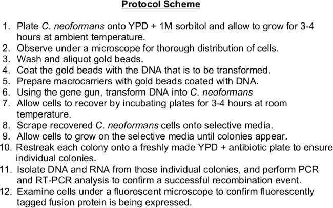
Figure 1. Protocol scheme.
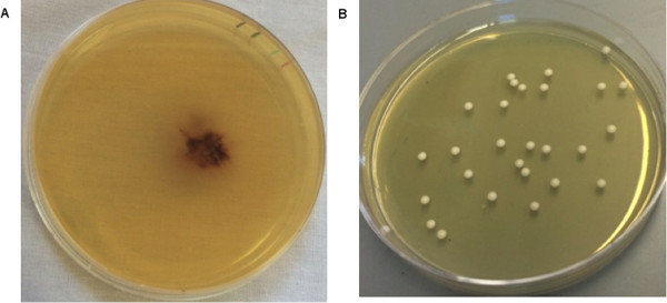
Figure 2A. DNA-coated gold beads successfully shot onto a YPD + 1M sorbitol plate. An orange patch seen in the center of the YPD + 1M sorbitol plate is due to the DNA-coated gold beads, indicating proper gold preparation, as well as a successful shoot. Figure 2B. Transforming 2 µg of DNA results in 20-30 colonies per plate. If the cells were diluted as mentioned in the protocol, approximately 20-30 colonies are expected prior to plating on selective media. Please click here to view a larger version of this figure.
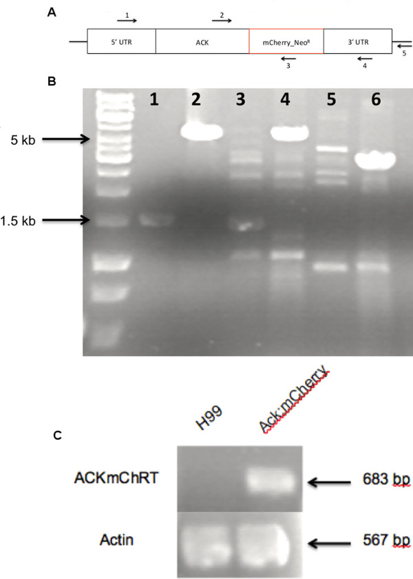
Figure 3A. Schematic of the ACK:mCherry:Neo construct and primer design. Figure 3B. PCR used to confirm successful homologous recombination. Lanes 1 and 2: PCR products obtained using primers 2 and 5 (Table 1) with genomic DNA from wild type C. neoformans H99 (lane 1) and the ACK:mCherry transformed strain, (lane 2). Expected sizes are 1511 and 5622 bp, respectively. Lanes 3 and 4 are the DNA products of the C. neoformans H99 (expected size 1443 bp) and the ACK:mCherry (expected size 5552 bp) strains, respectively, using primers 2 and 4 in Table 1. Lanes 5 and 6 are the DNA products of the C. neoformans H99 (should not anneal) and ACK:mCherry (expected size 3016 bp) strains, respectively, using primers 1 and 3. Figure 3C. RT-PCR confirmation of expression of the mCherry tag. The top lanes are the cDNA products of the ACKmCherry fusion product (expected size 683 bp) amplified from the C. neoformans H99 and the ACK:mCherry strains using primers 2 and 3 in Table 1. The actin gene was included as a control and was amplified under the same conditions as ACKmCherry (expected size 567 bp) using primers 6 and 7 in Table 1. Please click here to view a larger version of this figure.
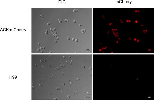
Figure 4. Fluorescence of the mCherry tagged Ack. Microscopic analysis of strains producing Ack fused to a mCherry tag with an excitation optimum at 587 nm and an emission optimum at 610 nm. Please click here to view a larger version of this figure.
1 | KI003 | 5’ – GTA GCG AGG TCT GGA AGC CAC – 3’ |
2 | ACKmChRT-F | 5'- GCT TTG GCC GGT ACT ACC AAC -3 |
3 | ACKmChRT-R | 5'- GAC AGC TTC AAG TAG TCG GGG -3' |
4 | KI004 | 5’ – GAC TTG GGG AAG AGG AAT TC – 3’ |
5 | KI0032 | 5' – CGG GGT ACC ATC AAT AAA AGC TTT CTT CAC TCC – 3' |
6 | Actin 1 | 5’- CGC TAT CCT CCG TAT CGA TCT TGC -3’ |
7 | Actin 2 | 5’- CAG CTG GAA GGT AGA CAA AGA GGC -3’ |
Table 1. PCR and RT-PCR primers.
Utilizing this protocol, biolistic transformation can be accomplished in which linear DNA is integrated into a desired locus in the Cryptococcus neoformans genome by homologous recombination. Certain steps in the protocol can have a dramatic effect on the effectiveness/efficiency of the transformation. For a successful transformation, it is imperative that the DNA utilized in the shoot has a concentration of at least 1 µg. However, the volume of DNA added to the gold beads can be increased in the chance the DNA yield is lower than 1 µg (Step 3.4).
Another important step is in the DNA coating of the gold beads. Insufficient numbers of beads in the DNA preparation sample, due to an error in the preparation, leads to a decreased amount of DNA shot onto the plate. After the DNA has been loaded onto the gold beads, they are pipetted onto the biolistic disc and allowed to dry. When dry, a visible gold circle about 1 cm in diameter should be present on the disc. The absence of this circle suggests that the concentration of the gold beads is not high enough. Another clue that the gold bead concentration is too low is following the shoot. There should be a gold ring visible on the plate (Figure 2A), and if no gold ring is visible and the rupture disc burst, this could indicate that the concentration of gold beads used in the preparation was not high enough.
The typical yield using the biolistic transformation method is 20-30 colonies. Fewer colonies may indicate that the technique or gene gun set-up is not 100% efficient. One reason for the fewer colonies may be the amount of cells scraped off from step 5.2. Depending on the size of the pellet in step 5.3 and the number of colonies that appear from the previous experiments, the volume the pellet is resuspended in, in step 5.4, may need to be altered. From these colonies, DNA should be isolated, and PCR conducted to confirm a larger size gene product compared to WT, indicating the presence of the tag. RNA should be isolated and RT-PCR performed to confirm that there are transcripts of the gene product being made, and if a fluorescent tag was inserted into the genome, then microscopy should be used to observe whether the tag is being expressed.
The main limitation to this protocol is the requirement for specialized equipment such as a gene gun and a fluorescent microscope. However, biolistic transformation is the best choice for introduction of linear DNAs for gene knockouts versus electroporation, which is used for introduction of episomes or Agrobacterium tumefaciens mediated transformation, which has been used for random insertional mutagenesis12. Biolistics may also prove to be a suitable method for rapid introduction of a wide variety of vital dyes into Cryptococcus. Lipophilic dyes are used to stain extracellular vesicles and the capsule of C. neoformans 14. Biolistic delivery of gold particles coated with lipophilic dye that imbed into the membranes of cells and organelles has been used to study the interconnection of neighboring cells 13. Therefore, biolistics may be a less time-consuming technique to visualize extracellular vesicles and organelles.
The authors have nothing to disclose.
This work was supported by awards from the National Science Foundation (Award# 0920274) and the South Carolina Experiment Station Project SC-1700340. This paper isTechnical Contribution No. 6283 of the Clemson University Experiment Station. The authors thank Dr. Lukasz Kozubowski for his helpful advice in development of this final protocol and Dr. Cheryl Ingram-Smith, Katie Glenn, and Grace Kisirkoi for their critical reading of the manuscript.
Product | Company | Catalog # | Website |
0.6 μm gold beads | Bio-Rad | 165-2262 | http://www.bio-rad.com |
Spermadine-free base | Sigma- Aldrich | S0266 | https://www.sigmaaldrich.com |
G418 – Sulfate (Neomycin) | Gold Biotechnology | G-418-10 | www.goldbio.com |
Hygromycin | Gold Biotechnology | H-270-1 | www.goldbio.com |
1350 psi Rupture Discs | Bio-Rad | 165-2330 | http://www.bio-rad.com |
Stopping Screens | Bio-Rad | 165-2336 | http://www.bio-rad.com |
Macrocarriers discs | Bio-Rad | 165-2335 | http://www.bio-rad.com |
YPD Broth | Becton Dickinson & Co. | 242820 | www.bd.com |
Agar | Becton Dickinson & Co. | 214530 | www.bd.com |
Sorbitol | Fisher Scientific | BP439 | http://www.fishersci.com |
PDS-1000/He System | Bio-Rad | 165-2257 | http://www.bio-rad.com |
Microscope | Zeiss | Axio | http://www.zeiss.com/microscopy |
KOD One Step PCR Kit | EMD Millipore | 71086-4 | http://www.emdmillipore.com |
One Step RT-PCR Kit | Qiagen | 210212 | www.qiagen.com |
Wizard Genomic DNA Purification Kit | Promega | A1120 | www.promega.com |
RNeasy Mini Kit | Qiagen | 74104 | www.qiagen.com |
Mini Beadbeater – 1 | BioSpecs | 3110BX | http://www.biospec.com |
Microfuge 18 Centrifuge | Beckman Coulter | F241.5P | www.beckmancoulter.com |
Microplate Spectrophotometer | BioTek | EPOCH | www.biotek.com |