Here, we present a protocol for synthesizing virus-like particles using either baculovirus or mammalian expression systems, and ultracentrifugation purification. This highly customizable approach is used to identify viral antigens as vaccine targets in a safe and flexible manner.
Virus-like particles (VLPs) and subviral particles (SVPs) are an alternative approach to viral vaccine design that offers the advantages of increased biosafety and stability over use of live pathogens. Non-infectious and self-assembling, VLPs are used to present structural proteins as immunogens, bypassing the need for live pathogens or recombinant viral vectors for antigen delivery. In this article, we demonstrate the different stages of VLP design and development for future applications in preclinical animal testing. The procedure includes the following stages: selection of antigen, expression of antigen in cell line of choice, purification of VLPs/SVPs, and quantification for antigen dosing. We demonstrate use of both mammalian and insect cell lines for expression of our antigens and demonstrate how methodologies differ in yield. The methodology presented may apply to a variety of pathogens and can be achieved by substituting the antigens with immunogenic structural proteins of the user's microorganism of interest. VLPs and SVPs assist with antigen characterization and selection of the best vaccine candidates.
Virus-like particles (VLPs) are an approved technology for human vaccination. In fact, some of the more contemporarily licensed vaccines, including the human papillomavirus (HPV) and hepatitis Β (HepΒ) vaccines employ this approach. VLPs are formed from structural proteins capable of self-assembly. The assembled particles mimic viral morphologies, but cannot infect or replicate because they lack viral genomes. VLPs can be expressed and purified from a number of prokaryotic and eukaryotic systems. A review of the literature revealed that different expression systems are employed at the following rates: bacteria – 28%, yeast – 20%, plant – 9%, insect – 28%, and mammalian – 15%1. Of note, HPV vaccines based on L1 capsid protein are produced in yeast (Gardasil) or in an insect cell system (Cervarix)2. HepΒ vaccines, Recombivax and Engerix-Β, are also produced in yeast, and are composed of HepΒ surface antigen3,4.
We use mammalian and insect cell expression systems to produce VLPs requiring co-expression of multiple structural proteins for assembly. Our work focuses on designing, producing, and purifying VLP-based vaccines against human pathogens: influenza virus, respiratory syncytial virus (RSV), dengue virus (DENV), and chikungunya virus (CHIKV). Our methods are flexible enough to allow for co-expression of the multiple structural proteins from multiple expression plasmids, or a single expression plasmid (Figure 1). Previously, we produced and purified H5N1 VLPs assembled from the co-expression of plasmids encoding influenza hemagglutinin (HA), neuraminidase (NA), and matrix (M1) in human embryonic kidney 293T cells5,6. The genes were codon-optimized for expression in mammalian cells and cloned into pTR600, a eukaryotic expression vector containing the cytomegalovirus immediate-early promoter plus intron A for initiating transcription of eukaryotic inserts and the bovine growth hormone polyadenylation signal for termination of transcription7. A similar approach using the three-plasmid co-expression of HA, NA (Figure 1A) and an alternative viral matrix protein, HIV Gag p55, was used for generation of human seasonal influenza subtype H3N2 VLPs in this study and has been shown to generate VLPs of similar size as influenza particles8. Although influenza vaccines predominantly elicit anti-HA antibodies, the addition of influenza neuraminidase mediates sialidase activity to enable VLP budding from transfected cells9 and also present additional immunogenic targets. To produce RSV VLPs, we also selected the unrelated core of HIV Gag to design prototypical vaccines that present exclusively RSV surface glycoproteins to further demonstrate flexibility of VLP formation using HIV Gag, as previously described and characterized by electron microscopy6,10. Others have previously shown that VLPs presenting RSV glycoproteins can be assembled using various viral components from Newcastle Disease virus (NDV)11, and influenza matrix12. Full-length surface glycoprotein sequences were utilized in this study to retain native conformations that may be necessary for functional receptor binding and antibody recognition assay through enzyme-linked immunosorbent assay (ELISA).
Our examples for use of single plasmid expression systems to generate particles are DENV and CHIKV. In the case of DENV, we can produce subviral particles (SVPs) with no capsid in 293T cells from a single plasmid containing a prM/E structural gene expression cassette13. The term SVP is used to denote the lack of a core or capsid protein in the assembly of viral structural proteins. CHIK VLPs can also be produced using a single plasmid containing a CHIKV structural gene cassette, encoding capsid and envelope proteins, or in insect cells by infecting with a recombinant baculovirus encoding the same structural gene cassette optimized for insect cell expression (Figure 1B–C).
The end result of the expression approaches discussed above is the release of VLPs into cell culture medium that can then be purified via ultracentrifugation through a 20% glycerol cushion. In this report, we present methods to express and purify these VLPs from mammalian and insect cell systems.
1. Mammalian Expression System for Generation of Influenza H3N2 VLP
2. CHIK VLP Expression Using Baculovirus/Insect Cell System
3. Sedimentation/Purification of VLP/SVPs
4. Determining Protein Yields and Specific Antigen Yields
VLP yields were variable by viral antigen construct design. In this protocol, we have demonstrated use of insect and mammalian cells for production of SVP or VLPs in supernatant and purification by ultracentrifugation. Four subtypes of DENV prM/E structural gene expression cassettes were used to construct the versions of DENV SVPs (demarcated as 1-4) in Table 1 and demonstrate a range between 1.1-2.6 mg of total protein in 0.6 ml volumes. For VLPs that require a three gene constructs, we found the optimized DNA transfection ratio of 1:1:2 for HA:NA:Gag respectively to synthesize influenza VLPs; in contrast, a single plasmid expressing multiple chikungunya viral proteins was adequate for the generation of recombinant baculovirus and mammalian CHIK VLP. Additionally, a dual-plasmid transfection procedure is also possible with mammalian cells, as such with RSV F and Gag transfected at a 1:1 ratio, and yields approximately 0.01 mg/ml of total cell culture supernatant. As shown in the Table 1, CHIK SVP yield from Sf9 cells can range between 0.008-0.016 mg total protein/ml of supernatant volume, while production through mammalian 293T yields a 10-fold reduction of protein. Between the two versions of H3N2 VLPs using different wild-type, full-length HA plasmids, the H3N2 VLP Sample 2 had a reduced yield of both total protein and specific HA content. Other sample VLP HA quantities are shown in Figures 2 and 3 and illustrate how HA and ELISA are used to estimate surface content on the VLPs. As with a conventional direct ELISA method, a standard curve is generated using known concentrations of a recombinant HA to compare ELISA reactivity to sample H3 VLPs loaded onto an ELISA plate. ELISA or similar immunoassay are recommended for quantification of VLPs that have readily available monoclonal antibodies with known crossreactivity to a well-conserved epitopes, but may not be available for all antigens. RSV F is well-characterized, therefore an ELISA using monoclonal anti-RSV F antibody is also applicable for quantification of surface F on pelleted RSV F VLPs. In addition to antigen functionality and antigen recognition by specific antibodies, VLP preparations can be structurally assessed by electron microscopy. Figure 4 is a representative image showing that HA (Gag-core) influenza VLPs are successfully expressed, assembled, and purified as VLPs.
Description | Culture Vol (ml) | Harvesta | Total Volume | Total Protein Yield (mg) | Yield (mg)/Vol (ml) | Specific antigen content |
DENV1 SVP | 293T186 | 4 d | 0.6 ml | 1.115 | 0.006 | n.d. |
DENV2 SVP | 293T/186 | 4 d | 0.6 ml | 2.5 | 0.0134 | n.d. |
DENV3 SVP | 293T/186 | 3 d | 0.6 ml | 1.536 | 0.0083 | n.d. |
DENV4 SVP | 293T/186 | 4 d | 0.6 ml | 2.613 | 0.014 | n.d. |
CHIK VLP | Sf9/186 | 3 dpi | 0.6 ml | 1.503 | 0.0081 | n.d. |
CHIK VLP | Sf9/500 | 4 dpi | 2.0 ml | 8.276 | 0.0166 | n.d. |
CHIK VLP | 293T/186 | 3 d | 0.6 ml | 0.296 | 0.0016 | n.d. |
H3N2 VLP 1 | 293T/216 | 3 d | 0.6 ml | 1.25 | 0.0058 | 1.9 µg HA/μl |
H3N2 VLP 2 | 293T/216 | 3 d | 0.6 ml | 0.956 | 0.0044 | 0.96 µg HA/μl |
H3N2 VLP 3 | 293T/216 | 3 d | 0.6 ml | 1.6 | 0.0074 | 1.2 µg HA/μl |
H3N2 VLP 4 | 293T/216 | 3 d | 0.6 ml | 1.54 | 0.0071 | 0.98 µg HA/μl |
RSV F VLP | 293T/216 | 3 d | 0.6 ml | 2.3 | 0.0106 | 0.15 µg RSV F/μl |
a d = days post-transfection, dpi = days post-infection |
Table 1: Subviral and virus-like particle yields by construct. Representative data of VLP volumes, total protein yield (determined by conventional BCA assay) or specific antigen content (determined by ELISA) is shown among a variety of SVP/VLP constructs. Yields are variable due to difference in SVP/VLP assembly and cell type, and maximum yields may require user optimization. The various versions of either DENV1-4 differ based on serotype, while the CHIK VLPs use the same sequence but preparations differ in either volume or cell culture type. The different versions of H3N2 VLPs (1-4) express four unique HA sequences that share a HA-specific ELISA reactive epitope and were co-expressed with the same NA and Gag core. The RSV F VLP, which is generated from a dual-plasmid transfection of RSV F and Gag, was synthesized and RSV F content was approximated using RSV F specific ELISA assay.
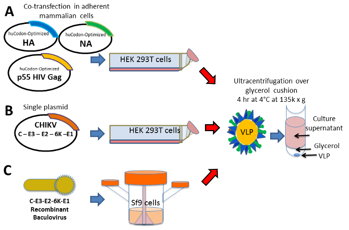
Figure 1: Approach – VLP design, synthesis, and purification. Schematic representation of plasmid gene designs for expression of viral structural proteins that will form VLPs: (A) co-expression of multiple structural proteins by co-transfection of three plasmids in 293T cells, (B) expression of structural proteins from a single gene cassette encoded in a single plasmid in 293T cells, and (C) recombinant baculovirus encoding CHIK structural proteins is used to infect Sf9 cells cultured in spinner flasks to promote high density cell growth in suspension. The VLPs are harvested from the cell culture media, clarified of cell debris, and separated from non-particles via sedimentation by ultracentrifugation on a 20% glycerol cushion. Please click here to view a larger version of this figure.
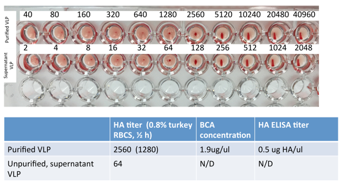
Figure 2: Example Hemagglutination assay of H3 VLPs. A conventional hemagglutination assay was performed on a representative sample of H3N2 VLP and its corresponding clarified supernatant; H3N2 HA assay can be performed using 0.8% turkey red blood cells or guinea pig red blood cells (not shown). Total protein content is estimated using the standard BCA assay on the purified VLP and was not performed or recommended for clarified supernatant. ELISA was performed on the purified VLP and HA content was estimated using a recombinant HA standard of known concentration. Please click here to view a larger version of this figure.
Figure 3: Example H3 ELISA for quantification of H3 content in H3 VLPs. An ELISA plate was coated with serial dilutions of a recombinant HA standard of known concentration and diluted H3N2 VLP samples generated from different HA sequences (Samples 1-9). Correlating the concentration (µg/µl) of the standard to the optical density values obtained at 492 n.m. (O.D.), concentrations are obtained by deducing the x-value that intersects the linear portion of the standard curve. Dilutions of recombinant HA standard and unknown samples were loaded in duplicate in wells A& B or C & D, respectively. The final concentration of the VLP sample is determined by multiplying the concentration by the dilution factor of the VLP sample. Please click here to view a larger version of this figure.
Figure 4: Electron microscopy of HA-VLPs using Gag-core. Representative electron microscopy image of purified influenza HA VLPs. VLPs assemble into spherical particles coated with HA. Please click here to view a larger version of this figure.
We have used the techniques described above to successfully express and purify SVPs and VLPs composed of multiple structural proteins for various pathogens. In general, we use mammalian expression systems to generate our VLPs. However, in our hands, mammalian-cell derived CHIK VLP yields were low. CHIK VLP yield was more robust when using A recombinant baculovirus-insect cell system. In general, baculovirus-insect cell systems yield higher amounts of recombinant proteins, which may result from the higher cell densities that can be cultured in spinner flasks versus tissue culture flasks, and also due to high level of expression of foreign genes while under the control of the baculovirus polyhedrin promoter.16 However, a disadvantage of using baculovirus is the very presence of baculovirus in VLP preparations, which has adjuvant activity, and skew immunological results in vaccine studies17 We also found variability in the yield of influenza VLPs depending on the HA and NA combinations used; H3N2 VLP2 had relatively reduced yield compared to H3N2 VLP1, potentially due to less efficient VLP assembly or NA-compatibility15 and remains to be further investigated. Optimization of DNA constructs and transfection ratios remain critical steps for ensuring optimal VLP yields. We opted to use a mammalian vector pTR600 consistently among our mammalian-based VLPs because expression is generally efficient and subcloning is convenient with multiple restriction enzymes. Ratios of plasmids may be adjusted for user's requirements to improve VLP yields. However, as discussed, current limitations of the mammalian VLP production remain with cost of transfection and the cell growth capacity in current tissue culture flasks. Although not performed in this protocol, we have observed additional successful production of VLPs in transfected cells when media is replenished with fresh growth media (e.g., OptiMEM) after the first 72 hr harvest and subsequent collection of VLPs after an additional 72 hr, although yields may be slightly reduced as compared to the first collection of VLPs.
We demonstrate a procedure for expressing viral structural proteins in either insect or mammalian cell cultures to release VLPs in the supernatant for semi-purification by ultracentrifugation. The SVP/VLPs are generally stable for use in antigen immunoassays and vaccination studies, and pose fewer safety threats as compared to live viral particles, particularly for select agents or high-pathogen avian influenza (HPAI) strains8,18, which require Biosafety Level 3 (BSL-3) laboratory conditions19. VLP production is relatively simple and often yields particles that may be recognized by antibodies elicited against native viral structural proteins, not exclusively denatured linear epitopes. VLPs serve as a useful platform for eliciting specific immune responses to the antigen and may be a promising candidate in vaccine development.
This protocol demonstrates two approaches for transfecting viral genes in the mammalian system. In this illustration, two influenza genes, HA and NA, are co-transfected with the HIV Gag to generate a VLP that has a Gag internal core, with HA and NA assembled on the surface. The substitution of Gag instead of influenza Matrix 1 (M1) demonstrates flexibility of Gag-based VLPs with unrelated viral glycoproteins, such as RSV F and potentially others. Alternatively, the synthesis of CHIKV utilizes a structural gene cassette, which expresses the necessary components for SVP self-assembly, with CHIKV E proteins presented on the surface of a center composed of C protein. These two approaches demonstrate the flexibility of VLP synthesis, which requires minimal structural viral proteins to assemble from transfected cells.
Finally, while we presented general protocols and representative results for expression and purification of H3N2, RSV, DENV, and CHIKV, these procedures are easily amenable for generation of VLPs using other viral proteins. These procedures have been successfully used by our group in the generation of other influenza virus subtypes including H1, H5, and H7 viruses. Moreover, the single plasmid, prM/E design used for generation of DENV SVPs can be adapted for generation of SVPs for other members of the Flavivirus family that include West Nile and Japanese encephalitis viruses. Similarly, methods of CHIK VLP production can be used for other members of the Togaviridae family, including the Venezuelan, East, and West equine encephalitis viruses.
The authors have nothing to disclose.
We wish to acknowledge the prior members of the Ross lab who have helped optimize the protocol for maximal efficiency and yields.
Plasmid Maxi Kit | Qiagen | 12163 | |
DMEM | Cellgro | 10-013 | |
Lipofectamine | Life Technologies | L3000075 | Lipofectamine 2000, Lipofectamine 3000 |
Opti-MEM I | Life Technologies | 31985062 | |
Bac-to-Bac Baculovirus Expression System | Life Technologies | 10359-016 | |
Cimarec I 6 multipoint stirrer plate | ThermoFisher Scientific | 50094596 | |
Polyclear ultracentrifuge tubes | Seton | 7025 | Confirm appropriate tubes for ultracentrifuge and bucket size |
Micro BCA Protein Assay Kit | ThermoFisher Scientific | 23235 | |
Phosphate citrate buffer | Sigma | P4922 | |
o-phenylenediamine dihydrochloride | Sigma | P8287 | |
0.22 μm vacuum filter top (500 mL) | Nalgene | 569-0020 | |
Glycerol | Sigma | G5516 | |
H2SO4 | Sigma | 339741 | |
Sf-900 II SFM | ThermoFisher Scientific | 10902-096 |