We collected, stained and imaged cells from the conjunctiva of the inner eyelid margin of human subjects. By characterizing cell morphology and metabolic activity, this method may further our understanding of dry eye and the role that friction between the ocular surfaces may play in perceiving ocular discomfort.
Few reports on the cellular anatomy of the lid wiper (LW) area of the inner eyelid exist and only one report makes use of cytological methods. The optimization of a method of collecting, staining and imaging cells from the LW region using impression cytology (IC) is described in this study. Cells are collected from the inner surface of the upper eyelid of human subjects using hydrophilic polytetrafluoroethylene (PTFE) membranes, and stained with cytological dyes to reveal the presence of goblet cells, mucins, cell nuclei and various degrees of pre- and para-keratinization. Immunocytochemical dyes show cell esterase activity and compromised cell membranes by the use of a confocal scanning laser microscope. Up to 100 microscopic digital images are captured for each sample and stitched into a high-resolution, large scale image of the entire IC span. We demonstrate a higher sensitivity of IC than reported before, appropriate for identifying cellular morphologies and metabolic activity in the LW area. To our knowledge, this is the first time this selection of fluorescent dyes was used to image LW IC membranes. This protocol will be effective in future studies to reveal undocumented details of the LW area, such as assessing cellular particularities of contact lens wearers or patients with dry eye or lid wiper epitheliopathy.
The human upper eyelid executes around 10,000 blinks every day1, with just a 1 – 2 mm narrow conjunctival region opposing the ocular globe. Due to its wiping motion during the blink, this portion of the lid margin has been termed the "lid wiper" region2. It is assumed that friction is increased in this region during habitual blinking, due to the lid wiping over the globe. This may play a significant role in ocular comfort. Contact lens wearers in particular experience ocular discomfort and this is one of the primary reasons for ceasing lens wear3. When a contact lens is placed on the eye, the friction coefficient between the lens material and the ocular surface has been shown to change4. There is evidence to suggest that dryness symptoms could be related to this altered friction2,5,6.
This association may also be reflected in clinically observable phenomena, notably lid wiper epitheliopathy (LWE), also called upper lid margin staining (ULMS)6. LWE is an early sign of ocular irritation and a potential predictor for dry eye disease. It is observed and measured by the vital staining of the upper and lower lid margin regions. While this grading system2 and its clinical validity (i.e., correlation with subjective symptoms) are still under debate, ocular discomfort remains a conundrum for clinicians and scientists alike and, most importantly, an inconvenience for patients.
To-date, little is known about clinically-relevant variations in the (sub-)cellular anatomy and physiology of the lid wiper area7 and only one report makes use of cytological methods8. Impression cytology (IC) has been employed for over 40 years to collect cells from the epithelial surface of the bulbar or tarsal conjunctiva by application of a membrane9,10. Upon removal, the adherent cells undergo cytological staining and microscopic imaging. Due to the anatomic and cellular differences in the lid wiper region, this technique requires optimization.
Ethics statement: Prior to collecting cells from human subjects, informed consent and ethics approval must be obtained.
1. Prepare Stain
NOTE: Prepare stain on day of experiment.
2. Collect Cells
3. Sample Processing
4. Sample Staining
5. Sample Imaging
The cell culture membrane spanned over the plastic holder is a convenient tool for handheld collection of epithelial cells from the lid wiper conjunctiva. This method eliminates the need for additional sterilized instruments typically used to prepare and handle IC membranes. Figure 1 depicts the IC of the lid wiper area using a cell culture insert. We optimized two different staining protocols that complement each other to characterize epithelial cells from the lid wiper area in a novel fashion. Fluorescent immunocytochemical dyes reveal esterase activity, an indicator of cell viability, and nucleic acids, the staining of which reflects cell membrane compromise, as shown in Figure 2. The cytological staining protocol allows for a fine distinction between keratinization degrees. Figure 3 shows the transition between low keratinization in the tarsal conjunctiva and advanced keratinization in the lid wiper conjunctiva and epidermis. Panoramic imaging of cytology-stained cell collections permits the analysis of the entire collection area, as opposed to a selected region of interest. Image resolution is adequate for zooming to nuclear level; cell morphology can be determined with precision. Figure 4 shows the final image of a cytologically stained cell collection, stitched together using approx. 100 separate high resolution files. These tools may be employed to assess the effects of friction and/or dry eye in a novel way.
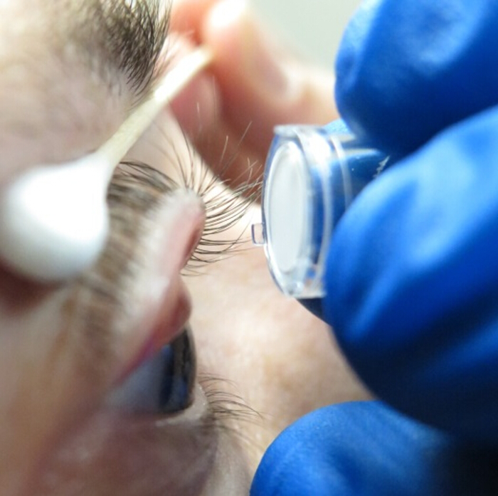
Figure 1. Impression Cytology of the Lid Wiper Area. Impression cytology performed on the lid wiper region exposed by eversion of the upper lid. Membrane used is the Cell Culture Insert, 12 mm, hydrophilic PTFE. Note the tabs of the membrane holder, which may be retained (unlike previous studies on the bulbar conjunctiva, in which they were removed prior to application) as they do not interfere with application of the membrane onto the narrow eyelid margin and may actually aid alignment. Please click here to view a larger version of this figure.
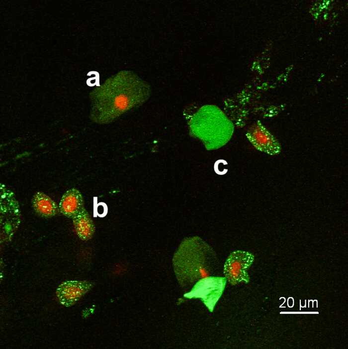
Figure 2. Fluorescent Cells from the Lid Wiper Region. Confocal laser scanning microscopy showing variation of cell morphology between large squamous cells (a) and the smaller columnar/cuboidal cells (b). Green fluorescence of Calcein AM indicates esterase activity in the cell body (i.e., cell viability). Red fluorescence of Ethidium reveals nucleic acids, indicating cell membrane compromise. Few cells show intense green and no red fluorescence, possibly indicative of cell membrane integrity (c). Please click here to view a larger version of this figure.
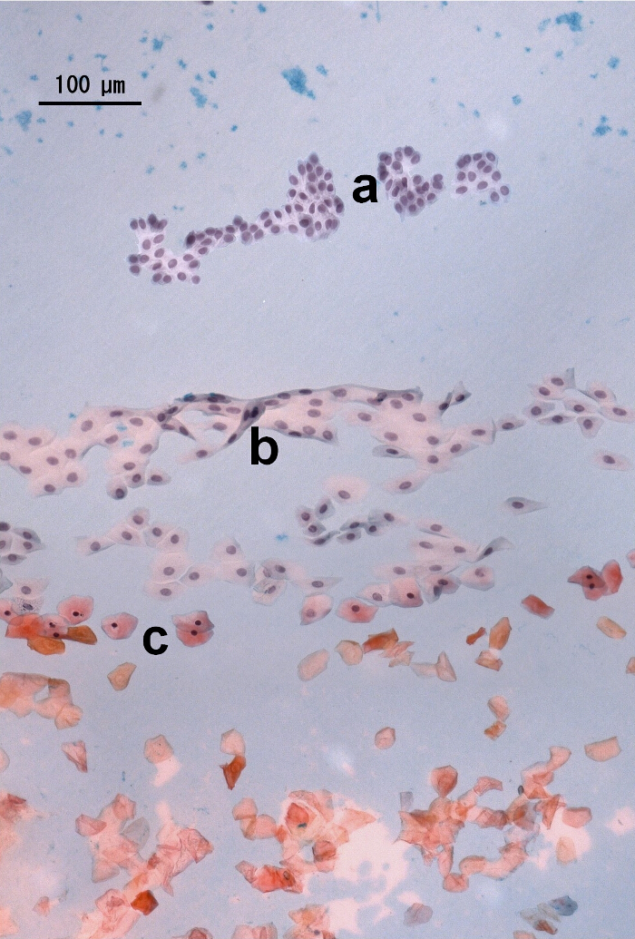
Figure 3. Cytological Staining of IC Sample. Cells of the lid wiper region show varying morphology and different keratinization degrees. Blue color of small, columnar and cuboidal cells with large nuclei, found in the marginal/tarsal conjunctiva, indicates no keratinization (a); red/orange stain of large squamous cells with condensed nuclei of the muco-cutaneous junction (MCJ/Marx' Line) and anuclear cells of the epidermis represents advanced keratinization (c). Transitional color (red/blue) and morphology of cells in the lid wiper conjunctival area suggest limited keratinization (b). Please click here to view a larger version of this figure.
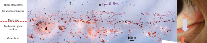
Figure 4. Composite Image of Lid Wiper Cell Collection. Impression cytology of the lid wiper area after cytological staining. Image stitched together using approx. 100 individual photos taken with a microscope and 20X objective. (a) Small columnar/cuboidal epithelial cells of the tarsal/marginal conjunctiva. Cells here exhibit blue/green/purple color indicating no keratinization; (b) cells of the lid wiper conjunctiva, transitional in morphology and stain color between regions (a) and (c); (c) large squamous cells of the muco-cutaneous junction/Marx' line. Orange stain color indicates some degree of keratinization, and red/pink indicate late stage squamous transition; (d) Meibum impression; (e) anuclear, cornified cells of the epidermis; (f) Goblet cell impression or tear film mucins. Downward arrow indicates tarsal conjunctival area, upward arrow indicates outer lid. Please click here to view a larger version of this figure.
Impression cytology is typically performed on the bulbar conjunctiva, using mixed cellulose ester membranes. Samples are cut to size from bulk material sheets, sterilized, applied and removed from the conjunctival surface using forceps. Using the same membrane material, a piston-controlled IC device was recently designed to match the surface geometry of the bulbar conjunctiva, and maintain consistent pressure between applications13. These approaches are inefficient for the narrow, prominently curved lid wiper region (see Figure 1). Additionally, mixed cellulose ester membranes do not provide the necessary transparency for setups where bright field microscopy is necessary to align samples prior to fluorescent imaging. The cell culture inserts proposed in this protocol are convenient because they are sterilized, ideally sized and can be handled manually, without further equipment, ensuring rapid collections. The membranes are made from hydrophilic PTFE, which provides adequate transparency for confocal microscopy analysis.
Overall, morphological features of collected cells coincide with previous literature reports7,8,14,15. Replacing the commonly used (and chromatically very intense) periodic acid-Schiff (PAS) stain with alcian blue allows for subsequent Papanicolaou dyes to display finer chromatic variation, enabling a more detailed perspective on the cellular keratinization level, than reported before7,8.
Microscopic imaging of cytological samples usually involves visual inspection through the oculars, and high magnification photography of structures deemed "of interest". With this approach, "bigger picture" aspects of the entire collection may be lost. Low magnification shots are usually of inferior optical quality (due to vignetting, aberrations, etc.) and the resolution is insufficient to depict cellular details. The panoramic stitching protocol proposed here, while slightly more time consuming, is advantageous, as it provides an overview of the full membrane, as well as the ability to zoom in to nuclear detail, all in one image. Quantitative metrics such as cell count, nuclear-cytoplasmic (NC) ratio16 and distances can be computed here.
There are three critical steps within the protocol that require special attention: correct application of the membrane on the conjunctiva (section 2.4), timing between sample processing steps (sections 2.6 and 3 through 5) and consistent imaging parameters for optimal panoramic stitching (section 5.2.6 – 5.2.8). Unlike the relatively flat bulbar conjunctiva, the lid wiper region has a pronounced curvature, spanning a narrow width of only 1 – 2 mm between epidermis and tarsal sulcus. It is essential that the membrane be applied at the correct angle, as this can greatly influence the type of cells collected. Secondly, membrane pressure ought to be consistent between applications. The investigator should therefore develop the necessary dexterity for ensuring repeatable collections. Once the samples are collected using the impression cytology technique, the timing of processing becomes crucial. Samples should never be permitted to dry out, i.e., the membrane should not become opaque (sections 2.6, 3 through 5). While the sample to undergo cytological staining can be left in the fixative for a prolonged time (20 min – 2 hr), the immunocytochemical stains should be added and imaged without delay, to assess cell viability as accurately as possible. Consistency of timing is also essential for comparable results between samples; one should aim to keep fixing and staining times equal for all samples to be compared. Finally, in order to obtain qualitative panoramic images of the cytology sample, all imaging parameters (microscope light intensity, camera exposure, contrast, white balance, etc.) should be calibrated to a representative region of the sample, with the highest diversity of features of interest (i.e., cell types), and all automated metering be disabled for subsequent shots (section 5.2.6). The investigator should also aim to orientate the cell collection in line with either the X or Y axis of the microscope stage. This will ease the image acquisition and stitching process. Aim to locate and use distinctive features on the sample (e.g., cell patches, meibum, debris, etc.) in the overlapping regions between adjacent pictures. The microscope focus should be the only adjustment to be corrected between shots.
The limitation of this technique lies in the variability of cell collections, as originally observed by Jalbert8, who reported a 67% success rate in obtaining confluent patches of lid wiper cells. The application angle dictates the number and type of cells collected. At this stage it is uncertain whether collection quality (i.e., number of cells on the sample) is correlated with ocular health. Clinical trials with larger sample sizes are necessary to prove the validity of this technique. A further drawback lies in the inherent invasiveness of the IC technique itself. The superior cell layers are forcefully removed from the conjunctiva and this may induce damage. While the immunocytochemical stains are intended to reflect cell viability (and esterase activity is present in all collections), it is unclear what the red fluorescence of cell nuclei in our collections indicate. Occasionally, meibum (i.e., lipids secreted at the lid margin) and other components from the ocular surface or tear film will tend to cover cellular features and make analysis difficult or impossible. The protocol could be modified with an additional rinse in Xylene to remove meibum lipids. Lastly, the effect of anesthetic drops on the conjunctival cells is unknown.
Because confocal microscopy is time consuming, a sufficient number of images is not practically obtainable for panoramic stitching. To this end, regular epifluorescence microscopy may instead be employed to reduce imaging time at the expense of resolution.
Obtaining data of the cellular structure of the lid wiper region will help verify the correlation between the friction that occurs between these cells and the ocular surface, and subjective comfort. This will provide valuable knowledge and permit future clinical trials to explore the cellular particularities of contact lens wearers, subjects with lid wiper epitheliopathy, dry eye or dryness symptoms, in contrast to asymptomatic subjects, to hopefully shed light on the topic of ocular discomfort.
The authors have nothing to disclose.
We would like to acknowledge our colleague Dr. Jalaiah Varikooty who provided insight and expertise on the topic of the lid wiper, that greatly assisted this research.
Alcian Blue, 1% in 3% Acetic Acid | Sigma | B8438-250ML | |
Hematoxylin, Gill 1 | Sigma | GHS116-500ML | |
Papanicolaou stain, OG-6 | Sigma | HT40116-500 ml | |
Papanicolaou stain, Modified EA | Sigma | HT40232-1L | |
Live/Dead Viability/Cytotoxicity Kit | Life Technologies | L-3224 | contains ethidium homodimer-1 and Calcein AM dyes |
Alcaine (0.5% proparacaine hydrochloride) | Alcon | ||
Millicell Cell Culture Insert, 12 mm, hydrophilic PTFE, 0.4 µm | EMD Millipore | PICM01250 | |
35 mm Glass Bottom Dishes | MatTek Corporation | P35G-0-20-C |