There is a significant liver donor shortage, and criteria for liver donors have been expanded. Normothermic ex vivo liver perfusion (NEVLP) has been developed to evaluate and modify organ function. This study demonstrates a rat model of NEVLP and tests the ability of pegylated-catalase, to mitigate liver preservation injury.
There is a significant shortage of liver allografts available for transplantation, and in response the donor criteria have been expanded. As a result, normothermic ex vivo liver perfusion (NEVLP) has been introduced as a method to evaluate and modify organ function. NEVLP has many advantages in comparison to hypothermic and subnormothermic perfusion including reduced preservation injury, restoration of normal organ function under physiologic conditions, assessment of organ performance, and as a platform for organ repair, remodeling, and modification. Both murine and porcine NEVLP models have been described. We demonstrate a rat model of NEVLP and use this model to show one of its important applications — the use of a therapeutic molecule added to liver perfusate. Catalase is an endogenous reactive oxygen species (ROS) scavenger and has been demonstrated to decrease ischemia-reperfusion in the eye, brain, and lung. Pegylation has been shown to target catalase to the endothelium. Here, we added pegylated-catalase (PEG-CAT) to the base perfusate and demonstrated its ability to mitigate liver preservation injury. An advantage of our rodent NEVLP model is that it is inexpensive in comparison to larger animal models. A limitation of this study is that it does not currently include post-perfusion liver transplantation. Therefore, prediction of the function of the organ post-transplantation cannot be made with certainty. However, the rat liver transplant model is well established and certainly could be used in conjunction with this model. In conclusion, we have demonstrated an inexpensive, simple, easily replicable NEVLP model using rats. Applications of this model can include testing novel perfusates and perfusate additives, testing software designed for organ evaluation, and experiments designed to repair organs.
There are 14,578 patients on the waiting list for liver transplantation and approximately 7,000 transplants are performed per year1,2. In response to this significant donor shortage, the criteria for liver donors have expanded; these are often referred to as marginal organs or extended criteria donors and are expected to perform less well after transplantation than standard criteria allografts, with higher rates of primary graft dysfunction and delayed graft function3,4,5,6. As a result, NEVLP has been introduced as a method to evaluate and modify organ function6,7. We have designed a rat model of NEVLP and used this model to demonstrate one of its important potential applications – the testing of novel molecule additives to liver perfusate.
NEVLP has been evaluated in both murine (rat) and porcine models, as well as in discarded human organs6,8,9. The results of the first human trials of NEVLP have also recently been published10. Although hypothermic machine perfusion has clearly become the standard for kidney preservation, the temperature at which liver machine perfusion should occur is still controversial. NEVLP has many proposed advantages in comparison to hypothermic and subnormothermic perfusion. These include reduced preservation injury, restoration of normal organ function under physiologic conditions, the ability to assess organ performance, and as a platform for organ repair, remodeling, and modification7,11,12,13,14,15,16,17.
A significant number of studies have been completed using porcine NEVLP models. Although these models are comparatively inexpensive when considering models using discarded human organs or human clinical trials, they are very expensive when compared to our small animal NEVLP model. A significant component of the per experiment cost is the perfusate. We are able to complete a 4 h perfusion with 300 mL of perfusate at a relatively low cost. Additionally, the cost of small animals including rats is very low in comparison to the cost of pigs.
In comparison to other models of NEVLP in the rat, the model presented here is relatively simple to implement and has a broad range of applications. The perfusion circuit can be seen in Figure 1. The perfusate starts in the perfusate reservoir (1), which is a water jacketed container. Perfusate is pulled from the reservoir by a roller pump (2) and pushed into a windkessel (3) and then the oxygenator (4). The oxygenator is set for countercurrent gas and perfusate flow to provide maximum gas exchange. The perfusate then proceeds to a heating coil (5) inside the perfusion chamber to ensure it is at physiologic temperature, and a bubble trap (6) to prevent perfusion of air bubbles There are pre-organ (7) and post-organ (8) sample ports, which allow the perfusate to be sampled. The perfusate then enters the liver through the portal vein cannula. The portal vein cannula is attached to a pressure monitor that charts the values on the data collection software. The perfusate then exits the liver through the IVC cannula and flows into the pressure equalizer block (9). Finally, the perfusate is pulled from the pressure block back through the roller pump and emptied into the reservoir. This model includes continuous perfusion to the portal vein and leaves out the pulsatile flow to the hepatic artery and dialysis used in some other models, each of which requires a separate and additional circuit, but have previously been demonstrated to not be required9,13.
To explore the addition of a novel therapeutic molecule to the perfusate, we chose the enzyme catalase. Catalase is an endogenous ROS scavenger that is part of the cells internal defense mechanism to mitigate the effects of ROS18. Catalase expression is increased in hepatic ischemia reperfusion injury19. Experimental addition of catalase has been demonstrated to decrease ischemia-reperfusion in the eye, brain, and lung20,21,22,23,24. Pegylation has been shown to target catalase to the endothelium and aid in catalase uptake into endothelial cells25. PEG-CAT has been administered systemically with limited efficacy in reducing hepatic ischemia-reperfusion injury; however, we hypothesized that adding PEG-CAT to an isolated organ perfusion circuit would lead to improved results26,27,28. Here, we add PEG-CAT to our base perfusate and demonstrate its ability mitigate liver preservation injury.
All procedures were performed according to the guidelines of the Institutional Animal Care and National Research Council’s Guide for the Humane Care and Use of Laboratory Animals (IACUC) and has undergone approval by the Ohio State University IACUC committee.
1. Initial Set-up
2. Induction of Anesthesia
3. Procurement Procedure
4. Ex Vivo Normothermic Liver Perfusion
NOTE: The perfusate used here was prepared in protocol step 1.1.1.
5. Post-experiment Analysis
A sample size of three rats per group was used. ALT was measured at 0, 30, 60, 90, 120, 150, 180, 210, and 240 min of perfusion. We used Student's t-tests to compare results between the base perfusate and base perfusate plus PEG-CAT groups at each time point. In comparing the base perfusate and base perfusate plus PEG-CAT groups, there is significantly less (p <0.05) ALT in the base perfusate plus PEG-CAT group at 150, 180, 210, and 240 min (Figure 14A).
Liver tissue was procured in order to analyze tissue damage from both the base perfusate and base perfusate plus PEG-CAT groups. We used Student's t-tests to compare results between the base perfusate and base perfusate plus PEG-CAT groups. Tissue ATP was maintained in the base perfusate plus PEG-CAT group in comparison to the base perfusate alone group (Figure 14B, p <0.05). Tissue MDA production was significantly higher in the base perfusate group than in the base perfusate plus PEG-CAT group (Figure 14C, p <0.05). Total GSH was maintained in the base perfusate plus PEG-CAT group in comparison to the base perfusate alone group (Figure 14D, p <0.05).
To analyze apoptosis, liver tissue caspase 3/7 activity was compared between the groups. Fluorescence was measured in each well. We used Student's t-tests to compare results between the base perfusate and base perfusate plus PEG-CAT groups. Caspase 3/7 activity was significantly decreased in the base perfusate plus PEG-CAT group in comparison to the base perfusate alone group (Figure 15A, p <0.05). Terminal deoxynucleotidyl transferase (TdT) dUTP Nick-End Labeling (TUNEL) staining was used to compare apoptosis between the groups. The percentage of apoptotic cells was significantly less in the base perfusate plus PEG-CAT group in comparison to base perfusate alone group (Figure 15B, p <0.05).
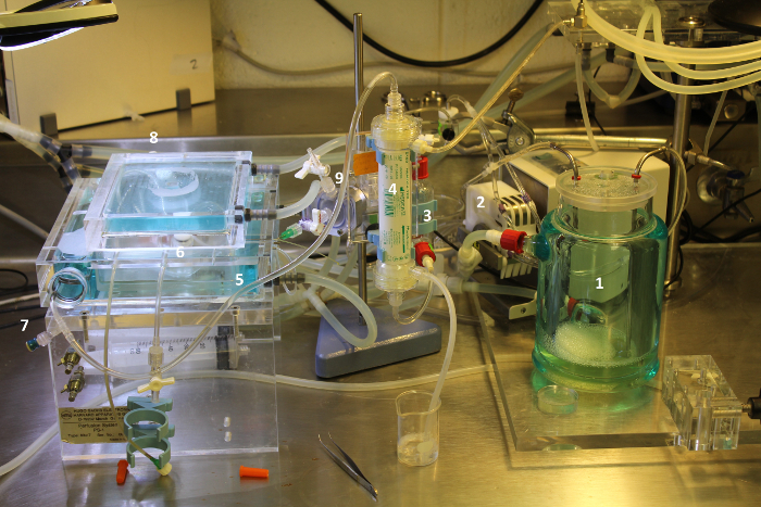
Figure 1: Perfusion Circuit. Components of the circuit are labeled. The perfusate starts in the perfusate reservoir (1), which is a water jacketed container. Perfusate is pulled from the reservoir by a roller pump (2) and pushed into a windkessel (3) and then the oxygenator (4). The oxygenator is set for countercurrent gas and perfusate flow to provide maximum gas exchange. The perfusate then proceeds to a heating coil (5) inside the perfusion chamber to ensure it is at physiologic temperature, and a bubble trap (6) to prevent perfusion of air bubbles. There are pre-organ (7) and post-organ (8) sample ports, which allow the perfusate to be sampled. The perfusate then enters the liver through the portal vein cannula. The portal vein cannula is attached to a pressure monitor, which regulates the pressure equalizer (9). Finally, the perfusate is pulled from the pressure block back through the roller pump and emptied into the reservoir. Please click here to view a larger version of this figure.
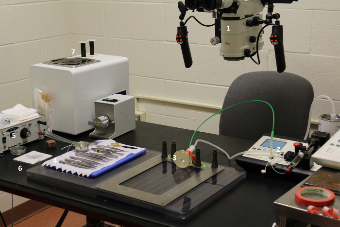
Figure 2: Operating room and surgical instrument set-up. The surgical microscope (1) should be adjusted to the appropriate height and magnification for the user. Isoflurane can be pre-loaded into the anesthesia machine (2). The animal's nose is placed in the nose cone (3). Surgical instruments should be laid out where they can be easily accessed (4). Having electrocautery (5) nearby is helpful. Sutures (6) should be pre-cut so pieces can be obtained quickly when needed, and extra should be available (7). Please click here to view a larger version of this figure.
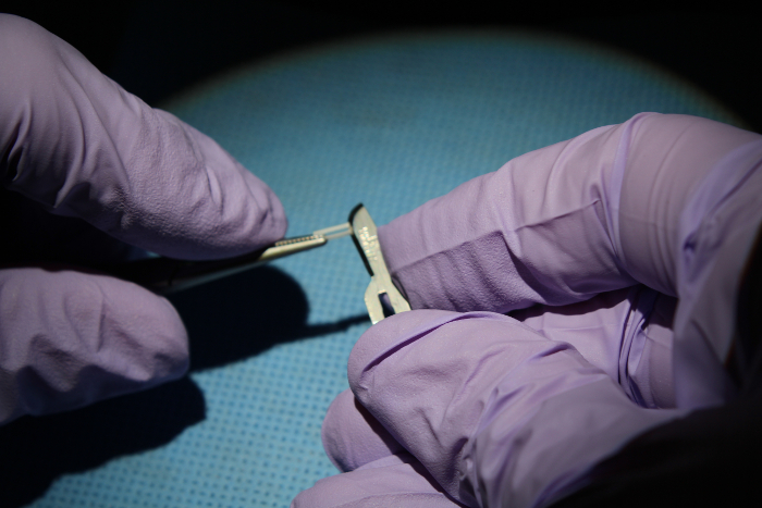
Figure 3: Prepare the 16 G portal vein cuff. Begin with a 16 G angiocatheter. Cut a 7 mm section of tubing. Determine the midpoint of the 7 mm section by measuring 3.5 mm. Incise here and remove the anterior half of the tubing. Use a hemostat to crush this now flat portion. Use a lighter to burn the other end of the angiocatheter to create a lip. Do not place the tip directly into the flame or it will ignite. Please click here to view a larger version of this figure.
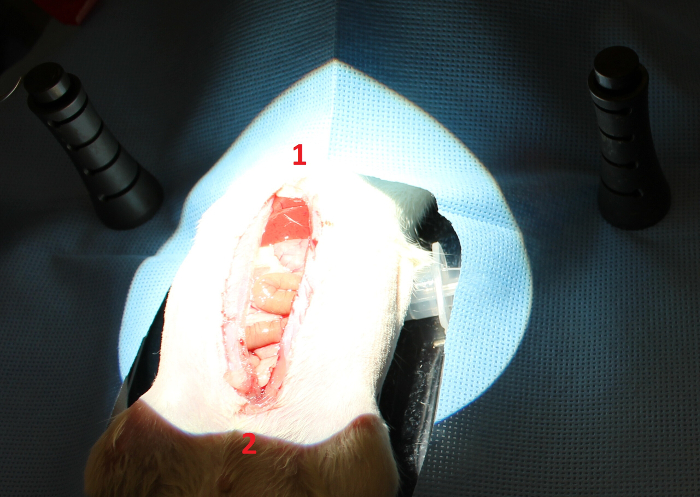
Figure 4: Midline incision. Make a midline incision from the xiphoid (1) to the pubis (2) using sharp scissors and extending through the skin and muscle. Please click here to view a larger version of this figure.
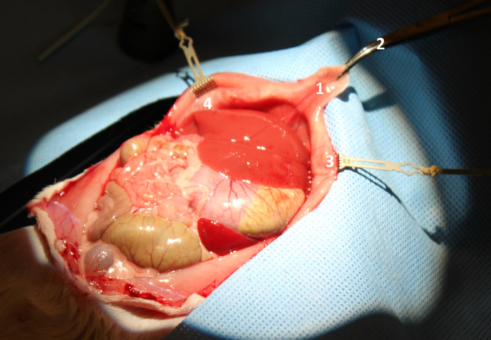
Figure 5: Obtain adequate retraction. Retract the xiphoid process (1) using a curved mosquito clamp (2) and the rib by placing rib retractors (3, 4). Please click here to view a larger version of this figure.
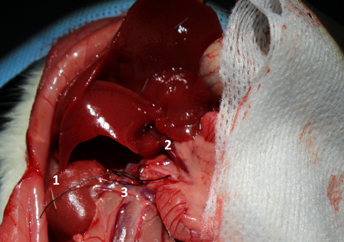
Figure 6: Inferior vena cava (IVC) dissection. Flip the liver up to expose the right kidney (1) and portal vein (2). Dissect around the IVC (3) and place a loop of 7-0 suture for future use. Please click here to view a larger version of this figure.
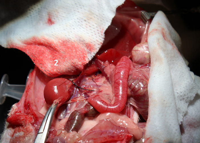
Figure 7: Ligation of the right adrenal vein. Retract the right kidney (1) to provide exposure to the right adrenal vein. Tie off the right adrenal vein and cut across it. A moistened gauze (2) can be used to protect the liver during this maneuver. Please click here to view a larger version of this figure.
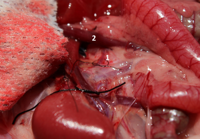
Figure 8: Hepatic artery dissection. Dissect around and place a tie around the hepatic artery (1) near where it passes under the portal vein (2). Please click here to view a larger version of this figure.
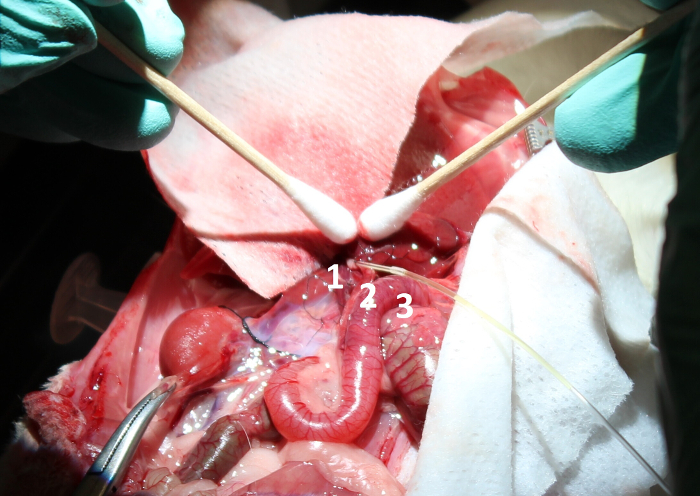
Figure 9: Bile duct cannulation. Cannulate the bile duct (1) using the 27 G angiocatheter (2) connected to the 27 G tubing (3). This will help to collect the bile during perfusion. Please click here to view a larger version of this figure.
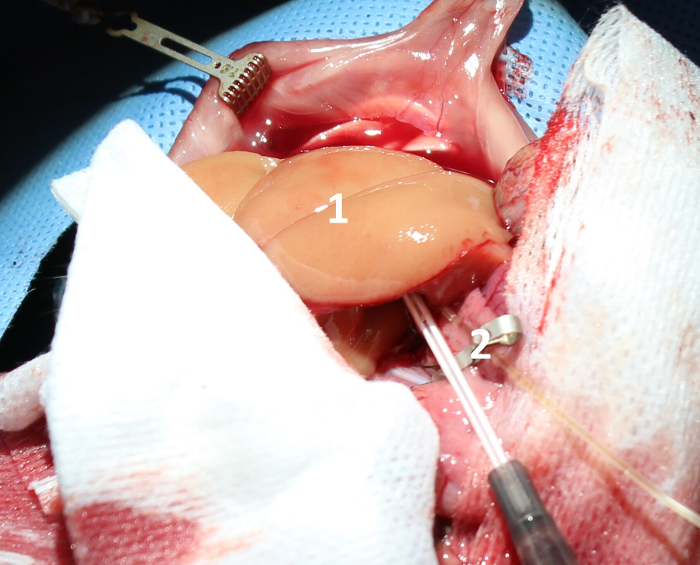
Figure 10: Liver flush. Flush the liver (1) with 60 cc of cold 0.9% normal saline with 100 U (1 mL) of heparin using a 16 G angiocatheter (2). Please click here to view a larger version of this figure.
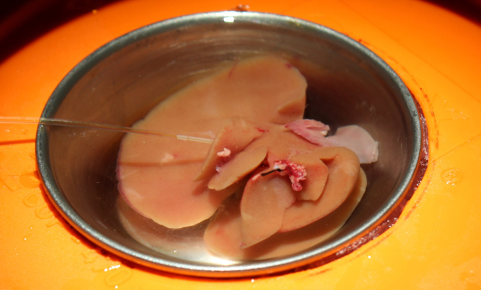
Figure 11: After the hepatectomy. Perform a hepatectomy and place the liver in cold saline. Take care not to dislodge the bile duct cannula. Please click here to view a larger version of this figure.
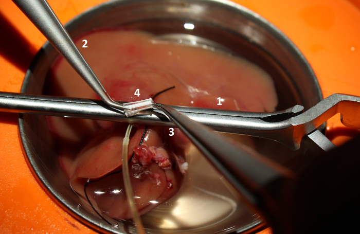
Figure 12: Portal vein cuffing. Locate the portal vein. Use a large clamp (1) to hold up the vein leaving a several millimeter-lip of vein above the clamp. Use microsurgical forceps (2, 3) to place a 16 G vascular cuff (4) in the portal vein. Please click here to view a larger version of this figure.
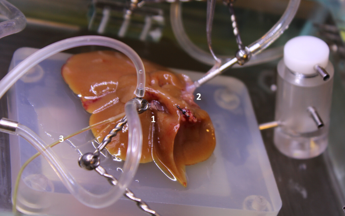
Figure 13: Portal vein cuff and superior IVC cannulation. Cannulate the portal vein cuff (1) and superior IVC (2). Great care must be taken not to dislodge the bile duct cannula (3). Additionally, take care not to twist the superior IVC. Please click here to view a larger version of this figure.
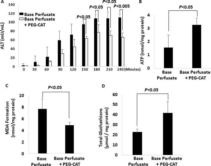
Figure 14: Analysis of tissue damage in base perfusate-only and base perfusate and plus PEG-CAT groups (N = 3/group). Error bars represent standard deviation. (A) Alanine aminotransferase (ALT) Levels. In comparing ALT levels between the base perfusate and base perfusate plus pegylated-catalase (PEG-CAT) group, there is significantly less ALT in the base perfusate plus PEG-CAT group at 150, 180, 210, and 240 min (p <0.05). (B) Adenosine Triphosphate Levels. Tissue adenosine triphosphate (ATP) was maintained in the base perfusate plus PEG-CAT group in comparison to the base perfusate alone group (p <0.05). (C) Malondialdehyde Levels. Tissue malondialdehyde (MDA) production was significantly higher in the base perfusate group than in the base perfusate plus PEG-CAT group (p <0.05). (D) Glutathione Levels. Total glutathione (GSH) was maintained in the base perfusate plus PEG-CAT group in comparison to the base perfusate alone group (p <0.05). Please click here to view a larger version of this figure.
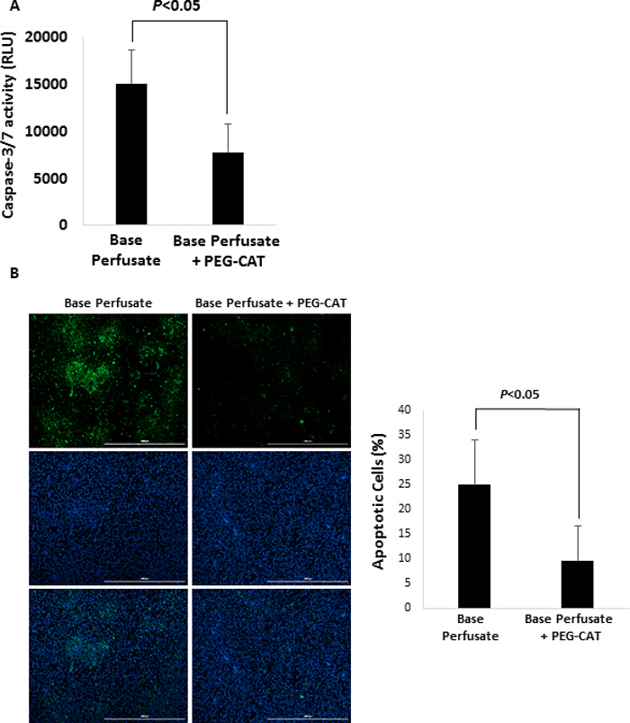
Figure 15: Analysis of apoptosis in base perfusate-only and base perfusate and plus PEG-CAT groups (N = 3/group). Error bars represent standard deviation. (A) Caspase-3/7 Activity.Caspase 3/7 activity was significantly decreased in the base perfusate plus PEG-CAT group in comparison to the base perfusate alone group (p <0.05). (B) Terminal deoxynucleotidyl transferase (TdT) dUTP Nick-End Labeling (TUNEL) staining. Images were taken using a 4X fluorescent microscope. The percentage of apoptotic cells was significantly less in the base perfusate plus PEG-CAT group in comparison to base perfusate (p <0.05). Green: apoptotic cells. Blue: nuclear. Scale bars = 1,000 µm. TUNEL positive cells were quantified by counting cells from 4 random microscopic fields. Please click here to view a larger version of this figure.
There is a significant shortage of liver allografts available for transplantation and in response donor criteria have been expanded1,2,3,4,5. As a result of the donor shortage, NEVLP has been introduced as a method to evaluate and modify organ function6,7. We have designed a rat model of NEVLP. Furthermore, we have used this model to demonstrate one of its important potential applications — the testing of novel molecule additives to liver perfusate. Here, we added PEG-CAT to the base perfusate and demonstrated its ability to mitigate liver preservation injury.
Critical Steps
The liver perfusion circuit was purchased and used without modification. The circuit can be visualized in Figure 1. The perfusate reservoir used is a water-jacketed container that is used to keep the perfusate at physiological temperature. From the reservoir perfusate is pulled out by a roller pumped and pushed into a windkessel chamber. This chamber helps to dampen the pulsatile flow of the perfusate and make it more laminar for entry into the organ. After the windkessel chamber the perfusate flows to the oxygenator. The oxygenator is set for countercurrent gas and perfusate flow providing maximum gas exchange to the perfusate with 95% oxygen. The perfusate then proceeds to a heating coil to ensure that it is still at physiological temperature. A bubble trap right before the organ prevents perfusion of air bubbles. The perfusate is then pumped out of the bubble trap and through the portal vein cannula into the liver. The portal vein cannula has a small branch off of it for the pressure monitor. The tubing to the sensor should be primed with fluid not air so there is no loss of pressure to the sensor. After oxygenating the organ, the perfusate flows out of the liver through the inferior vein cannula to a pressure equalizer. The pressure equalizer block helps prevent over pressurization of the circuit or organ. Finally, the perfusate is pulled from the pressure block back through the roller pump and emptied into the reservoir.
Before beginning each perfusion visual inspection of the circuit should be performed to identify any damage or buildup on circuit components or tubing. If there is a buildup of bacteria or other substances on the circuit, parts should be replaced or cleaned, if possible. Next, the detergent solution maintaining the internal components should be rinsed out. As the components are being rinsed out the pressure sensor and pressure line should be purged of any air bubbles with deionized water. Also, flow should be adjusted at regular intervals to make sure the pressure reading is responding appropriately to changes. If the pressure sensor is not responding appropriately, all items in the line to the sensor should be checked and recalibrated if necessary. At the start of a perfusion it is crucial to make sure the vessels of the liver do not become kinked or twisted when connecting the liver to the circuit. If this has occurred there will be an immediate pressure spike seen on the monitor in a logarithmic trend. The most common error is a kink in the portal vein with a poorly positioned cannula. This issue can be resolved by moving the vessel into a more natural position by pulling it slightly out and straightening the portal vein. The pressure monitor indicates resolution of this problem with a drop in pressure and improved consistency. Next, when connecting the portal vein cuff to the portal vein cannula the vessel can become twisted impeding perfusion of the organ. Adjusting the cuff and correcting this error will result in a sudden spike in portal vein pressure that should then immediately return to a lower pressure and level off at a consistent flow. A kinked or twisted IVC can quickly be identified by no flow from the cannula and a bulging of the vessel. Both of these errors in the vena cava will also result in an increased pressure, however unlike portal vein troubles this pressure is exerted on the organ and should be resolved quickly. This issue should be resolved within 10 min or the experiment should be canceled. An immediate indication for canceling the experiment is seeing clear edema in the organ within the first 20 min.
If there is a leak from the liver or from one of the cannula connections it will be important to monitor perfusate reservoir level. Running out of perfusate and pumping air can be catastrophic to the experiment. Once air is pumped into the lines it is not possible to pause the experiment and re-prime the tubing lines. The only possible correction is for the bubble trap to capture the injected air.
Modifications and Troubleshooting
Once the circuit is flushed the oxygenator can be put in line and then the circuit can be primed with perfusate. Properly purging air from the oxygenator can take a few min but is a crucial step in making sure an air embolism is not generated in the middle of a perfusion. After the oxygenator is fully primed with perfusate the bubble trap should be filled next to capture any air bubbles that do form. At this point the circuit flow should be set to a flow of 1 or 2 mL/min to keep the perfusate moving until the liver is ready for cannulation.
After cannulating the portal vein and the IVC of the liver, the pressure should increase and then level off. As flow is increased to a normal physiological pressure the recorded pressure should start to increase in a similar stepwise manner. Once the desired flow (8–16 mmHg) has been achieved the pressure should remain fairly constant. We aim for a pressure of 10 mmHg, and adjust the flow accordingly. The required flow to reach a pressure of 10 mmHg may vary by organ. There may be a slight leak of perfusate from the organ but this perfusate can be collect and returned to the reservoir.
The circuit should be cleaned after every perfusion to maintain the chamber and reservoir and to preserve the disposable tubing and ports. All perfusate should be removed from the circuit. The circuit should be immediately flushed with a minimum of 300 mL of deionized water. While the deionized water flushes the circuit tubing the external parts should be cleaned appropriately. External components should be rinsed off or gently wiped down and allowed to air dry. Circuit components are fragile and can be damaged easily. It is therefore of utmost importance to clean it gently. The internal circuit should be preserved in a 5% solution of alkaline detergent in deionized water when not in use. The detergent in the circuit helps to extend the life of the tubing and prevent buildup on other components, such as the bubble trap and pressure equalizer.
Most difficulties with the circuit can be prevented with thorough cleaning and maintenance of the circuit after each use. This helps to ensure there is no buildup of residual perfusate that can lead to clogged tubing or cannulas. Circuit components and tubing should be inspected regularly and replaced as needed before each use to make sure there is no contamination or restriction in flow.
Limitations
A limitation of this small animal NEVLP model is that it does not currently include post-perfusion transplantation. It is therefore impossible to evaluate liver graft function after transplantation. This is an important area for future research. Additionally, utilizing the small animal circuit requires both knowledge and skill.
Significance with Respect to Existing Models
Porcine and murine (rat) models of hypothermic, subnormothermic, and normothermic ex vivo liver perfusion have been described in the literature. Although controversy still exists regarding perfusion temperature, it has been shown that that machine perfusion can improve function of liver grafts regardless of temperature9. The NEVLP model presented here is simple, easily replicable, low cost, and has a broad range of applications. This model does not include dialysis or pulsatile flow to the hepatic artery, which are included in some other models, as they have been shown to be unnecessary9,13. Furthermore, the results of the first human trials using NEVLP have shown this to be an effective method of liver preservation — therefore, this model is ideal for testing future applications of ex vivo liver perfusion10.
Future Applications
A variety of future applications for NEVLP have been proposed in the literature. Each of these will need to be methodically tested in animal models prior to testing in discarded human organs and then in human livers. The model presented here is ideal for testing these novel future applications as it is easily replicable, eliminates extraneous steps, and is low cost. One of the most important potential applications of this model is the one demonstrated here – the testing of novel pharmacologic perfusate additives. Other proposed applications include repair of damaged organs, defatting of livers to allow transplantation of steatotic organs, introduction of hepatitis C viral resistance, mesenchymal stem cell therapy, gene modification, and perfusion with immunosuppressant agents11,31,32,33,34,35,36,37,38.
Conclusions
In conclusion, we have demonstrated an inexpensive, easily replicable NEVLP model using rats. Use of this model requires careful preparation, practice, and knowledge, but can be implemented at low cost. Applications of this model can include testing novel perfusate additives, as was demonstrated in the representative results. Additional applications of this model may include testing software designed for organ evaluation, different perfusates, and artificial or hemoglobin-based oxygen carriers and agents designed to repair organs.
The authors have nothing to disclose.
This work was supported by NIH T32AI 106704-01A1 and the T. Flesch Fund for Organ Transplantation, Perfusion, Engineering and Regeneration at The Ohio State University.
Perfusate | |||
8% Albumin | CLS Behring, King of Prussia, PA | 0053-7680-32 | |
Williams Media | Sigma Aldrich, St. Louis, MO | W1878 | |
Penicillin/Streptomycin | Sigma Aldrich, St. Louis, MO | P4333 | |
Insulin | Eli Lilly, Indianapolis, IL | 0002-8215-91 | |
Heparin | Fresnius Lab, Lake Zurich, IL | C504701 | |
L-glutamine | Sigma Aldrich, St. Louis, MO | G3126 | |
Hydrocortisone | Sigma Aldrich, St. Louis, MO | H0888 | |
THAM | Hospira, Inc, | 0409-1593-04 | |
Polyethylene Glycol – Catalase | Sigma Aldrich | S9549 SIGMA | |
Personal Protective Equipment | |||
Surgical Mask | Generic | N/A | |
Protective Gown | Generic | N/A | |
Surgical Gloves | Generic | N/A | |
Liver Procurement | |||
Sprague-Dawley Rat | Harlan Sprague Dawley Inc. | 250 -350 grams | |
Surgical Microscope | Leica | M500-N w/ OHS | |
Charcoal Canisters | Kent Scientific | SOMNO-2001-8 | |
Isoflurane | Piramal Healthcare | N/A | |
Pressure-Lok Precision Analytical Syringe | Valco Instruments Co, Inc. | SOMNO-10ML | |
Electrosurgical Unit | Macan | MV-7A | |
Warming Pad | Braintree Scientific | HHP2 | |
SomnoSuite Small Animal Anesthesia System | Kent Scientific | SS-MVG-Module | |
PhysioSuite | Kent Scientific | PS-MSTAT-RT | |
Isoflurane chamber | Kent Scientific | SOMNO-0530LG | |
SurgiVet | Isotec | CDS 9000 Tabletop | |
Oxygen | Praxair | 98015 | |
Rib retractors | Kent Scientific | INS600240 | |
GenieTouch | Kent Scientific | GenieTouch | |
Normal Saline | Baxter | NDC 0338-0048-04 | |
4×4 Non-Woven Sponges | Criterion | 104-2411 | |
Sterile Q-Tips | Henry Schein Animal Health | 1009175 | |
U-100 27 Gauge Insulin Syringe | Terumo | 22-272328 | |
5mL Syringe | BD | REF 309603 | |
4-0 Braided Silk Suture | Deknatel, Inc. | 198737LP | |
7-0 Braided Silk Suture | Teleflex Medical | REF 103-S | |
16 gauge Catheters | BBraun Introcan Safety | 4252586-02 | |
14 gauge Catheters | BBraun Introcan Safety | 4251717-02 | |
Bile Duct Cannular Tubing | Altec | 01-96-1727 | |
Liver Perfusion Circuit Components | |||
Water Bath Warmer | Lauda Ecoline Staredition | E103 | |
Data Collection Software | ADInstruments | Labchart 7 | |
Liver Perfusion Circuit | Harvard Apparatus | 73-2901 | |
Membrane Oxygenator | Mediac SPA | M03069 | |
Roller Pump | Ismatec | ISM827B | |
Gas (95% oxygen and 5% carbon dioxide) | Praxair | 98015 | |
Organ Chamber | Harvard Apparatus | ILP-2 | |
1.8 mL Arcticle Cryogenic Tube | USA Scientific | 1418-7410 | |
Mucasol | Sigma-Aldrich | Z637181 | |
Microsurgical Instruments | |||
Small Scissors | Roboz | RS-5610 | |
Large Scissors | S&T | SAA-15 | |
Forceps – Large Angled | S&T | JFCL-7 | |
Forceps – Small Angled | S&T | FRAS-15 RM-8 | |
Clip Applier | ROBOZ | RS-5440 | |
Scissors – non micro | FST 14958-11 | 14958-11 | |
Forceps – Straight Tip | S&T | FRS-15 RM8TC | |
Large Microsurgical Clip | Fine Scientific Tools | 18055-01 | |
Small Microsurgical Clip | Fine Scientific Tools | 18055-01 | |
Small Microsurgical Clip | Fine Scientific Tools | 18055-02 | |
Small Microsurgical Clip | Fine Scientific Tools | 18055-03 | |
Small Mosquito Clamps | Generic | N/A | |
Post-Experiment Analysis | |||
Alanine Aminotransferase (ALT) Activity Colorimetric/Fluorometric Assay Kit | BioVision | K752 | |
Adenosine Triphosphate (ATP) Colorimetric/Fluorometric Assay Kit | BioVision | K354 | |
Glutathione Assay Kit | Cayman Chemical | 703002 | |
Lipid Peroxidation (MDA) Assay Kit | Abcam | ab118970 | |
Caspase-Glo 3/7 Assay Systems | Promega | G8090 | |
POLARstar OMEGA Microplate Reader | BMG LABTECH | N/A |