This protocol describes a comprehensive hemocompatibility evaluation of blood-contacting devices using laser-cut neurovascular implants. A flow loop model with fresh, heparinized human blood is applied to mimic blood flow. After perfusion, various hematologic markers are analyzed and compared to the values gained directly after blood collection for hemocompatibility evaluation of the tested devices.
The growing use of medical devices (e.g., vascular grafts, stents, and cardiac catheters) for temporary or permanent purposes that remain in the body's circulatory system demands a reliable and multiparametric approach that evaluates the possible hematologic complications caused by these devices (i.e., activation and destruction of blood components). Comprehensive in vitro hemocompatibility testing of blood-contacting implants is the first step towards successful in vivo implementation. Therefore, extensive analysis according to the International Organization for Standardization 10993-4 (ISO 10993-4) is mandatory prior to clinical application. The presented flow loop describes a sensitive model to analyze the hemostatic performance of stents (in this case, neurovascular) and reveal adverse effects. The use of fresh human whole blood and gentle blood sampling are essential to avoid the preactivation of blood. The blood is perfused through a heparinized tubing containing the test specimen by using a peristaltic pump at a rate of 150 mL/min at 37 °C for 60 min. Before and after perfusion, hematologic markers (i.e., blood cell count, hemoglobin, hematocrit, and plasmatic markers) indicating the activation of leukocytes (polymorphonuclear [PMN]-elastase), platelets (β-thromboglobulin [β-TG]), the coagulation system (thombin-antithrombin III [TAT]), and the complement cascade (SC5b-9) are analyzed. In conclusion, we present an essential and reliable model for extensive hemocompatibility testing of stents and other blood-contacting devices prior to clinical application.
The in vivo application of implants and biomaterials, which interact with human blood, requires intense preclinical testing focusing on the investigation of various markers of the hemostatic system. The International Organization for Standardization 10993-4 (ISO 10993-4) specifies the central principles for the evaluation of blood-contacting devices (i.e., stents and vascular grafts) and considers the device design, clinical utility, and materials needed1.
Human blood is a fluid that contains various plasma proteins and cells, including leukocytes (white blood cells [WBCs]), erythrocytes (red blood cells [RBCs]), and platelets, which carry out complex functions in the human body2. The direct contact of foreign materials with blood can cause adverse effects, such as activation of the immune or coagulation system, which can lead to inflammation or thrombotic complications and serious issues after implantation3,4,5. Therefore, in vitro hemocompatibility validation offers an opportunity prior to implantation to detect and exclude any hematologic complications that may be induced upon contact of the blood with a foreign surface6.
The presented flow loop model was established to assess the hemocompatibility of neurovascular stents and similar devices by applying a flow rate of 150 mL/min in tubing (diameter of 3.2 mm) to mimic cerebral flow conditions and artery diameters2,7. Besides the need for an optimal in vitro model, the source of blood is an important factor in gaining reliable and unaltered results when analyzing hemocompatibility of a biomaterial8. The collected blood should be used immediately after sampling to prevent changes caused by prolonged storage. In general, a gentle collection of blood without stasis using a 21 G needle should be performed to minimize the preactivation of platelets and the coagulation cascade during blood drawing. Furthermore, donor exclusion criteria include those who smoke, are pregnant, are in a poor state of health, or have taken oral contraceptives or painkillers during the previous 14 days.
This study describes an in vitro model for the extensive hemocompatibility testing of stent implants under flow conditions. When comparing uncoated to fibrin-heparin-coated stents, results of the comprehensive hemocompatibility tests reflect improved hemocompatibility of the fibrin-heparin-coated stents9. In contrast, the uncoated stents induce activation of the coagulation cascade, as demonstrated by an increase in thombin-antithrombin III (TAT) concentrations and loss of blood platelet numbers due to the adhesion of platelets to stent surface. Overall, integrating this hemocompatibility model as a preclinical test is recommended to detect any adverse effects on the hemostatic system that are caused by the device.
The blood sampling procedure was approved by the Ethics Committee of the medical faculty at the University of Tuebingen (project identification code: 270/2010BO1). All subjects provided written, informed consent for inclusion before participation.
1. Preparation of Heparin-loaded Monovettes
2. Blood Sampling
3. Preparation of the Flow Loop
4. Performance of Hemocompatibility Testing
5. Whole Blood Count Analysis
6. Collection of Citrate Plasma
7. Collection of EDTA Plasma
8. Collection of CTAD Plasma
9. Measurement of Human TAT from Citrate Plasma
10. Measurement of PMN-elastase from Citrate Plasma
11. Measurement of Terminal Complement Complex (TCC) from EDTA Plasma
12. Measurement of β-thromboglobulin from CTAD Plasma
13. Sample Preparation for Scanning Electron Microscopy
14. Scanning Electron Microscopy
Briefly summarized, human whole blood was collected in heparin-loaded monovettes then pooled and used to evaluate the baseline levels of cell counts as well as plasmatic hemocompatibility markers.
Subsequently, the tubing containing the neurovascular implant samples was filled, and the blood was perfused for 60 min at 150 mL/min and 37 °C using a peristaltic pump. Again, the number of cells was analyzed in all groups, and the plasma samples were prepared for ELISA analyses (Figure 1). The quantification of the blood cells and blood parameters, such as hemoglobin and hematocrit, was performed directly after blood collection as well as after perfusion in the flow loop model for all sample types and the control. No changes were detected regarding the number of WBCs (Figure 2A), RBCs (Figure 2B), or the hematocrit values (Figure 2C). However, a decrease in hemoglobin levels was detected after the incubation of blood in the flow loop model when compared to the baseline values, which was due to the perfusion of blood in the flow loop system (Figure 2D). In addition, a decrease in platelet numbers was observed due to blood perfusion. Furthermore, this effect was increased when an uncoated stent was present in the tubing, indicating the adhesion of platelets to the biomaterial. Nonetheless, it was clearly demonstrated that the loss of platelets was significantly higher when the uncoated stent was incubated with blood, as opposed to the fibrin-heparin-coated stent (Figure 2E).
Potential alterations of the hematologic plasma markers were also investigated in the test groups after perfusion and compared to the baseline values of the freshly drawn blood. The TAT complex concentration, which reflects the activation status of the coagulation system, was mildly increased due to blood perfusion (Figure 3A). In the bare metal stent group, however, a significant increase in the TAT was detected, indicating a profound activation of the coagulation system. The fibrin-heparin-coated stent prevented the activation of the coagulation system, since no increase in the TAT was determined.
The perfusion led to an increased activation of the complement cascade, which was determined by measuring SC5b-9 (Figure 3B). However, incubation with uncoated or fibrin-heparin-coated stents did not further increase the SC5b-9 concentration. Similar results were obtained when analyzing the activation of the neutrophil granulocytes through the quantification of PMN-elastase concentrations (Figure 3C).
Visualization of the stent surface was performed using SEM. Clear differences between the two stent groups were detected after blood incubation. While on the surface of the uncoated stent a dense network of blood cells and proteins was present, no adhesion of proteins or cells was detected on the surface of the fibrin-heparin-coated stent (Figure 4).
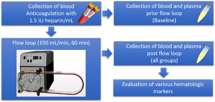
Figure 1: Schematic overview of the hemocompatibility evaluation of stents in a well-established flow loop model. Fresh human whole blood is collected from healthy donors in blood tubes containing heparin for anticoagulation. For each donor, an empty tube as well as tubes preloaded with the sample material are subsequently filled with fresh blood and incubated in the flow loop at a rate of 150 mL/min at 37 °C for 60 min. Additionally, plasma samples are prepared from freshly drawn blood to obtain the baseline values of each donor. After the incubation, the plasma samples from the test tubing, with and without sample materials, are prepared and analyzed using a specific ELISA. Please click here to view a larger version of this figure.
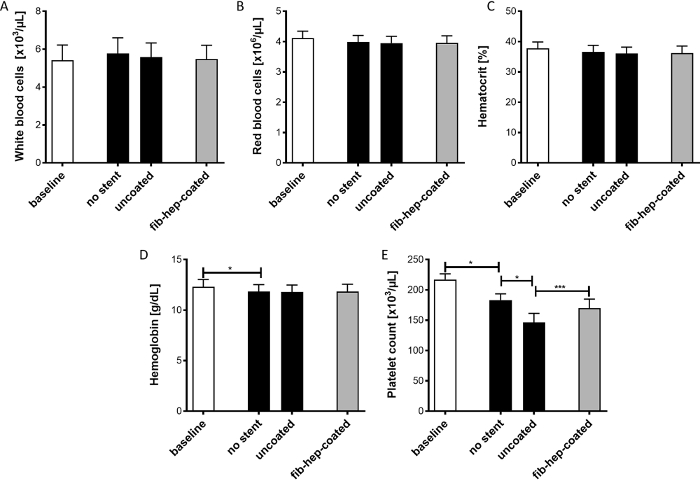
Figure 2: Analysis of different cell types and blood parameters before and after incubation of different stent implants in the flow loop model. The determination of white blood cells (A), red blood cells (B), hematocrit (C), hemoglobin (D), and platelets (E) was performed. The data are displayed as mean ± SEM (n = 5, p* < 0.5, p*** < 0.001). Please click here to view a larger version of this figure.
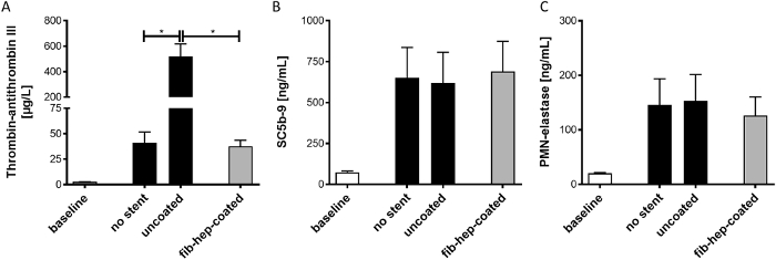
Figure 3: Determination of platelet or immune system activation markers before and after incubation with neurovascular implants. The markers for the (A) activation of blood coagulation (TAT), (B) complement system (SC5b-9), and (C) neutrophils (PMN-elastase) were quantified using ELISA. The analysis was performed on plasma samples gained from freshly drawn blood or blood incubated with different stents in the flow loop model. The data are displayed as mean ± SEM (n = 5, p* < 0.5). Please click here to view a larger version of this figure.
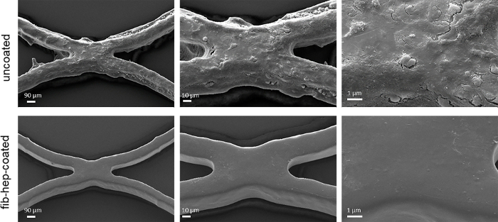
Figure 4: Scanning electron microscopic analysis of stents after incubation with blood. The aggregation of blood plasma proteins and platelets on uncoated stent material was observed. In comparison, stent materials with the fibrin-heparin coating did not demonstrate adhesion of cells or other blood components on the surface (magnification of 500-, 1,000-, and 2,500-fold). Please click here to view a larger version of this figure.
The presented protocol describes a comprehensive and reliable method for the hemocompatibility testing of blood-contacting implants in accordance with ISO 10993-4 in a shear flow model imitating human blood flow. This study is based on the testing of laser-cut neurovascular implants but can be performed with a variety of samples. The results demonstrate that this method enables the broad analysis of various parameters such as the blood cell count, prevalence of several hemocompatibility markers, and microscopic visualization of the device surface after blood contact. Using this protocol, potential differences regarding the hemocompatibility of different devices can be detected.
An alternative to in vitro hemocompatibility assessment consists of in vivo animal testing, which is associated with several disadvantages, such as higher variability and distortion of device-related effects due to the overwhelming short-term effects of tissue injury6.
For in vitro hemocompatibility testing, three types of models are available: (1) static blood incubation models, (2) agitated blood incubation models, and (3) shear flow models. The static model provides a simple and rapid method to determine thrombogenicity by incubating the device directly with blood, but it only leads to rudimentary results regarding hemocompatibility11. To overcome the main disadvantages of static models (i.e., sedimentation of blood cells and the large air-contacting surface), the agitating blood incubation model may be used, in which a test chamber containing the implants is filled with blood and incubated on a rocking platform12. However, these model types are still inferior compared to the existing shear flow models, such as the flow loop presented here. The quintessence of these models is that vascular human blood flow can be imitated; thus, a close depiction of the real interaction between the implant and blood cells can be displayed13. In addition to the flow loop model, models such as the Chandler loop or several perfused flow chamber exist14,15,16.
The Chandler loop is a closed tube system that is partly filled with air and clamped into a rotating device, resulting in blood circulation through the tubing17. In the present flow loop system, the tube is completely filled with blood, and the flow is forced by using a peristaltic pump. When using the Chandler loop model, operators face two major disadvantages due to the requirement of including air into the test tubing. First, it is known that the constant interaction of blood and air triggers the aggregation of leukocytes and platelets as well as protein denaturation18,19. Second, the blood circulation rate is limited, because the air always remains at the highest point of the loop20.
These drawbacks can be overcome when using the flow loop system. Since no air-liquid interface is present in the system, no platelet activation occurs. Thus, the model has a low background for thrombotic events so that a low concentration of anticoagulants, typically 1 IU/mL or 1.5 IU/mL of heparin, is sufficient to prevent clotting, even if high flow rates are applied6. The adjustable pump-regulated blood flow rate and the freely selectable tube diameter allow the operator to mimic the physiological conditions of a vein or artery, which correspond to the implant to be tested, and achieve relevant test results21. However, this advantage is at the same time a limitation, due to the mechanical stress applied to the blood through the pump, and the destruction of erythrocytes (i.e., hemolysis) may occur2. This arising intrinsic blood damage reduces the method sensitivity and impedes prolonged exposure to the blood21. Nevertheless, several studies have demonstrated the effective use of the flow loop model for hemocompatibility evaluation22,23,24.
However, the main gap between all in vitro models and the in vivo mechanisms includes the missing endothelium, which expresses cytokines, anti-thrombotic components, and adhesion molecules; therefore, this component plays a crucial role in the interaction of the implant and circulating blood25. In conclusion, the flow loop model is adjustable, efficient, reliable, and cost-effective to assess the hemocompatibility of implants before clinical use.
The authors have nothing to disclose.
For the performance of scanning electron microscopy, we thank Ernst Schweizer from the section of Medical Materials Science and Technology of the University Hospital Tuebingen. The research was supported by the Ministry of Education, Youth and Sports of the CR within National Sustainability Program II (Project BIOCEV-FAR LQ1604) and by Czech Science Foundation project No. 18-01163S.
aqua ad iniectabilia | Fresenius-Kabi, Bad-Homburg, Germany | 1088813 | |
beta-TG ELISA | Diagnostica Stago, Duesseldorf, Germany | 00950 | |
Centrifuge Rotana 460 R | Andreas Hettich, Tuttlingen, Germany | – | |
Citrat monovettes (1.4 mL) | Sarstedt, Nümbrecht, Germany | 6,16,68,001 | |
CTAD monovettes (2.7 mL) | BD Biosciences, Heidelberg, Germany | 367562 | |
EDTA monovettes (1.2 mL) | Sarstedt, Nümbrecht, Germany | 6,16,62,001 | |
Ethanol p.A. (1000 mL) | AppliChem, Darmstadt, Germany | 1,31,08,61,611 | |
Glutaraldehyde (25 % in water) | SERVA Electrophoresis, Heidelberg, Germany | 23114.01 | |
Heparin coating for tubes | Ension, Pittsburgh, USA | – | |
Heparin-Natrium (25.000 IE/ 5 mL) | LEO Pharma, Neu-Isenburg, Germany | PZN 15261203 | |
Multiplate Reader Mithras LB 940 | Berthold, Bad Wildbad, Germany | – | |
NaCl 0,9% | Fresenius-Kabi, Bad-Homburg, Germany | 1312813 | |
Neutral monovettes (9 mL) | Sarstedt, Nümbrecht, Germany | 2,10,63,001 | |
PBS buffer (w/o Ca2+/Mg2+) | Thermo Fisher Scientific, Darmstadt, Germany | 70011044 | |
Peristaltic pump ISM444B | Cole Parmer, Wertheim, Germany | 3475 | |
Pipette (100 µL) | Eppendorf, Wesseling-Berzdorf, Germany | 3124000075 | |
Pipette (1000 µL) | Eppendorf, Wesseling-Berzdorf, Germany | 3123000063 | |
Plastic container (100 mL) | Sarstedt, Nümbrecht, Germany | 7,55,62,300 | |
PMN-Elastase ELISA | Demeditec Diagnostics, Kiel Germany | DEH3311 | |
Polyvinyl chloride tube | Saint-Gobain Performance Plastics Inc., Courbevoie France | – | |
Reaction Tubes (1.5 mL) | Eppendorf, Wesseling-Berzdorf, Germany | 30123328 | |
neurovascular laser-cut implants | Acandis GmbH, Pforzheim | 01-0011x | |
SC5b-9 ELISA | TECOmedical, Buende, Germany | A029 | |
Scanning electron microscope | Cambridge Instruments, Cambridge, UK | – | |
Sealing tape (96 well plate) | Thermo Fisher Scientific, Darmstadt, Germany | 15036 | |
Syringe 10/12 mL Norm-Ject | Henke-Sass-Wolf, Tuttlingen, Germany | 10080010 | |
TAT micro kit | Siemens Healthcare, Marburg, Germany | OWMG15 | |
Waterbath Type 1083 | Gesellschaft für Labortechnik, Burgwedel, Germany | – |