A modified 2 kidney 1 clip (2K1C) Goldblatt mouse model was developed using polyurethane tubing to initiate renal artery stenosis, inducing an increase in renin expression and kidney injury. Here, we describe a detailed procedure of preparing and placing the cuff onto the renal artery to generate a reproducible and consistent 2K1C mouse model.
Renal artery stenosis is a common condition in patients with coronary or peripheral vascular disease where the renin angiotensin aldosterone system (RAAS) is overactivated. In this context, there is a narrowing of the renal arteries that stimulate an increase in the expression and release of renin, the rate-limiting protease in RAAS. The resulting rise in renin expression is a known driver of renovascular hypertension, frequently associated with kidney injury and end organ damage. Thus, there is a great interest in developing novel treatments for this condition. The molecular and cellular mechanism of renin control in renal artery stenosis is not fully understood and warrants further investigation. To induce renal artery stenosis in mice, a modified 2 kidney 1 clip (2K1C) Goldblatt mouse model was developed. The right kidney was stenosed in wild type mice and sham operated mice were used as control. After renal artery stenosis, we determined renin expression and kidney injury. Kidneys were harvested, and fresh cortices were used to determine protein and mRNA expression of renin. This animal model is reproducible and can be used to study pathophysiological responses, molecular and cellular pathways involved in renovascular hypertension and kidney injury.
Renal artery stenosis (RAStenosis) is an intractable problem affecting about 6% of people over 65 and in up to 40% of people with coronary or peripheral vascular disease1,2. Current treatments for the disease are limited; therefore, there is a critical need to develop new therapies to treat renovascular hypertension or resistant hypertension induced by RAStenosis. Renin angiotensin aldosterone system (RAAS) is the key pathway involved in the pathogenesis of RAStenosis induced hypertension or renovascular hypertension3,4. Known therapies targeting RAAS, such as ACE inhibitors or angiotensin receptor blockers, alleviate hypertension, but need close examining for kidney failure and hyperkalemia5,6,7. Renin catalyzes the rate-limiting step in RAAS; it converts angiotensinogen to angiotensin I. In atherosclerosis, plaque formation causes the narrowing of renal artery that drives renin secretion, resulting in renovascular hypertension and kidney damage8. A number of studies have reported increased levels of oxidative stress during renovascular hypertension in humans, which were corroborated with the two kidney one clip (2K1C) mice model as well as other hypertensive animal models2,9,10,11,12,13,14,15,16. The molecular mechanism of renin expression control during RAStenosis induced renovascular hypertension is not well understood and warrants further investigation.
Experimental animal models that reliably and reproducibly recapitulate RAStenosis are important in elucidating the cellular and molecular mechanisms of renin expression control for the development of novel therapies. The 2K1C mouse model is a well-established experimental model to study the pathogenesis of renovascular hypertension17,18,19,20. This model is generated by the constriction of the renal artery using a clip17,20,21, therefore producing renal artery occlusion that results in an increase in renin expression and hypertension17,19,20,21. However, there are no technical reports available, which describe a step by step procedure to generate renal artery stenosis in animal models.
Conventional U-shaped silver clips, polyurethane tubes and other clips have been used to constrict the renal artery to induce renal artery stenosis. Some studies have shown that the design and material of the clip are critical to obtaining reliable and reproducible data with the 2K1C animal model. According to Lorenz et al., the use of conventional U-designed silver clips induces a low success rate of hypertension (40-60%)21. Due to the clip design, the renal artery is press laterally, triggering a few constrictions and greater probability to be dislodged from the renal artery. Silver malleability and ductility may allow changes in clip widths; therefore, causing different hypertension levels among mice. Silver dioxides on the clip can cause perivascular inflammation, intimal proliferation, and tissue granulation, altering the renal artery diameter22. Due to the variability in the levels of hypertension obtained with the conventional U-design silver clip, Warner et al. and Lorenz et al. have successfully used a rounder-design polyurethane tubing to initiate renal artery stenosis in mice, generating a more reliable and consistent induction of the two kidney one clip animal model20,21.
In this report, we describe a surgical protocol to generate experimental RAStenosis in mice, using the polyurethane tubing to constrict the renal artery. The polyurethane round-design cuff is a more reproducible, reliable and low-cost clip to generate stenosis in mouse. The goal of this experimental model is to study and define the molecular and cellular mechanism of renin expression control during renal artery stenosis. We confirmed the success of RAStenosis mice model by measuring renin expression and kidney injury marker neutrophil gelatinase-associated lipocalin (N-GAL).
Mice were housed and cared at the Vanderbilt University Medical Center (VUMC) Division of Animal Care following the National Institutes of Health (NIH) guidelines and the Guide for the Care and Use of Laboratory Animals, US Department of Health and Human Services. All animal procedures were approved by the VUMC Institutional Animal Care and Use Committee prior to starting the experiments.
1. Animal preparation and dissection
2. Right renal artery stenosis
3. Post-operative care
4. Tissue harvest
5. Statistics
Renal artery constriction increases renin expression in the stenosed kidney while repressing expression in the contralateral kidney. The two kidney one clip (2K1C) or Goldblatt model of stenosis induces increased renin expression and kidney injury. This is recognized as the best representative model of unilateral renal artery stenosis in humans.
Expression of renin and prorenin (precursor of renin) were measured using immunoblotting. The data show that renin and prorenin expression increased in the stenosed kidney comparing to contralateral and sham kidneys, suggesting that the cuff was constricting the renal artery causing changes in renal perfusion (Figure 1). To visualize the localization of renin expression, IHC was performed. IHC corroborated immunoblotting data showing increased expression of renin in the clipped kidney (Figure 2). Moreover, juxtaglomerular (JG) cells recruitment along the afferent arteriole was seen in the stenosed kidney (Figure 2). To investigate the effect on renin mRNA expression levels, ISH was performed. The ISH data suggest increased renin mRNA and JG cells recruitment in the stenosed kidney when compared to contralateral and sham kidneys (Figure 3).
Another characteristic of renal artery stenosis is the upregulation of kidney injury markers due to changes in kidney perfusion, superoxide production and hypertension2,25,26. Neutrophil gelatinase-associated lipocalin (NGAL) is a well characterized acute injury marker and is overexpressed during kidney injury27,28. Therefore, acute kidney injury marker NGAL was measured using immunoblotting. Immunoblotting data showed that N-GAL was highly upregulated in the stenosed kidney when compared to the contralateral and sham kidneys (Figure 4).
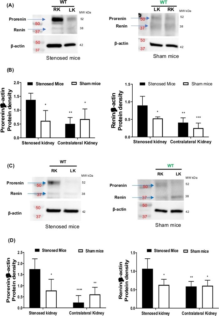
Figure 1: Renin expression. After 15- and 3-days of renal artery stenosis, mice were euthanized. Kidneys were harvested, and renin expression was determined by western blot. (A) is showing representative western blots images from 15-days stenosed (left panel) and sham mice (right panel). (B) is showing the densitometric analysis of prorenin (left panel) and renin (right panel) protein bands. Beta-actin was used as loading control. (C) is showing the representative western blots images from 3-day stenosed (left panel) and sham mice (right panel). (D) is showing densitometric analysis of prorenin (left panel) and renin (right panel) protein bands. Beta-actin was used as loading control. Data are presented as the mean ± SD. P value calculated with two-way ANOVA followed by Tukey post-hoc test. *P<0.05, **P< 0.01, ***P< 0.001, N=3-6. Please click here to view a larger version of this figure.
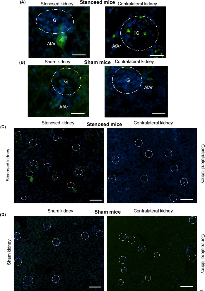
Figure 2: Immunohistochemistry analysis for visualization and localization of renin expression after renal artery stenosis. Kidneys were isolated after euthanizing mice from 3-day renal artery stenosis. One piece from longitudinal cut of the whole kidney was fixed with 4% neutral buffered formalin solution, after that dehydrated in a graduated ethanol series, and embedded in paraffin. Green staining represents renin protein expression; blue, nuclei. (A) Representative microscopy images of stenosed kidney (left side), contralateral kidney (right side) from stenosed mice. (B) Representative microscopy images of sham kidney (left side), and contralateral kidney (right side) from sham mice. Scale bar 30 microns. 90X magnification. (C) Representative microscopy images of stenosed kidney (left side), contralateral kidney (right side) from stenosed mice. (D) Representative microscopy images of sham kidney (left side), and contralateral kidney (right side) from sham mice. These images were mainly taken from cortex region. Scale bar 50 µm. 15x magnification. White dotted circles denote the location of glomeruli. G: Glomeruli, AfAr: Afferent arteriole, N=4. Please click here to view a larger version of this figure.
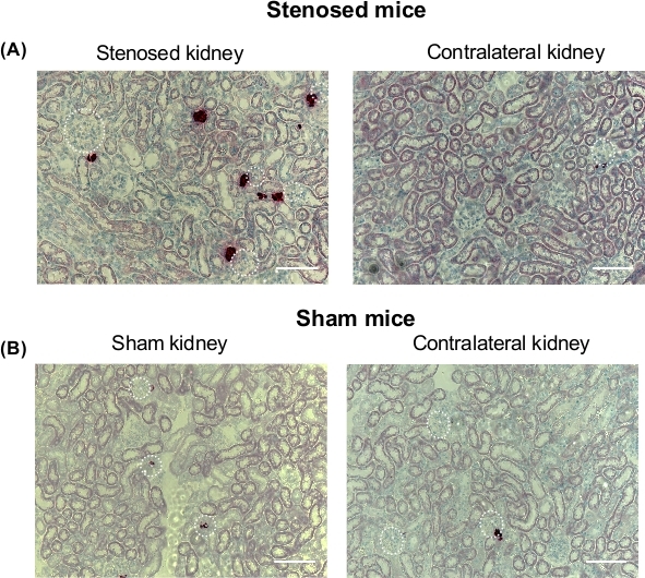
Figure 3: In situ hybridization analysis of renin mRNA expression after renal artery stenosis. After 3-days of renal artery stenosis, mice were euthanized and kidneys were isolated and perfusion-fixed with 4% neutral buffered formalin solution, dehydrated in a graduated ethanol series, and embedded in paraffin. In situ hybridization was performed following the manufacturer’s instructions. Dark red staining represents mRNA renin expression; blue, nuclei. (A). Representative microscopy image of stenosed kidneys (left side), and contralateral kidney (right side) from stenosed mice. (B). Representative microscopy image of sham kidney (left side), and contralateral kidney (right side) from sham mice. Scale bar 50 µm. White dotted circles denote glomeruli expressing renin. G: Glomeruli, AfAr: Afferent arteriole, N=4. Please click here to view a larger version of this figure.
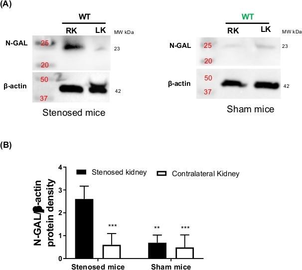
Figure 4: Neutrophil gelatinase-associated lipocalin (N-GAL) expression after renal artery stenosis. After 3-day renal artery stenosis, mice were euthanized and kidneys were harvested, and N-GAL expression measure by Western blot. (A) Representative Western blots images from 3-day stenosed (left panel) and sham mice (right panel). Beta-actin was used as loading control. (B) Densitometric analysis of N-GAL bands. Protein density values of N-GAL were normalized to β-actin. Data are presented as the mean ± SD. P value calculated with two-way ANOVA followed by Tukey post-hoc test. **P<0.01, ***P< 0.001, N=3-5. Please click here to view a larger version of this figure.
Renal artery stenosis is an important cause of secondary or resistant hypertension, and kidney injury1,29. The two kidney one clip (2K1C) Goldblatt model has been employed to study RAStenosis induced renovascular hypertension1,17,18,19. A number of previous studies using various animals models have shown that stenosis in the renal artery is a strong stimulator of renin overexpression and release, and kidney injury18,30,31,32,33,34,35. Moreover, this model is used to study immune cell infiltration, fibrosis, inflammation, and acute and chronic kidney injury markers29.
Here, we described a detailed and step by step procedure to generate reproducible, reliable and consistent renal artery stenosis model in mice. Earlier, metal clips have been employed to initiate renal artery stenosis36,37,38. As an alternative, we used polyurethane round tubing (MRE 025; internal diameter (ID) = 0.30 mm; outside diameter (OD) = 0.63 mm; wall thickness, (WT) = 0.16 mm). We used tubing, since placement of a polyurethane cuff would result constriction in two dimensions (constriction) rather than one (flattening), as with a metal clip. Also, using polyurethane round tubing provides an advantage of uniform constriction in the renal artery. The critical step and challenges are to cut the right size of polyurethane tubing, which requires extreme attention to details that must be performed using a microscope. Another critical criterion is to keep mice between 18-22 g to fit the tubing onto the renal artery. Mice within this weight range normally have a renal artery outer diameter (OD) that is consistently within the range of the tubing cuff diameter. A limitation of the method is that heavy (above 25 g) or small (below 16 g) mice are difficult to perform surgery on because of the size of the tube and cuff made in it. However, when required, changes in the polyurethane tubing can be made to accommodate younger or older mice.
We have conducted 3-day and 15-day studies to initiate renal artery stenosis in mice with about 95% success rate. In our experience, induction of the renal artery stenosis using this method produced reliable, reproducible, and consistent results among the mice regardless of the sex. To confirm the constriction of renal artery, we measured renin expression and kidney injury. Our data suggest that renin expression significantly increased in the stenosed kidney.
The authors have nothing to disclose.
Research was supported by NHLBI Research Scientist Development Grant (1K01HL135461-01) to JAG. Thank you to David Carmona-Berrio, and Isabel Adarve-Rengifo for their technical assistance.
Diet Gel | Clear H2O | Diet-Gel 76A | Surgery recovery diet |
EMC Heated Hard pad | Hallowell | 000A2788B | Heating pads were used to keep mice warm |
Ethilon Nylon Suture | Ethicon | 662G | 4-0 (1.5 metric), This suture was used to close the peritoneum, and skin |
Ethilon Nylon Suture | Ethicon | 2815 G | 8-0 (0.4 metric), This suture was used to close cuff to tie and constrict the artery |
Germinator 500 | Braintree Scientific Inc. | GER 5287 | Sterilize surgical tools between surgeries |
Ketoprofen | Zoetis | Ketofen | Painkiller |
Polyurethane | Braintree Scientific Inc. | MRE-025 | This tube was used to initiate stenosis |
Povidone-iodine antiseptic swabsticks | Medline | MDS093901 | It was applied after hair removal and surgery on the skin |
Reflex 7 Clip Applier | Roboz Surgical Instrument Co | 204-1000 | This clip applier was used to apply clip in case one or more sutures went off |
Sterile towel drapes | Dynarex | 4410 | It was used as a bedsheet for mice during surgery |
Triple antibiotic ointment | Medi-First | 22312 | |
Water pump | Stryker | T/pump Professionals | Used to warm and circulate water in the heating hard pad to keep mice warm during and post-surgery |