Described here is a method for utilizing zebrafish embryos to study the ability of functionalized nanoparticles to target human cancer cells in vivo. This method allows for the evaluation and selection of optimal nanoparticles for future testing in large animals and in clinical trials.
Developing nanoparticles capable of detecting, targeting, and destroying cancer cells is of great interest in the field of nanomedicine. In vivo animal models are required for bridging the nanotechnology to its biomedical application. The mouse represents the traditional animal model for preclinical testing; however, mice are relatively expensive to keep and have long experimental cycles due to the limited progeny from each mother. The zebrafish has emerged as a powerful model system for developmental and biomedical research, including cancer research. In particular, due to its optical transparency and rapid development, zebrafish embryos are well suited for real-time in vivo monitoring of the behavior of cancer cells and their interactions with their microenvironment. This method was developed to sequentially introduce human cancer cells and functionalized nanoparticles in transparent Casper zebrafish embryos and monitor in vivo recognition and targeting of the cancer cells by nanoparticles in real time. This optimized protocol shows that fluorescently labeled nanoparticles, which are functionalized with folate groups, can specifically recognize and target metastatic human cervical epithelial cancer cells labeled with a different fluorochrome. The recognition and targeting process can occur as early as 30 min postinjection of the nanoparticles tested. The whole experiment only requires the breeding of a few pairs of adult fish and takes less than 4 days to complete. Moreover, zebrafish embryos lack a functional adaptive immune system, allowing the engraftment of a wide range of human cancer cells. Hence, the utility of the protocol described here enables the testing of nanoparticles on various types of human cancer cells, facilitating the selection of optimal nanoparticles in each specific cancer context for future testing in mammals and the clinic.
The development of nanoparticles that are capable of detecting, targeting, and destroying cancer cells is of great interest to both physicists and biomedical researchers. The emergence of nanomedicine led to the development of several nanoparticles, such as those conjugated with targeting ligands and/or chemotherapeutic drugs1,2,3. The added properties of nanoparticles enable their interaction with the biological system, sensing and monitoring biological events with high efficiency and accuracy along with therapeutic applications. Gold and iron oxide nanoparticles are primarily used in computed tomography and magnetic resonance imaging applications, respectively. While the enzymatic activities of gold and iron oxide nanoparticles allow the detection of cancer cells through colorimetric assays, fluorescent nanoparticles are well suited for in vivo imaging applications4. Among them, ultrabright fluorescent nanoparticles are particularly beneficial, due to their ability to detect cancers early with fewer particles and reduced toxicities5.
Despite these advantages, nanoparticles require experimentation using in vivo animal models for the selection of suitable nanomaterials and optimization of the synthesis process. Additionally, just like drugs, nanoparticles rely on animal models for preclinical testing to determine their efficacy and toxicities. The most widely used preclinical model is the mouse, which is a mammal whose upkeep comes at a relatively high cost. For cancer studies, either genetically engineered mice or xenografted mice are typically used6,7. The length of these experiments often spans from weeks to months. In particular, for cancer metastasis studies, cancer cells are directly injected into the circulatory system of the mice at locations such as tail veins and spleens8,9,10. These models only represent the end stages of metastasis when tumor cells extravasate and colonize distant organs. Moreover, due to visibility issues, it is particularly challenging to monitor tumor cell migration and nanoparticle targeting of tumor cells in mice.
The zebrafish (Danio rerio) has become a powerful vertebrate system for cancer research due to its high fecundity, low cost, rapid development, optical transparency, and genetic conservations11,12. Another advantage of the zebrafish over the mouse model is the fertilization of the fish eggs ex utero, which allows the embryos to be monitored throughout their development. Embryonic development is rapid in zebrafish, and within 24 hours postfertilization (hpf), the vertebrate body plane has already formed13. By 72 hpf, eggs are hatched from the chorion, transitioning from the embryonic to the fry stage. The transparency of the zebrafish, the Casper strain in particular14, provides a unique opportunity to visualize the migration of cancer cells and their recognition and targeting by nanoparticles in a living animal. Finally, zebrafish develop their innate immune system by 48 hpf, with the adaptive immune system lagging behind and only becoming functional at 28 days postfertilization15. This time gap is ideal for the transplantation of various types of human cancer cells into zebrafish embryos without experiencing immune rejections.
Described here is a method that takes advantage of the transparency and rapid development of zebrafish to demonstrate the recognition and targeting of human cancer cells by fluorescent nanoparticles in vivo. In this assay, human cervical cancer cells (HeLa cells) genetically engineered to express a red fluorescent protein were injected into the vascularized area in the perivitelline cavity of 48 hpf embryos. After 20-24 h, HeLa cells had already spread throughout the embryos through the fish circulatory system. Embryos with apparent metastasis were microinjected with ~0.5 nL of a nanoparticle solution directly behind the eye, where the rich capillary bed is located. Using this technique, the ultrabright fluorescent silica nanoparticles can target HeLa cells as quickly as 20-30 min postinjection. Due to its simplicity and effectiveness, the zebrafish represents a robust in vivo model to test a variety of nanoparticles for their ability to target specific cancer cells.
All animal procedures were approved by the Institutional Animal Care and Use Committee (IACUC) at Boston University School of Medicine under the protocol #: PROTO201800543.
1. Generation of Casper zebrafish embryos
2. Preparation of human cancer cells for transplantation
3. Transplantation of human cancer cells
4. Injection of nanoparticles or vehicle
5. Imaging and tracking of nanoparticles and cancer cells
The protocol schematic in Figure 1 illustrates the overall procedures for this study. Transparent Casper male and female adult fish were bred to generate embryos (section 1). RFP+ HeLa cells were injected into the vascularized area under the perivitelline cavity of the zebrafish embryos at 48 hpf, with uninjected embryos as controls (section 3). For individuals experienced in microinjection, the survival rate of embryos is often high, with at least 50% of embryos transplanted with cancer cells surviving in the 35.5 °C incubator, a temperature suboptimal for zebrafish embryos but required for the survival and migration of human cancer cells. HeLa cells are highly invasive and can intravasate and spread to the tail region of the embryos as quickly as 8 h postinjection. By 20-24 h post-transplantation, ~50% of embryos transplanted showed signs of metastatic spread of HeLa cells. Those embryos with cancer cell tail metastases were selected for downstream experiments. At 72 hpf, these embryos were subsequently injected behind the eyes either with blue fluorescent nanoparticles (section 4 and Figure 1B) or solely with the vehicle as controls (Figure 1A). Age-matched embryos injected with nanoparticles but without cancer cell transplantation were the second group of controls (Figure 1C). For more detailed information on nanoparticle synthesis, preparation, and characterization see Peerzade et al.18.
At 0, 30, 60, 90, 120, 180, 210 min postinjection of nanoparticles, the injected embryos were monitored by imaging to determine the interaction of nanoparticles with RFP+ HeLa cells, using the vehicle-injected embryos as controls. Specifically, the zebrafish tail areas where RFP+ HeLa cells had spread to were imaged at red, blue, and brightfield illumination using a fluorescent microscope (section 5). The detailed characterization of the ability of the ultrabright nanoparticles to target xenografted cancer cells in zebrafish over time is shown in Figure 5 of Peerzade et al.18. The red dots seen in the tail of the embryos are metastatic human cervical cancer cells that were visible in both vehicle- and nanoparticle-injected embryos (Figure 2A, D; Figure 3A, D). As expected, no specific blue fluorescent signals were detected in embryos with the vehicle-only injection (Figure 2B, E). Additionally, when the images captured in the red and blue channels were merged, only red cancer cells in the tail region without any blue signals were observed (Figure 2C, F). However, in embryos that were injected with ultrabright fluorescent silica nanoparticles, there were blue dots in the tails, concentrated near and around the cancer cells at 3.5 h (Figure 3B, E). In the overlaid images captured from both red and blue channels, red HeLa cells and blue nanoparticles colocalized, seen as pink dots (Figure 3C, F). In those embryos that were injected solely with nanoparticles but not transplanted with HeLa cells, the blue fluorescent particles did not concentrate into any particular cells or areas, but distributed relatively evenly into the circulatory system of the embryos, highlighting blood vessels (Figure 4B, E). As expected, no specific red fluorescent signals were detected in these embryos despite some weak background fluorescent signals (Figure 4A, C, D, F).
This protocol was subsequently used to test different types of nanoparticles18,19,20. Colocalization of cancer cells with certain types of nanoparticles were observed as early as 30 min postinjection depending on the properties of the nanoparticle tested. By 120 min, there was >80% targeting of cancer cells by these nanoparticles in the tail region of the fish. However, for other nanoparticles, minimal targeting of cancer cells was observed, consistent with their lack of cancer-specific ligand. The detailed results and analysis are included in Peerzade et al. (see Figure 3 and Figure 4, Supplementary Figures S12-S16, and Supplementary Table S6)18. These results demonstrated differential targeting of nanoparticles to xenografted HeLa cells in zebrafish. Thus, using this protocol, one should be able to efficiently select nanoparticles based on their ability to recognize and target metastatic human cancer cells in vivo.
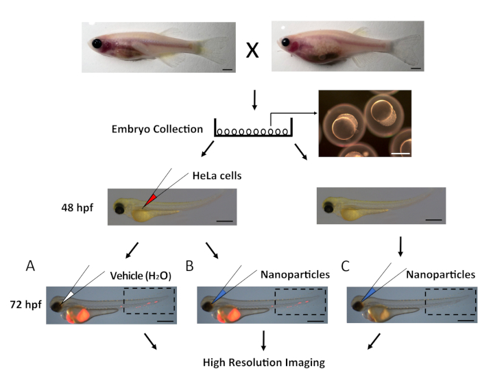
Figure 1: Protocol schematic for studying the ability of nanoparticles to target human cancer cells. Transparent Casper embryos were generated through breeding male and female adult fish. Fertilized embryos were collected in a Petri dish. At 48 hpf, RFP+ HeLa cells were injected into zebrafish embryos at the perivitelline cavity, leaving some age-matched embryos uninjected as controls. At 72 hpf, embryos with metastatic RFP+ HeLa cells were selected and split into two groups: (A) injected with vehicle (H2O) as control and (B) injected with nanoparticles suspended in H2O. The third group was age-matched embryos that were injected with nanoparticles alone (C). All three groups were imaged under a fluorescent microscope. The boxed area shown is where images were captured (see Figure 2-Figure 4). Scale bars for adult fish = 1 mm and for embryos = 500 µm. Please click here to view a larger version of this figure.
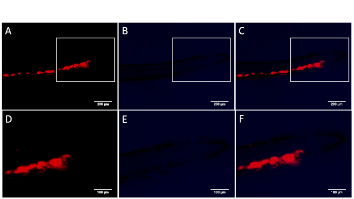
Figure 2: Zebrafish transplanted with metastatic HeLa cells without nanoparticles. Only red fluorescent HeLa cells were visible in the individual (A, D) or overlaid images of the red channel and blue channel (C, F). No specific blue fluorescent signals were detected in the embryo with vehicle injection control (B, E). Images in (A-C) show the fish tail region boxed as in Figure 1A. Images in (D-F) are enlarged views of the boxed areas in (A-C). Scale bars in (A-C) = 200 µm and in (D-F) = 100 µm. Please click here to view a larger version of this figure.
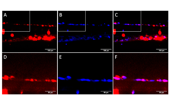
Figure 3: Colocalization of red fluorescent HeLa cells and blue fluorescent nanoparticles in zebrafish. The zebrafish tails were imaged at both low (A-C) and high (D-F) magnification in the red and blue channel. Red fluorescent signals revealed metastatic HeLa cells (A, D), whereas blue fluorescent signals showed the nanoparticles (B, E). The overlaid images from both red and blue channels (C, F) show colocalization of HeLa cells and nanoparticles. The images were taken after 3.5 h from injection with ultrabright silica nanoparticles. Images in (A-C) show the fish tail region as boxed in Figure 1B. Images in (D-F) are enlarged views of the boxed areas in (A-C). Scale bars in (A-C) = 100 μm and in (D-F) = 50 μm. Please click here to view a larger version of this figure.
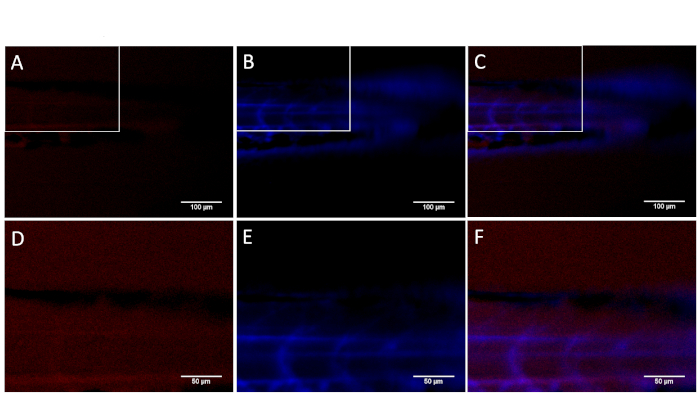
Figure 4: Zebrafish injected with nanoparticles without human HeLa cells. Blue fluorescent nanoparticles were distributed into the circulatory system of the embryos in the individual (B, E) and overlaid images of the red and blue channel (C, F). No specific red fluorescence was visible at either low or high magnification (A, D) except some background fluorescence common to zebrafish embryos. Images in (A-C) show the fish tail region as boxed in Figure 1C. Images in (D-F) are enlarged views of the boxed areas in (A-C). Scale bars in (A-C) = 100 µm and in (D-F) = 50 µm. Please click here to view a larger version of this figure.
The protocol described here utilizes the zebrafish as an in vivo system to test the ability of nanoparticles to recognize and target metastatic human cancer cells. Several factors can impact the successful execution of the experiments. First, embryos need to be fully developed at 48 hpf. The correct developmental stage of the embryos enables them to endure and survive the transplantation of human cancer cells. Embryos younger than 48 hpf have a significantly lower survival rate compared to older and more developed embryos. Second, cancer cells should be kept as healthy as possible by ensuring they are: 1) in the exponential growth phase21, 2) freshly harvested 30 min-1 h immediately before transplantation, and 3) kept warm at all times. Third, the needle must not be clogged. Pipette the HeLa cells up and down at least 20x before loading the cell mixture into the needle. Fourth, different types of needles must be used for transplantation of human cancer cells and injection of nanoparticles. The needle for human cell transplantation is relatively wide, with an angle to avoid cell clogging, whereas the needle for nanoparticle injection is sharp and thin. Fifth, the location of the injection differs. The location for transplantation of human cells is the perivitelline cavity, but for nanoparticle injection, the needle should be inserted behind the eye, where there are enriched capillaries. Finally, the skill of the individual who performs transplantation matters. An experienced individual can accurately inject HeLa cells into the perivitelline cavity space, while an inexperienced person often injects tumor cells into the yolk area where tumor cells barely spread into the fish body. Similarly, the embryos' survival rate is much higher when handled by an experienced individual, with at least 50% of embryos transplanted with cancer cells surviving.
Although zebrafish embryos are usually incubated at 28.5 °C, human cancer cells require higher temperatures to survive and migrate22,23. To allow the survival of both fish embryos and human cancer cells, the embryos transplanted with human cancer cells are incubated at 35.5 °C instead. Although it may be easier to deliver cancer cells into the yolk sac, they barely spread into the circulatory system. Therefore, it is critical to inject the cancer cells into the vascularized area under the perivitelline cavity to ensure intravasation and spread of cancer cells. Additionally, one must take care not to add too much or too concentrated MS222 when anesthetizing the embryos during injection and imaging. To aid in imaging and visualization of nanoparticles, the Casper zebrafish was chosen instead of AB fish. The Casper fish is a double mutant for Nacre and Roy that lacks melanocytes and iridophores, resulting in increased transparency compared to AB fish14. The transparency of the Casper zebrafish enables monitoring the spread of nanoparticles in circulation and the targeting of the nanoparticles to cancer cells. The challenge of this protocol is the relatively high mortality rate of the embryos if an unexperienced individual performs the transplantation of human cancer cells. Interestingly, the injection of nanoparticles slightly behind the eyes is relatively well tolerated. This is likely due to the use of thinner needles compared to the needles used for tumor cell transplantation. To avoid damaging human cancer cells, one must use needles with wide openings, but these can lead to damage of the embryos if handled inappropriately.
This protocol utilizes the Casper zebrafish to visualize targeting of metastatic cancer cells with functionalized nanoparticles in vivo. A major advantage of this assay is that it allows the researchers to perform real-time imaging over the course of zebrafish development to monitor the interaction of cancer cells with nanoparticles. In fact, due to its high fecundity and rapid development, the zebrafish allows the researcher to obtain results in just afew days18,19,20,24. Moreover, this assay also allows the elimination of toxic nanoparticles from further research if most embryos die after injection of a particular type of nanoparticle. Although the zebrafish is not a mammal, it facilitates the selection of a large number of nanoparticles in a rapid and economical manner, providing useful information for downstream studies in large animals and clinical testing. Taken together, the zebrafish is making an impact in nanomedicine and nanotechnology by helping select suitable nanoprobes for early detection and potential destruction of cancer cells through cancer-specific targeting.
The authors have nothing to disclose.
The authors thank Ms. Kaylee Smith, Ms. Lauren Kwok, and Mr. Alexander Floru for proofreading the manuscript. H.F. acknowledges grant support from the NIH (CA134743 and CA215059), the American Cancer Society (RSG-17-204 01-TBG), and the St. Baldrick's Foundation. F.J.F.L. acknowledges a fellowship from Boston University Innovation Center-BUnano Cross-Disciplinary Training in Nanotechnology for Cancer (XTNC). I.S acknowledges NSF support (grant CBET 1605405) and NIH R41AI142890.
Agarose | KSE scientific | BMK-A1705 | |
Borosilicate glass capillaries | World Precision Instruments | 1.0 mm O.D. x 0,78 mm | |
Computer and monitor | ThinkCentre | X000335 | |
DMEM (Dulbecco's Modified Eagle's Medium) | Corning | 10-013-CV | sold by Fisher |
Fetal Bovine Serum | Sigma-Aldrich | F0926 | |
Fish incubator | VWR | 35960-056 | |
Hemocytometer | Fishersci brand | 02-671-51B | |
Magnetic stand | World Precision Instruments | M10 | |
Microloader tip | Eppendorf | E5242956003 | sold by Fisher |
Micromanipulator | Applied Scientific Instrumentation | MMPI-3 | |
Needle Puller | Sutter instruments | P-97 | |
Olympus MVX-10 fluorescent microscope | Olympus | MVX-10 | |
P200 tip | Fishersci brand | 07-200-293 | |
PBS (Dulbecco's Phosphate-Buffered Salt Solution 1X) | Corning | 21-030-CV | sold by Fisher |
Petri dish | Corning | SB93102 | sold by Fisher |
Plastic pipette | Fishersci brand | 50-998-100 | |
pLenti6.2_miRFP670 | Addgene | 13726 | |
Pneumatic pico pump | World Precision Instruments | SYSPV820 | |
Pronase | Roche-Sigma-Fisher | 50-100-3275 | Roche product made by Sigma- sold by Fisher |
Razor blade | Fishersci brand | 12-640 | |
SZ51 dissection microscope | Olympus | SZ51 | |
Tricaine methanesulfonate | Western Chemicals | NC0872873 | sold by Fisher |
Trypsin-EDTA | Corning | MT25053CI | sold by Fisher |
Tweezer | Fishersci brand | 12-000-122 |