The ovaries were obtained from U. S. Meat Animal Research Center16. As stated previously16, all procedures were approved by the U.S. Meat Animal Research Center (USMARC) Animal Care and Use Committee in accordance with the guide for Care and Use of Agricultural Animals in Agricultural Research and Teaching. The ovaries were brought to the University of Nebraska-Lincoln Reproductive Laboratory where they were processed and cultured.
1. Preparation of required media
- Waymouth MB 752/1 medium
- Fill a 1 L tissue culture bottle with 900 mL of sterile water. While the water is gently stirring on a stir plate, gradually add the powdered medium. Once the powdered medium is dissolved, add 2.24 g of sodium bicarbonate followed by 1.25 g of bovine serum albumin (BSA). Use a pH meter and adjust the pH to 7.25-7.35. Add additional sterile water to bring the final volume to 1 L.
- Move to a biological safety cabinet and add penicillin-streptomycin sulfate at a concentration of 0.1% v/v of the medium. Filter the medium with a 0.22 µm pore 33.2 cm2 500 mL bottle top filter.
- Pour off the filtered medium into several 50 mL conical tubes. Add 0.5 mL of Insulin-Transferrin-Selenium per 50 mL of aliquoted medium.
- Wrap the conical tubes and stock bottle of the medium in aluminum foil and store at 4 °C. This medium is light sensitive.
NOTE: Waymouth medium can be stored for up to 1 month.
- Leibovitz's L-15 (LB-15) medium
NOTE: LB-15 medium is used to clean tissue in preparation for culture.- Fill a 1 L tissue culture bottle with 900 mL of sterile water. While the sterile water is gently stirring on a stir plate, gradually add the prepared powdered medium. Use a pH meter and adjust the pH to 7.25-7.35. Add additional sterile water to bring the final volume to 1 L.
- Move to biological safety cabinet. Make 1 L of LB-15 with 0.1% antibiotic (see Table of Materials). Filter the medium into two 500 mL tissue culture bottles using a 0.22 µm pore 33.2 cm2 500 mL bottle top filter. Wrap bottles in aluminum foil as LB-15 medium is light-sensitive and store at 4 °C.
NOTE: LB-15 medium can be stored for up to 1 month.
- Phosphate Buffered Saline (PBS)
- Make PBS in the lab or purchase sterile PBS without calcium or magnesium (Table of Materials). To make PBS in the lab, begin with 800 mL of distilled water and add 8 g of sodium chloride (NaCl) to it. Then, add 0.2 g of potassium chloride (KCl), 1.44 g of sodium phosphate dibasic (Na2HPO4), and 0.24 g of potassium phosphate dibasic (KH2PO4). Adjust pH to ~7.4 and adjust total volume to 1 L. Sterilize the solution by autoclaving.
- Make 1 L PBS with 0.1% antibiotic (see Table of Materials) while in a biological safety cabinet.
2. Ovarian cortical culture protocol
NOTE: Ovaries were obtained from spring born USMARC heifers at 13 months of age. Ovaries were rinsed thoroughly, and all blood and other fluid were removed with PBS containing antibiotic (0.1%) and transported at 37 °C23 to University of Nebraska-Lincoln Reproduction Laboratory UNL (1.5 h away). (For comments on temperature of ovaries during transport please see Discussion)
- Prepare the ovarian tissue on a clean bench (Figure 1).
- Disinfect the clean bench with 70% ethanol. Place a fresh absorbent pad on the benchtop. Ensure that the clean bench blower is turned on half an hour prior to dissection along with UV light to sterilize anything in the clean bench, including absorbent pad and make sure appropriate PPE is used.
- Arrange the Petri dishes (60 x 15 mm) for tissue washes. Three Petri dishes are required for PBS wash, three for PBS with antibiotic, and three for LB-15 washes. An additional LB-15-containing Petri dish with accompanying lid will be utilized for final placement of pieces after washes.
- Fill each Petri dish with approximately 10 mL of appropriate fluids, either PBS or LB-15.
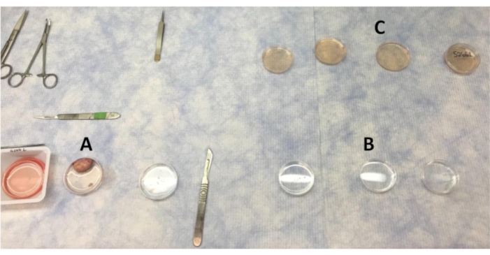
Figure 1: Layout of plates for washing the ovary and cortex pieces in the clean bench. (A) PBS used for washing the ovary as sections of the cortex are removed. (B) PBS with antibiotic washes that cortex pieces are moved through. (C) Ovarian cortex pieces are washed four times in LB-15 before moving to the biosafety cabinet for final wash in LB-15. Please click here to view a larger version of this figure.
- Remove the prepared Waymouth and LB-15 medium from the refrigerator and warm to room temperature.
- Autoclave all tools to ensure sterilization prior to use.
- Maintain ovaries at 37 °C until ovarian cortex is ready to be collected.
- Using forceps with serrated jaws, pick up the ovary and thoroughly wash in the first PBS-filled Petri dish. Transfer the ovary to the second PBS wash and thoroughly cleanse once more.
NOTE: The ovary will stay in the second PBS wash while ovarian cortical strips are removed. - Using serrated jaw forceps, secure the ovary and slice in half. At this time, the ovarian cortex will cut away from the medulla. Using a ruler, make sure that no more than 1–2 mm of depth of surface of ovary is removed away from the medulla16. Remove transverse sections of the ovarian cortex from medulla, cut 3–4 thin strips of ovarian cortex (Figure 2) with a scalpel (#11 scalpel blade; #3 handle), and place the strips in the third PBS-filled Petri dish.
NOTE: At this time, additional ovarian cortical tissue can be collected for RNA extraction or fixed and collected for histology of initial non-cultured cortex pieces. When removing strips of ovarian cortex, avoid areas with visible antral follicles or corpora lutea. In addition, avoid collecting medullary tissue. The histology of the medulla is very different as shown previously16. If the ovarian cortex is not cut to more than a 1–2 mm depth, then the medulla should not be obtained. Distinct histology allows for landmarks between the cortex and the medulla. - Cut the ovarian cortex strips in the third PBS wash into small, square pieces (~0.5–1 mm3) with a #21 scalpel blade. Use a ruler underneath the Petri dishes to ensure the pieces are of similar size and thickness to make consistent ovarian cortex pieces. Use forceps to secure the strips while cutting the pieces with a scalpel.
NOTE: The number of tissue pieces cut is dependent on the experiment. Four pieces of ovarian cortex is the minimum amount of tissue necessary for culture. Other methods for ensuring appropriate length and depth include using special slicers26 or precut plastic pieces as templates27. - Wash ovarian cortical pieces through all three PBS with antibiotic-filled Petri dishes. Use a curved tip forceps to move pieces between washes.
- Move cortex pieces through the series of LB-15 washes and place in final LB-15-filled Petri dish. Label the lid with animal ID and ovary side (left or right).
NOTE: Fully submerge the ovarian cortex pieces in each wash for thorough cleaning. - Collect four ovarian cortex pieces per ovary and fix for day zero histology. Additional pieces can also be flash frozen for RNA. The remaining tissue pieces will be used for culture. Wipe down dissecting tools with 70% ethanol after each tissue collection.
- Prepare a biological safety cabinet for final tissue wash and culture preparation. Sanitize supplies with 70% ethanol before placing in the biological safety cabinet. Use the aseptic technique when working in the biological safety cabinet.
- Move all ovarian cortex intended for culture to the biological safety cabinet and wash once more in an LB-15-filled Petri dish.
- In a 24-well tissue culture plate, pipette 350 µL of Waymouth medium per well.
- Place uncoated culture well inserts into each well using forceps. Ensure that no bubbles are formed under the base of the insert as this would result in the tissue drying out. The medium must be touching the inserts to allow for the medium to be absorbed up and surround the ovarian cortex pieces.
- Carefully position four ovarian cortex pieces onto the mesh of each insert (Figure 2). The forceps can puncture the mesh if the tissue pieces are not delicately placed. The tissue pieces should not be touching each other or the side of the insert.
- Incubate the tissue at 37 °C with 5% CO216.
NOTE: Others have used 38.8 °C28. However, no difference has been observed in the integrity of the tissue nor in the ability of follicles to progress in 37 °C tissue nor have others39,30. Thus, at this point any of these temperatures should be conducive to experiment success. Others have used 400 µL of medium. Either amount is fine as long as one is consistent, and the tissue is partially submerged allowing for adequate surface tension to allow for hydration of tissue (media surrounding ovarian cortex pieces). Fill empty wells with 500 µL of sterile water to help reduce evaporation from other wells.
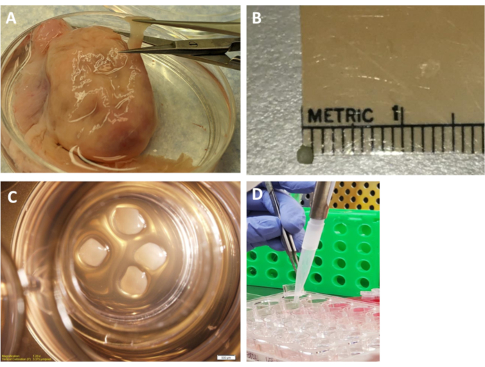
Figure 2: Ovarian cortex pieces and culture plate. (A) An ovarian strip being cut from the cortex of the ovary. (B) Ruler and cortex piece shown side by side. (C) Four cortex pieces (~0.5-1 mm3) resting on the insert in the culture medium in the plate. (D) Lifting the insert to collect the culture medium from the well. Collect and replace all the culture medium daily (250 µL) to maintain proper pH.Approximately 250 µL is obtained from each well each day (about 70% of the initial culture medium). Please click here to view a larger version of this figure.
3. Media collection
- Change ovarian cortex culture medium daily for 7 days. Medium changes should be as close to 24 h apart as possible to prevent large pH and color changes in medium. Warm Waymouth medium to 37 °C prior to medium change. Approximately 250 µL is obtained from each well each day (about 70% of initial culture medium).
- During medium changes, use forceps to gently lift the insert out of the well. Collect the cultured Waymouth medium in 0.5 mL tubes (approximately 250 µL /day). Set the insert back in well and add 350 µL of fresh culture medium by dispensing the medium between the side of the insert and well.
NOTE: Change most of the media daily to obtain enough media to measure all the steroids, cytokines, and chemokines necessary to determine ovarian microenvironment. Also, daily medium changes are important to prevent large pH changes (indicated by color change) in the medium. Drops of medium were retained surrounding the ovarian cortex pieces to ensure the pieces remained wet. No problems were observed with cultured tissue due to changing 70% of the media. - Store the collected medium from tissue culture at -20 °C.
4. Imaging and downstream processing
- After 7 days of culture at 37 °C with 5% CO2, image the ovarian cortex pieces using a dissection microscope with an attached camera and a computer imaging software program.
NOTE: A dark room is usually best for achieving the best picture quality for imaging. - After imaging, fix two ovarian cortex pieces per well in Bouins for histology and flash freeze two ovarian cortex pieces in liquid nitrogen to obtain RNA for cDNA. Repeat this step for all the wells with tissue. Collect the medium from day 7 and store at -20 °C.
- Let the ovarian cortex pieces remain immersed in Bouins (picric acid 750 mL, glacial acetic acid 50 mL, and 37%–40% formalin 250 mL) for approximately 1.5 h before being washed with 70% ethanol three times. The tissue will remain in 70% ethanol and be cleared daily until the solution is no longer yellow.
NOTE: Fixatives other than Bouins as well as paraformaldehyde can be used. In this experiement Bouins is used as it is the fixative to achieve optimal morphology. If more tissue is required for other analysis, additional wells of media and pieces of ovarian cortex can be obtained from each animal.
This bovine cortex culture procedure can be used to determine a wide variety of hormone, cytokine, and histology data from small pieces of the ovary. Staining, such as hematoxylin and eosin (H&E), can be used to determine ovarian morphology through follicle staging16,23,31 (Figure 3). Briefly, follicles were classified as primordial, which is an oocyte surrounded by a single layer of squamous pre-granulosa cells (0); transitional follicle or early primary, which is an oocyte surrounded by mostly squamous pre-granulosa cells and some cuboidal granulosa cells (1); primary follicle, which is an oocyte surrounded by 1-1.5 layers of cuboidal granulosa cells (2); secondary follicle, which is an oocyte surrounded by two or more cuboidal granulosa cells (3); antral follicle, which is no larger than 1 mm in diameter and surrounded by two or more layers of granulosa cells containing a distinct antrum (4)16,23 (Figure 3). Follicle staging can be conducted on ovarian cortex fixed prior to and following culture to assess folliculogenesis (Figure 4). We took three images per slide from three different slides stained with H&E. Then, the follicles were staged and counted by three individuals and averaged to determine the number of follicles at each stage16,23. The area of the field of view for an image (three per slide) at 400x magnification is 0.4mm2. Thus, 30% of the area of the ovarian cortex pieces were counted to determine follicle stages. The initial folliclenumber (before culture) (Figure 4A) is used to normalize the follicles counted after culture (Figure 4B).
Additionally, differences in morphology as determined by collagen deposition (Picro Sirus Red staining) can indicate fibrosis in ovarian cortex from Stair Step or Control heifers (Figure 5). Daily collection of culture medium can be pooled over 3 days to assess varied steroid hormone production by RIA (using 200 µL medium sample per animal; Figure 6) or steroid metabolites using high performance liquid chromatography mass spectrometry (HPLC-MS; 220 µL medium sample pooled over 4 days per animal; Table 1) and cytokine production (Table 2). Therefore, several replicates of one animal may be required to ensure enough cortex medium to perform all the desired assays.
Because Stair-Step heifers have increased primordial follicles at the beginning of the culture we expected these to progress in culture and obtain greater number of secondary follicles, which we observed in the results. Also, due to increase in secondary follicles, we would expect greater concentrations of steroids. We did see tendency for increases in androgens, glucocorticoid metabolites, and progesterone metabolites, which would support this in the current manuscript. Our lab has also evaluated effects of different VEGFA isoforms on follicle progression, steroidogenesis, and activation of different signal transduction molecules in the KDR (also known as Vascular Endothelial Growth Factor Receptor 2; VEGFR2) using signal transduction array plates16. To reduce animal variation, we use 4-6 animals per treatment and for other experiments depending on power analysis and variability we have used as many as 11.
Repeatability of results from bovine ovarian cortex culture is most affected by contamination of ovarian cortex pieces. Additionally, negative, or subpar results can occur if the medium is not changed regularly within a 24-h period. The medium when originally added is pink in color, but when the medium is collected, and the color appears to be orange or bright yellow this could indicate a change in pH that could be detrimental to the tissue. Also, tissue pieces that are cut too large might develop degeneration in the middle that would not be observed until tissue sectioning for histology. This degeneration will limit the use of the tissue for analysis. The data presented in this paper has been analyzed using nonparametric tests and a general linear model analysis in a statistical software program. The number of primordial, primary, secondary, and antral follicles per section before and after culture were analyzed using a generalized linear mixed model. Significance was determined at P < 0.05 and a tendency was reported at 0.14 ≤ P ≥ 0.05.
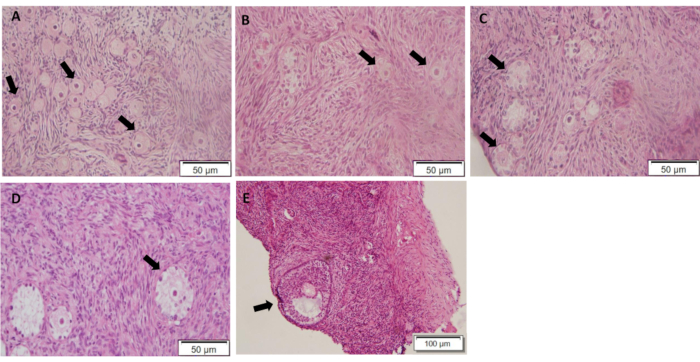
Figure 3: Hematoxylin and Eosin staining for follicle staging of ovarian cortex. Different stages of follicles are indicated by arrows. (A) Primordial follicles (stage 0); (B) Early primary follicles (stage 1); (C) Primary follicles (stage 2); (D) Secondary follicle (stage 3); (E) Antral follicle (stage 4). Area of the field of view for an image at 400x magnification is 0.4 mm2. We count 30% of the area of the ovarian cortex pieces to determine the follicle stages. Please click here to view a larger version of this figure.
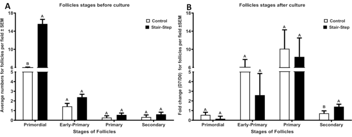
Figure 4: Average number of follicles at different follicular stages in Control (n=6) and Stair-Step (n=6) heifers. (A) Before culture Primordial P = 0.001, Early Primary P = 0.12, Primary P = 0.31, Secondary P = 0.22. (B) After 7 days of culture, Primordial P = 0.37, Early Primary P = 0.84, Primary P = 0.69, Secondary P = 0.02. Error bars are representative of SEM. Please click here to view a larger version of this figure.
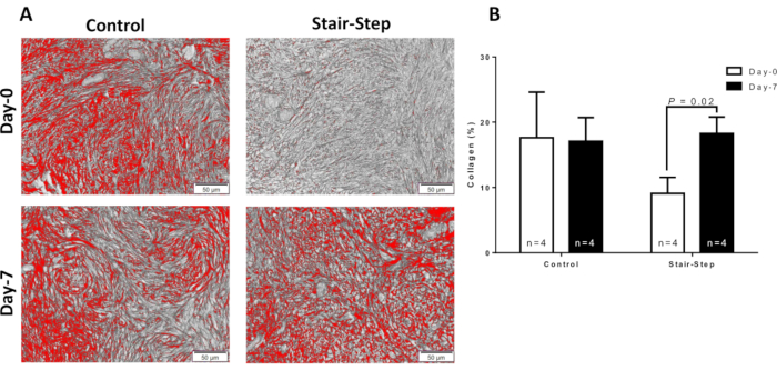
Figure 5: Collagen (Picro Sirius Red; PSR) staining in ovarian cortex. PSR in (A) Control and Stair-Step heifers from Day 0 and Day 7. (B) Graph comparing the average area of PSR-positive staining per ovarian cortex field (pixels/µm2) between Control (n = 4) and Stair-Step (n = 4) heifers. Error bars are representative of SEM. Area of the field of view for an image at 400x magnification is 0.4mm2. Please click here to view a larger version of this figure.
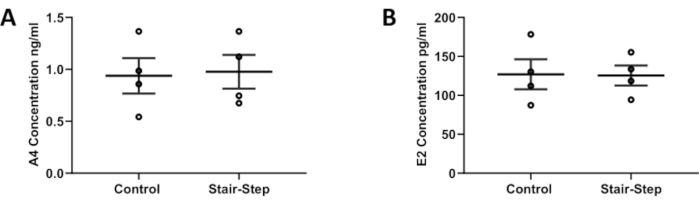
Figure 6: Concentrations of A4 and E2. (A) Concentration of A4 and (B) concentration of E2 pooled over 3 days of culture in ovarian cortex media of Control and Stair-Step heifers as measured by RIA's. n = 4 for each group. Error bars are representative of SEM. Please click here to view a larger version of this figure.
| Hormones | Control | Stair-Step | P-Value | |||
| n | 4 | 4 | ||||
| ng/mL | Mean | SEM ± | Mean | SEM ± | ||
| DOC | 0.33 | 0.28 | 0.72 | 0.48 | 0.15 | |
| INN | 0.61 | 0.60 | 4.93 | 3.99 | 0.08 | |
| CORT | 0.003 | 0.003 | 0.01 | 0.01 | 0.85 | |
| 17OHP | 0.59 | 0.53 | 1.88 | 0.99 | 0.08 | |
| A4 | 1.46 | 1.43 | 5.74 | 3.27 | 0.08 | |
| AN | 0.15 | 0.09 | 0.54 | 0.32 | 0.08 | |
| DHEAS | 4.50 | 2.60 | 7.59 | 0.85 | 0.56 | |
| E2 | 0.05 | 0.05 | 0.13 | 0.08 | 0.32 | |
| P4 | 5.05 | 2.78 | 6.52 | 1.66 | 0.39 | |
| T | 0.33 | 0.33 | 1.33 | 0.96 | 0.14 | |
| DHT | 0.07 | 0.02 | 0.01 | 0.01 | 0.09 | |
| DOC – 11-Deoxycorticosterone, INN – 11-Deoxycortisol, CORT – Corticosterone, 17OHP – 17-Hydroxyprogesterone, A4- Androstenedione, AN – Androsterone, DHEAS – dehydroepiandrosterone sulfate, E2 – Estradiol, P4 – Progesterone, T – Testosterone, DHT – Dihydrotestosterone | ||||||
Table 1: Steroid and steroid metabolites measured in ovarian cortex culture medium from one well for each animal pooled over 4 days of culture. Data presented with mean ± SEM. Blue indicates P < 0.1 and has a tendency to be different.
| Cytokines | Control | Stair-Step | P-Value | |||
| n | 4 | 4 | ||||
| pg/mL | Mean | SEM ± | Mean | SEM ± | ||
| ANG1 | 27.29 | 11.71 | 63.66 | 25.06 | 0.39 | |
| CD40L | 4527.01 | 2986.34 | 3537.59 | 3537.59 | 0.74 | |
| DCN | 1307.68 | 320.87 | 996.54 | 282.96 | 0.77 | |
| INFβ | 0.36 | 0.36 | 0.002 | 0.002 | 0.85 | |
| IL18 | 0.00 | 0.00 | 1832.89 | 1265.33 | 0.13 | |
| LIF | 97.45 | 88.12 | 337.58 | 231.25 | 0.54 | |
| RANTES | 698.06 | 322.59 | 1254.13 | 811.19 | 0.56 | |
| INFγ | 4.49 | 1.37 | 3.68 | 0.65 | 0.77 | |
| IL13 | 501.21 | 285.07 | 810.81 | 159.67 | 0.25 | |
| IL21 | 24.38 | 9.51 | 18.52 | 6.17 | 0.56 | |
| IL1F5 | 5.07 | 3.85 | 5.40 | 1.70 | 0.56 | |
| TNFα | 61.45 | 35.00 | 44.91 | 13.41 | 0.77 | |
| ANG1 – Angiopoietin 1, CD40L – CD40 Ligand, DCN – Decorin, IFNβ – Interferon Beta 1, IL18 – Interleukin-18, LIF – Leukemia inhibitory factory, RANTES – Regulated on Activation, Normal T Cell Expressed and Secreted, IFNγ – Interferon Gamma, IL13 – Interleukin 13, IL21 – Interleukin 21, IL1F5 – Interleukin 1 family member 5, TNFα – Tumor Necrosis Factor alpha | ||||||
Table 2: Cytokine and chemokines measured in ovarian cortex culture medium from one well for each animal pooled over 4 days of culture. Data presented with mean ± SEM. Blue indicates P < 0.1-0.14 and has a tendency to be different.
