Here, we present a protocol to achieve precise quad-zygomatic implant placement in patients with severely atrophic maxilla using a real-time dynamic navigation system.
Zygomatic implants (ZIs) are an ideal way to address cases of a severely atrophic edentulous maxilla and maxilla defects because they replace extensive bone augmentation and shorten the treatment cycle. However, there are risks associated with the placement of ZIs, such as penetration of the orbital cavity or infra-temporal fossa. Furthermore, the placement of multiple ZIs makes this surgery risky and more difficult to perform. Potential intraoperative complications are extremely dangerous and may cause irreparable losses. Here, we describe a practical, feasible, and reproducible protocol for a real-time surgical navigation system for precisely placing quad-zygomatic implants in the severely atrophic maxilla of patients with residual bone that does not meet the requirements of conventional implants. Hundreds of patients have received ZIs at our department based on this protocol. The clinical outcomes have been satisfactory, the intraoperative and postoperative complications have been low, and the accuracy indicated by infusion of the designed image and postoperative three-dimensional image has been high. This method should be utilized during the entire surgical procedure to ensure ZI placement safety.
In the 1990s, Branemark introduced an alternative technique for bone grafting, the zygomatic implant (ZI), which has also been called the zygomaticus fixture1. It was initially used for the treatment of trauma victims and patients with tumor resection where there was a defect in the maxillary structure. After maxillectomy, many patients retained anchorage only in the body of the zygoma or in the frontal extension of the zygomatic bone1,2,3.
More recently, the ZI technique has been widely used in edentulous and dentate patients with a severely resorbed maxilla. The main indication for ZI implants is an atrophic maxilla. The use of four ZIs in an immediate loading system (fixed prosthodontics) is practical for surgeons with broad clinical experience, and it appears to represent an excellent alternative method to bone graft techniques2,4. However, there are risks when placing ZIs, either by freehand or using a surgical template for guidance. Risks include inaccurate placement within the alveolus, penetration of the orbital cavity or infra-temporal fossa, and inappropriate placement within the zygomatic prominence5. The placement of multiple ZIs makes this surgery risky and difficult to perform. Hence, improving the precision of ZI placement is critical to its clinical use and safety.
The real-time surgical navigation system provides a different approach. It provides real-time and completely visualized trajectories through the analysis of preoperative and intraoperative computed tomography images. With the real-time navigation system, both precision and safety have been improved with sophisticated surgery and treatment5,6. A practical, feasible, and reproducible protocol was developed using the real-time surgical navigation system to precisely place ZIs in the severely atrophic maxilla5,7,8,9,10. With this protocol, we have treated hundreds of patients with satisfactory clinical outcomes5,6,7,8,9,10. Here, we present the protocol with the detailed information on the treatment procedure.
All of the clinical protocols were approved by the Medical Ethics Review Committee of the Shanghai Ninth People's Hospital, Shanghai Jiao Tong University, School of Medicine (SH9H-2020-T29-3).
1. Patient selection
2. Mini-screw implantation
3. Preoperative CBCT scanning for planning
4. Setting registration points
5. Planning for quad-ZI surgery
NOTE: This protocol requires the navigation system.
6. Surgical procedure
7. Postoperative medication
8. Immediate restoration
9. Image integration
The enrolled patient was a 60-year-old woman without any systematic diseases (Figure 1A–D, F). After CBCT scanning, the alveolar ridge in the anterior maxilla was less than 2.9 mm, while the residual bone height in the posterior maxilla region was less than 2.4 mm (Figure 1E, G and Table 1). The width and thickness of the zygomatic bone were approximately 22.4-23.6 mm and 6.1-8.0 mm (Figure 2, Table 3), respectively. According to the Zygoma Anatomy-Guided Approach, the entrance of the anterior ZI was at the level of the canine region, and the posterior ZI was in the second premolar (Figure 3E). The distance between the margin of the anterior ZI and the orbit was 5.2 mm on the right and 3.6 mm on the left, while the distance between the margin of the posterior ZI and the pterygopalatine fossa was 2.9 mm on the right and 4.3 mm on the left (Figure 3F–K).
The surgery was performed using the navigation system (Figure 4A–G). After surgery, the patient received a temporary restoration within 3 days, which addressed both the aesthetic and pronunciation issues (Figure 5C–G). Postoperative CBCT scanning and image integration showed that the errors of the entrance from the left posterior ZI to the left anterior ZI, then to the right anterior ZI, and last to the right posterior ZI were 1.25 mm, 1.35 mm, 1.35 mm, and 1.85 mm, respectively. The errors of the target from the left posterior ZI to the right posterior ZI were 2.25 mm, 1.55 mm, 2.40 mm, and 1.20 mm, respectively. The errors of the ZI angle were 3.50°, 3.59°, 3.20°, and 2.15°, respectively (Figure 5H–I, Table 4).
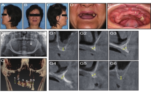
Figure 1: Preoperative examination. (A,C) Preoperative profile view. (B) Preoperative frontal image. (D) Frontal image of the smile line. (E) Intraoral view of the maxilla. (F) Preoperative panoramic radiograph. (G1–6) CBCT curve section. Please click here to view a larger version of this figure.
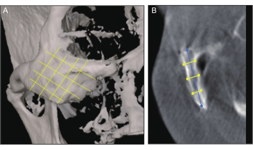
Figure 2: CT measurement. (A) Skull frontal view showing the zygoma divided into superior, middle, and inferior parts by the cross line. (B) Longitudinal tomography showing the measurements of the zygomatic thickness (yellow line) and length (blue line). Please click here to view a larger version of this figure.
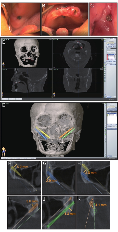
Figure 3: Preoperative planning. (A–C) Eight miniscrews were dispersedly implanted in the remaining maxilla to be registered. (D) Preoperative registration point settings on the navigation software. (E) Preoperative implant planning on the navigation software. (F–K) Distances for ZI planning. Please click here to view a larger version of this figure.
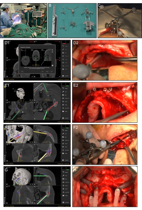
Figure 4: Navigation surgery. (A) Navigation surgery scene. (B) Navigation surgical tools. (C) Cephal bracket mounted to the patient's head for the purpose of tracking. (D1) Screen view of the navigation probe registration application in the sagittal coronal axial. (D2) Intraoral view of the navigation probe application. (E1) Screen view of the entry point location procedure using the navigation probe. (E2) Intraoral view of the procedure using the navigation probe. (F1,F2) Constant visualization of the drilling trajectory displayed on the screen in real-time. The entire procedure from the entry point to the exit point. (G) Screen view of the ZI position verification using the navigation probe. (H) Accomplishment of ZI placement. Please click here to view a larger version of this figure.
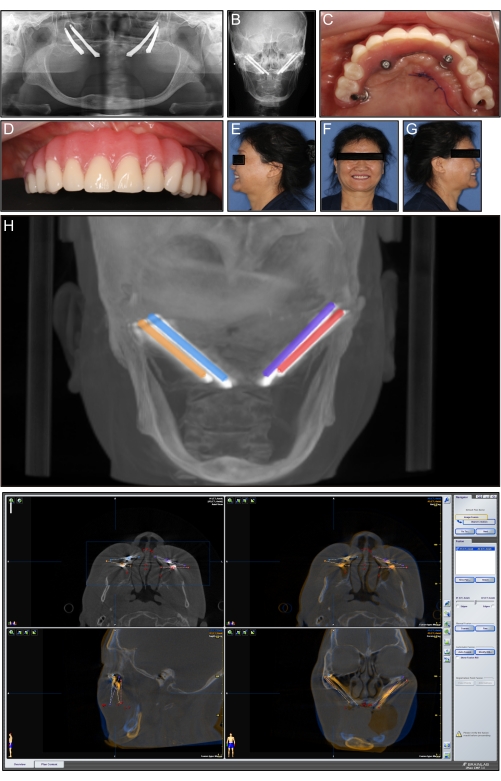
Figure 5: Postoperative view and image infusion. (A) Postoperative panoramic radiograph. (B) Postoperative frontal cephalometrics. (C) Intraoral view of the immediate temporary restoration. (D) Anterior view of the immediate temporary restoration. (E) Postoperative profile view after the immediate temporary restoration. (F) Frontal image after the immediate temporary restoration. (G) Postoperative profile view after the immediate temporary restoration. (H) Preoperative image integrated with the postoperative image, and measurement of the planned-placed deviations of implants. (I) Postoperative CBCT image integration observed in sagittal, coronal, and axial view. Please click here to view a larger version of this figure.
Inclusion criteria | Exclusion criteria |
1. Completely edentulous maxilla or be going to be edentulous maxilla | 1. Sufficient bone for conventional implant treatment |
2. Severe atrophy of the maxilla | 2. Bone graft was considered more appropriate |
3. Age range from 18-80 | 3. Untreated maxillary sinusitis |
4. Insufficient width for anterior maxilla to place regular implants of at least 3.75 mm | 4. Local or systemic contraindications for oral surgery |
5. Maxillary posterior bone height ranging from 1 to 3 mm in the premolar and molar regions | 5. A medication history of bisphosphonates |
6. The bone thickness for placing the apex of the ZI was at least 5.75 mm |
Table 1: Patient inclusion and exclusion criteria.
Anterior region width (mm) | Pre-molar region width (mm) | Molar region height (mm) | |
Left | 2.8 | 2.5 | 2.4 |
Right | 2.9 | 2.9 | 2.2 |
Table 2: Differences in alveolar bone thickness at points on the anterior region, and residual alveolar bone height at points on the premolar region and molar regions.
Zygomatic bone thickness (mm) | Zygomatic bone width (mm) | |||||
Superior | Middle | Inferior | Superior | Middle | Inferior | |
Left | 7.4 | 5.3 | 7.8 | 23 | 23.6 | 24.1 |
Right | 8 | 6.1 | 5.7 | 22.4 | 23.1 | 25.9 |
Table 3: Differences in zygomatic thicknesses at points on the superior, middle, and inferior areas.
Starting position error (mm) | Teminal position error (mm) | Angular deviation (°) | |
Distal of the left ZI | 1.25 | 2.25 | 3.5 |
Mesial of the left ZI | 1.35 | 1.55 | 3.95 |
Mesial of the right ZI | 1.35 | 2.4 | 3.2 |
Distal of the right ZI | 1.85 | 1.2 | 2.15 |
Table 4: Resulting deviation of four zygomatic implants.
Reconstructive rehabilitation of the atrophic maxilla using grafts is difficult because it requires good surgical technique, coverage of high-quality soft tissue over the graft, a significant amount of patient cooperation, and patients with health favorable for the finial restoration17,18. The placement of dental implants for reconstruction in patients with maxillary atrophy represents a significant clinical challenge. The pattern of facial bone resorption is associated with age and is especially evident in the edentulous maxilla, and particularly more noticeable in those using complete removable prostheses19,20. Thus, the development of the ZI represents an effective alternative for cases involving tumors, trauma, and ectodermal dysplasia. The main advantage of this technique is that it only requires one surgical approach, thus reducing the number of treatment stages and achieving the goal of immediate restoration. The immediate loading procedure also results in greater esthetic and functional patient satisfaction because in this technique, there is no edentulous mouth phase. After implant placement, restoration is performed immediately21. It also avoids the need for further bone-harvesting surgeries in donor sites22,23. It achieves stable bone anchorage in the zygomatic bone within the posterior maxilla region, which has bone quality type IV that does not allow the insertion of standard implants, through the addition of two to four standard implants in the anterior region or via the quadruple ZI approach24,25. Presently, the indication of this surgical technique has been applied to cases of trauma, severe periodontitis, and ectodermal dysplasia.
The ZI margin should be at a safe distance from important anatomic landmarks, such as the orbit and pterygopalatine fossa, and also for the interval between the two ZIs to ensure adjacent tissue impregnability andstabile implant osseointegration22. In some cases, computer-based guides, which are tailored planned for each patient, may have flaws that reduce their accuracy2,26. A real-time surgical navigation system could be applied to guide the drilling and placement of ZIs. With the assistance of the surgical navigation system, the incision extent can be limited, to some extent, to around the operated area. Moreover, drilling along the trajectory can avoid neighboring critical structures, such as the orbital cavity and infratemporal fossa, decreasing the risk of intraoperative complications and simplifies the operation.
In this case report, a passive optical dynamic navigation system was utilized requiring the use of fiducial markers firmly attached to the patient's dental arch during CBCT scanning27. A large number of implant placement-related studies, including the quad-approach for ZI placement and three ZIs studies, have shown the effective minimization of planned-placed deviations indirectly by reporting the reduction of intraoperative and post-operative complications with the assistance of the real-time surgical navigation system8,10,28,29,30,31. However, in these previous studies, more than six fiducial markers with a polygonal distribution were implemented by the operators before surgery. This meant that the bilateral maxillary tuberosity, the midline palatine suture, and both sides of the anterior nasal spine were selected as the area for titanium mini-screw anchorage32. Furthermore, all of the fiducial markers were recommended to be bone anchored to more than one titanium screw in each of the regions to ensure precise registration accuracy. It also avoided screws splitting off or moving during the open-flap surgery.
Another important procedure is the verification of error. The importance of the precision verification throughout the entire surgery cannot be overemphasized. Verification can be divided into four levels. The first level is the verification after the navigation registration procedure. The second level is the verification when locating the entry point on both the alveolar crest and the zygomatic bone. The third level is the verification during the drilling procedure with the zygomatic handpiece. The fourth level is the verification after ZI implementation to ensure accurate ZI position and direction. Furthermore, throughout the procedure, navigation calibration is also very important. Finally, both the operator and the surgical assistant should pay attention to the reference frame to ensure its stability, because any slight touch will likely affect the surgical navigation.
In the present case report, deviations generally appeared to be greater when the implants were placed in distal locations or with the placement of long implants33,34. Throughout the process, it was easy to locate the ZIs and it was safer to implant them by means of the real-time navigation system. Although the entry deviation, exit deviation, and angle deviations were limited under the guidance of the real-time surgical navigation system for ZI placement, it should be utilized during the entire surgical procedure to ensure safety.
The authors have nothing to disclose.
The authors thank Dr. Shengchi Fan for kindly providing valuable navigation technical support. This case report was funded by the Key project of China's Ministry of Science and Technology (2017YFB1302904), the Natural Science Foundation of Shanghai (No. 21ZR1437700), the Clinical research plan of SHDC (SHDC2020CR3049B), and the Combined Engineering and Medical Project of Shanghai Jiao Tong University (YG2021QN72).
Bistoury scalpel | Hufriedy Group | 10-130-05 | |
Branemark system zygoma TiUnite RP 35mm | Nobel Biocare AB | 34724 | TiUnite implant with overlength to place from the maxilla to the zygoma |
Branemark system zygoma TiUnite RP 40mm | Nobel Biocare AB | 34735 | TiUnite implant with overlength to place from the maxilla to the zygoma |
Branemark system zygoma TiUnite RP 42.5mm | Nobel Biocare AB | 34736 | TiUnite implant with overlength to place from the maxilla to the zygoma |
Branemark system zygoma TiUnite RP 45mm | Nobel Biocare AB | 34737 | TiUnite implant with overlength to place from the maxilla to the zygoma |
Branemark system zygoma TiUnite RP 47.5mm | Nobel Biocare AB | 34738 | TiUnite implant with overlength to place from the maxilla to the zygoma |
Branemark system zygoma TiUnite RP 50mm | Nobel Biocare AB | 34739 | TiUnite implant with overlength to place from the maxilla to the zygoma |
Branemark system zygoma TiUnite RP 52.5mm | Nobel Biocare AB | 34740 | TiUnite implant with overlength to place from the maxilla to the zygoma |
CBCT | Planmeca Oy,Helsinki, Finland | Pro Max 3D Max | |
connection to handpiece | Nobel Biocare AB | 29081 | the accessories to connect the intrument |
Drill guard | Nobel Biocare AB | 29162 | the accessories to protect the lips and soft tissue during the surgery |
Drill guard short | Nobel Biocare AB | 29162 | the accessories to protect the lips and soft tissue during the surgery |
Handpiece zygoma 20:1 | Nobel Biocare AB | 32615 | the basic instrument for implant drill |
Instrument adapter array size L | BRAINLAB AG | 41801 | |
Instrument adapter array size M | BRAINLAB AG | 41798 | |
Instrument calibration matrix | BRAINLAB AG | 41874 | a special tool for drill to calibration |
I-plan automatic image fusion software STL data import/export for I-plan VectorVision2®, (I-plan CMF software) | BRAINLAB AG | inapplicability | the software for navigation surgery planning |
Multi-unit abutment 3mm | Nobel Biocare AB | 32330 | the connection accessory between the implant and the titanium base |
Multi-unit abutment 5mm | Nobel Biocare AB | 32331 | the connection accessory between the implant and the titanium base |
Periosteal elevator | Hufriedy Group | PPR3/9A | the instrument for open flap surgery |
Pilot drill | Nobel Biocare AB | 32630 | the drill for the surgery |
Pilot drill short | Nobel Biocare AB | 32632 | the drill for the surgery measuring the depth of the implant holes |
Pointer with blunt tip for cranial/ENT | BRAINLAB AG | 53106 | |
Reference headband star | BRAINLAB AG | 41877 | |
Round bur | Nobel Biocare AB | DIA 578-0 | the drill for the surgery |
Screwdriver manual | Nobel Biocare AB | 29149 | |
Skull reference array | BRAINLAB AG | 52122 | a special made metal reference for navigation camera to receive the signal |
Skull reference base | BRAINLAB AG | 52129 | |
Suture vicryl 4-0 | Johnson &Johnson, Ethicon | VCP310H | |
Temporary copping multi-unit titanium (with prosthetic screw) | Nobel Biocare AB | 29046 | the temporary titanium base to fix the teeth |
Titanium mini-screw | CIBEI | MB105-2.0*9 | the mini-screw for navigation registration |
Twist drill | Nobel Biocare AB | 32628 | the drill for the surgery |
Twist drill short | Nobel Biocare AB | 32629 | the drill for the surgery |
Zygoma depth indicator angled | Nobel Biocare AB | 29162 | |
Zygoma depth indicator straight | Nobel Biocare AB | 29162 | the measurement scale for |
Zygoma handle | Nobel Biocare AB | 29162 | the instrument for zygomatic implant placement |