Portal vein injection of colorectal cancer (CRC) organoids generates stroma-rich liver metastasis. This mouse model of CRC hepatic metastasis represents a useful tool to study tumor-stroma interactions and develop novel stroma-directed therapeutics such as adeno-associated virus-mediated gene therapies.
Hepatic metastasis of colorectal cancer (CRC) is a leading cause of cancer-related death. Cancer-associated fibroblasts (CAFs), a major component of the tumor microenvironment, play a crucial role in metastatic CRC progression and predict poor patient prognosis. However, there is a lack of satisfactory mouse models to study the crosstalk between metastatic cancer cells and CAFs. Here, we present a method to investigate how liver metastasis progression is regulated by the metastatic niche and possibly could be restrained by stroma-directed therapy. Portal vein injection of CRC organoids generated a desmoplastic reaction, which faithfully recapitulated the fibroblast-rich histology of human CRC liver metastases. This model was tissue-specific with a higher tumor burden in the liver when compared to an intra-splenic injection model, simplifying mouse survival analyses. By injecting luciferase-expressing tumor organoids, tumor growth kinetics could be monitored by in vivo imaging. Moreover, this preclinical model provides a useful platform to assess the efficacy of therapeutics targeting the tumor mesenchyme. We describe methods to examine whether adeno-associated virus-mediated delivery of a tumor-inhibiting stromal gene to hepatocytes could remodel the tumor microenvironment and improve mouse survival. This approach enables the development and assessment of novel therapeutic strategies to inhibit hepatic metastasis of CRC.
Colorectal cancer (CRC) is a major cause of cancer mortality worldwide1. More than half of the CRC patients develop hepatic metastasis that occurs through the portal vein dissemination1. Currently, there are no effective therapeutics that can cure advanced liver metastasis, and most patients succumb to metastatic disease.
The metastatic niche or tumor microenvironment plays a key role in engraftment and growth of disseminated CRC cells2. Cancer-associated fibroblasts (CAFs), a prominent component of the tumor microenvironment, promote or restrain cancer progression through secreting growth factors, remodeling the extracellular matrix (ECM), and modulating immune landscapes and angiogenesis3,4,5. CAFs also confer resistance to chemotherapies and immunotherapies3. Moreover, CAFs regulate initiation and progression of CRC liver metastasis and predict prognosis in patients with CRC3,6,7,8. Thus, CAF-related factors could be exploited for the development of therapeutic strategies to inhibit CRC liver metastasis. However, the lack of satisfactory mouse models to study the metastatic tumor stroma has been a major obstacle to developing stroma-targeted therapies.
Currently, animal models to study CRC liver metastasis include primary CRC models that spontaneously develop hepatic metastasis and cancer cell transplantation models into the liver. Primary CRC mouse models, such as genetically engineered mouse models and colonic injection of cancer cells, rarely show metastasis to the liver9,10,11,12. Moreover, even if a liver metastasis is observed, these models show long latency from the primary tumor induction to metastasis, and potentially die of primary tumor burden12. To efficiently generate CRC liver metastases, cultured CRC cells are transplanted into the liver using three injection approaches: intra-splenic injection, direct intra-parenchymal injection into the liver, and portal vein injection. Intra-splenically injected cancer cells spread into the splenic vein, the portal vein, and ultimately to the liver13,14. However, the intra-splenic injection yields a lower tumor take ratio compared with other transplantation models15,16. With intra-splenic injection, surgical removal of the spleen is performed to avoid cancer growth in the spleen, which can potentially compromise immune cell maturation17. Furthermore, intra-splenic injection can also result in unintended tumor growth in the spleen and abdominal cavity18, complicating liver metastasis analyses. Direct intra-parenchymal injection into the liver efficiently induces hepatic metastasis16,19,20. Nonetheless, this approach does not fully recapitulate a biological step of liver metastasis that naturally occurs through portal vein dissemination. Using direct injection into the liver, entry of cancer cells into a non-portal, but systemic circulation can also result in multiple large lung metastases16. Although a majority of patients with CRC liver metastasis show multiple tumor nodules in the liver21, direct injection into a specific liver lobe generates a single tumor mass19,20. Portal vein injection or mesenteric vein injection, though technically challenging, allows efficient delivery of tumor cells into the liver in a manner that recapitulates the growth patterns seen in patients17. This strategy can minimize the possibility of secondary-site metastases and enables rapid growth of cancer cells in the liver, simplifying mouse survival analyses.
Historically, colorectal cancer cell lines such as mouse MC-38, human HT-29, and SW-620 were used to generate mouse models of hepatic metastasis22,23. However, these colorectal cancer cell lines do not induce a desmoplastic stromal reaction. Low stromal content in the tumors makes it difficult to investigate the biological roles of cancer-associated fibroblasts. Recent advances in CRC organoids and their transplantation have offered useful platforms to assess vital roles of the stroma in cancer progression24. Liver transplantation of CRC organoids generates a fibroblast-rich tumor microenvironment and has provided novel insights into stromal research6,25. Currently, portal or mesenteric vein injection of organoids has become a gold standard approach to generate CRC liver metastasis6,25,26,27,28. Nonetheless, to our knowledge, no previous papers have described detailed methods for the portal vein injection of colorectal tumoroids. Here, we present a methodology for using portal vein injection of CRC organoids to develop novel adeno-associated virus (AAV)-mediated stroma-directed therapy.
Hepatocytes are an important constituent of the metastatic tumor microenvironment in the liver and play a critical role in metastatic cancer progression29. Inspired by the success of AAV gene therapy approaches to induce protein expression in hepatocytes in non-neoplastic patients30,31, we investigated a similar approach but aimed at modifying the liver tumor microenvironment in CRC25. As such, we also describe herein the tail vein injection of AAV8 to induce expression of anti-tumorigenic proteins to modify the liver tumor microenvironment. The AAV8 serotype, designated by the choice of viral capsid protein during virus production, leads to high transduction efficiency specifically of hepatocytes (i.e., targeted gene expression in the liver tumor microenvironment)32. We have previously shown that Islr (immunoglobulin superfamily containing leucine-rich repeat) is a CAF-specific gene that induces bone morphogenetic protein (BMP) signaling, reduces CRC tumoroid growth, and promotes Lgr5+ intestinal stem cell differentiation25. We tested whether AAV8-mediated overexpression of the cancer-restraining stromal gene, Islr, in hepatocytes could attenuate hepatic metastasis progression by performing portal vein injection of CRC tumoroids in AAV8-Islr-treated mice.
In this paper, we first describe the tail vein injection procedure of liver tropic AAV. Then, we describe a method for tumoroid cell preparation and portal vein injection into the AAV-treated mice. Finally, we present approaches to monitor metastatic tumor progression to assess the efficacy of stroma-directed therapeutics.
All animal procedures in this article were reviewed and approved by the South Australian Health and Medical Research Institute Animal Ethics Committee (Approval number, SAM322).
1. Tail vein injection of adeno-associated virus
NOTE: Adeno-associated virus (AAV) should be handled as a biohazard under Biosafety Level 1 guidelines. Please refer to the published protocol for AAV preparation, purification, and titration33. Hepatocyte-tropic AAV, AAV834, encoding the cytomegalovirus (CMV) promoter-Islr gene, was used in this study25. To induce AAV-mediated overexpression, AAV dosing might require optimization depending on the promoter activity, gene, and mouse weight.
2. Cell preparation for colorectal cancer organoids
NOTE: CRC organoids used for this experiment solely contain epithelial cells. Culture and generation of CRC organoids has been previously described25,35. In short, normal colonic epithelial cells were isolated from the colon of a Rosa26-Cas9 mouse using a crypt isolation buffer (5 mM EDTA (ethylenediaminetetraacetate) in ice-cold PBS), and then embedded in basement membrane matrix medium, and cultured in organoid growth medium as described in reference35. Then, Apc and Trp53 mutations were introduced to the colonic epithelial cells by overexpressing single-guide RNAs that target Apc and Trp53 using lentivirus expression protocol. Single organoid clones were handpicked25. ApcΔ/Δand Trp53Δ/Δ colon cancer organoids (AP tumoroids), were injected as 5.0 x 10⁵ single cells in 100 µL of PBS with 10 µM Y-27632 into the portal vein per mouse, with organoid culture and single-cell preparation described below.
3. Portal vein injection of CRC organoids
NOTE: All surgical instruments and surgical gauzes must be autoclaved or sterilized before surgery. This protocol is modified from a previous protocol17. In this experiment25, portal vein injection was performed using Rosa26-Cas9 mice treated with AAV-mRuby2 or AAV-Islr in step 1.
4. Assessment of tumor growth kinetics by in vivo bioluminescent imaging
NOTE: If Firefly-expressing tumoroids are used for injection, metastatic tumor progression can be monitored weekly by in vivo imaging as described38,39. Luciferase expressed by cancer cells could elicit immune responses against the cancer cells and limit tumor growth40. Thus, caution is warranted in analyzing immune phenotypes and cancer progression in a mouse model using luciferase-expressing tumor cells.
5. Survival analysis and tissue collection
To induce AAV-mediated overexpression of a tumor-restraining stromal gene, Islr4,25,43,44, in hepatocytes, we intravenously injected Islr-encoding AAV8. 1.0 x 10¹¹ viral genomes (vg) of AAV8-Islr, or as a control, AAV8-mRuby2, was injected into the adult mouse tail vein (Figure 1A). Two weeks after the tail vein injection, livers were harvested to validate the overexpression of Islr in hepatocytes. We performed RNAscope in situ hybridization45 and confirmed that 3,3'-Diaminobenzidine (DAB)+ signals were observed throughout the liver (Figure 1B). No DAB+ signals were detected in the liver from an AAV8-mRuby2-treated mouse.
We practiced the portal vein injection procedure by injecting India ink (1:1000 dilution in PBS). After making an incision into the upper abdomen, the intestines were gently taken out from the abdominal space to allow visualization of the portal vein (Figure 2A). Portal vein injection of India ink delivered the ink throughout the liver, but not to the lung (Figure 2B). If the ink is mistakenly injected into other vessels such as the inferior vena cava (IVC) or abdominal aorta, systemic circulation of the ink alters the lung color to black. India ink also helps identify the amount of leakage from the portal vein, which indicates pancreatic or peritoneal dissemination.
Two weeks after the tail vein injection of AAV-mRuby2, ApcΔ/Δ and Trp53Δ/Δ colon cancer organoids (AP tumoroids) were dissociated to single cells and injected into the mouse portal vein (Figure 3A). To confirm metastatic tumor growth and evaluate histology, we collected the livers 3-4 weeks after the portal vein injection (i.e., at a timed point rather than at a humane endpoint), before prominent necrosis confounds histopathological analyses. Macroscopically, the portal vein injection of tumoroids resulted in multiple white tumor nodules in the liver (Figure 3B). We found that intra-splenic injection of the same cell number of tumoroids did not generate a large metastatic tumor mass. This suggests that the portal vein injection approach more efficiently induced liver metastases compared to the intra-splenic injection model. Hematoxylin and eosin staining of the CRC hepatic metastases induced by the portal vein injection demonstrated histopathology of moderately differentiated tubular adenocarcinoma accompanied with a desmoplastic stromal reaction and necrosis (Figure 3C). This stroma-rich histology faithfully recapitulated that of human CRC liver metastases (Figure 3C), making this model suitable for translational research investigating the metastatic tumor stroma. Moreover, immunohistochemistry for alpha-smooth muscle actin (αSMA), a well-established marker for CAFs, showed that about 7% of tumor areas were αSMA-positive, confirming the presence of CAFs in this mouse model (Figure 3D,E). Picro-Sirius red staining that stains collagen46 demonstrated abundant ECM in the tumor mesenchyme with approximately 13% of tumor areas positive for collagen (Figure 3D,E). Immunohistochemistry for EPCAM, an epithelial lineage marker, revealed that the metastatic CRC showed tumor budding (a single tumor cell or a cell cluster of up to four tumor cells) that is a characteristic of poor prognosis colorectal cancer47 (Figure 3F). Ki67 labeling index in the metastatic CRC was approximately 80%, indicating that most tumor cells are mitotically active (Figure 3G,H).
We performed mouse survival and tumor growth kinetics analyses in this preclinical model. The mice in our first pilot cohort showed a median survival of 57 days after tumoroid injection into the portal vein (N = 4 mice; Figure 4A). This was later replicated in larger control AAV8-mRuby2-injected groups25. At humane endpoint (as shown in NOTE, step 5.2), 3 out of 4 mice in the pilot group demonstrated ascites, which is also observed in patients with advanced liver metastasis48. To assess the tumor growth, in vivo imaging system (IVIS) was used to measure tumoroid-derived luminescence (Figure 4B,C). The bioluminescence signals were observed within the upper abdomen, suggesting liver-specific tumor growth (Figure 4B). If IVIS signals are observed in the lower abdomen, this indicates peritoneal dissemination or secondary metastases to abdominal organs. The weekly in vivo imaging allowed for a longitudinal assessment of tumor growth in each mouse (Figure 4C), making it easier to monitor a therapeutic effect in our subsequent larger study. The tumor take rate was 100% (4/4 mice) as assessed by IVIS signals. In this experiment, no macroscopically apparent lung metastasis was observed at the time of tissue collection (0/4 mice).
Finally, we investigated whether AAV-mediated hepatocyte-directed delivery of a cancer-restraining CAF gene, Islr4,25, could inhibit CRC hepatic metastasis growth in this preclinical mouse model. Notably, AAV8-Islr-treated mice showed improved mouse survival and decreased IVIS signals from tumors (Figure 5A–C)25. Immunohistochemistry for phosphorylated Smad1/5/8 demonstrated that hepatocyte overexpression of ISLR, a BMP signaling potentiator, augmented BMP signaling in metastatic tumors (Figure 5D). Treatment with AAV8-Islr decreased the number of Ki67+ proliferating cells in the CRC hepatic metastasis (Figure 5E). For full description of the results, refer to Kobayashi et al., Gastroenterology, 202125. Our collective data indicate that AAV8-mediated delivery of a tumor-inhibitory gene to hepatocytes, a vital constituent of the liver metastatic tumor stroma29, could be an effective preventive/therapeutic approach for CRC liver metastases (Figure 5F).
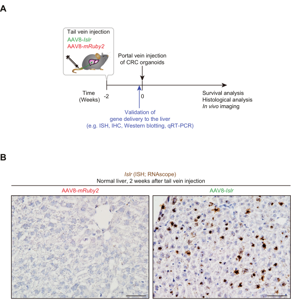
Figure 1: Tail vein-administered AAV8-Islr generates Islr overexpression in the liver. (A) Experimental scheme showing the tail vein injection of AAV8, followed by portal vein injection of colorectal cancer (CRC) organoids. ISH, in situ hybridization; IHC, immunohistochemistry; qRT-PCR, quantitative real-time polymerase chain reaction. (B) Representative pictures. RNA in situ hybridization for Islr was performed using livers from AAV8-Islr or AAV8-mRuby2-treated mice. The livers were collected 2 weeks after tail vein injection. Scale bars, 50 µm. Please click here to view a larger version of this figure.
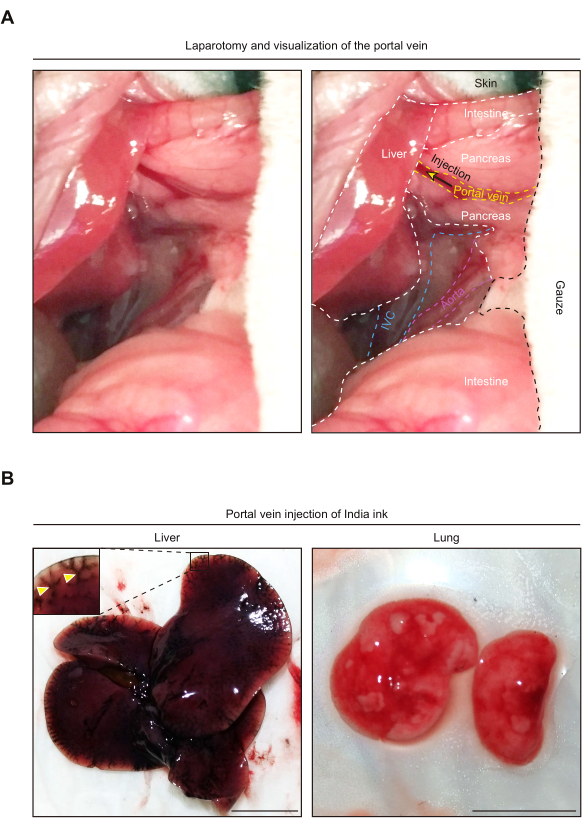
Figure 2: Portal vein injection of India ink results in its delivery to the liver, but not the lung. (A) Representative pictures showing anatomy of the upper abdomen after laparotomy. Note that, for visualization of the portal vein, the intestines are taken outside the abdominal cavity. The right-hand side picture shows an anatomical annotation of each organ and vessel. The yellow arrow denotes the injection site. IVC, Inferior vena cava. (B) Representative pictures of the liver and lung following portal vein injection of India ink. The yellow arrowheads indicate vessels stained with the India ink. Scale bars, 1 cm. Please click here to view a larger version of this figure.
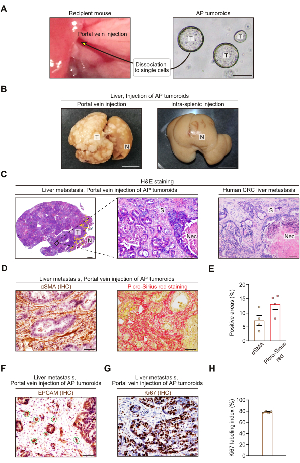
Figure 3: Portal vein injection of colorectal cancer organoids generates stroma-rich liver metastases. (A) Experimental scheme showing portal vein injection of ApcΔ/Δ and Trp53Δ/Δ colon cancer organoids (AP tumoroids). Scale bar, 200 µm. T, tumoroids. (B) Representative macroscopic pictures of the livers that were collected 3-4 weeks after portal vein injection (left) or intra-splenic injection (right). 5.0 x 10⁵ single cells from AP tumoroids were injected into the portal vein or spleen. Intra-splenic injection was performed as described13. White nodules indicate tumors. T, Tumor; N, Normal liver. (C) Representative hematoxylin and eosin (H&E) staining pictures of the CRC liver metastases from the portal vein injection mouse model (left and middle) and human (right). T, Tumor; N, Normal liver; S, Stroma; Nec, Necrosis. The yellow dotted line indicates a border between the tumor and normal liver (left). (D and E) Immunohistochemistry (IHC) for alpha-smooth muscle actin (αSMA; left) and Picro-Sirius red staining (right). (D) Representative pictures. (E) Quantification of αSMA-positive areas (left) and Picro-Sirius red-positive areas (right). N = 4 mice, 5 HPFs (High Power Fields; 400x)/mouse. (F) Representative picture showing immunohistochemistry for EPCAM, an epithelial cell marker. The green dotted lines denote tumor budding. (G and H) Immunohistochemistry for Ki67, a cell proliferation marker. (G) Representative picture. (H) Percentage of Ki67+ cells in total epithelial cells. Epithelial cells were visualized by hematoxylin counterstaining. N = 4 mice, 5 HPFs/mouse. In (B)-(H), all mouse liver tissues were collected 3-4 weeks after injection of AP tumoroids. Mean ± S.E.M. Each dot represents an average value of 5 HPFs from a mouse (E and H). Scale bars represent 1 cm (B), 1 mm (C; left), 100 µm (C; middle and right), and 50 µm (D, F, and G). Note that the study with human tissues was approved by the Ethics Committee of Nagoya University Graduate School of Medicine (2017-0127). Please click here to view a larger version of this figure.
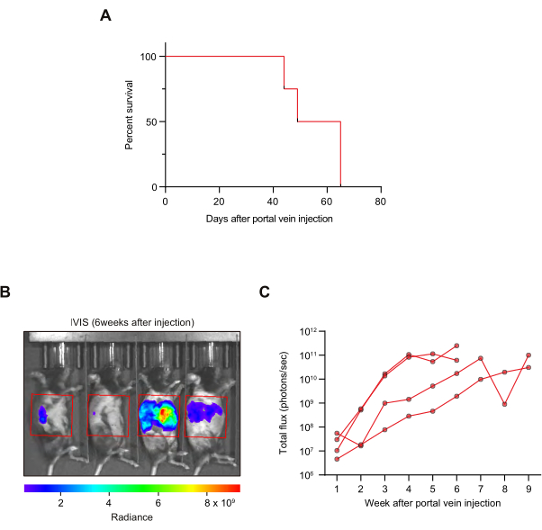
Figure 4: Survival analysis and tumor growth kinetic analysis by in vivo imaging. (A) Kaplan-Meier survival curves. N = 4 mice. (B,C) In vivo imaging system (IVIS) was used to evaluate tumor growth kinetics. (B) A representative picture. The area in the red box was used for quantification. (C) Growth kinetics. Luciferase signals from each mouse are shown. N = 4 mice. Please click here to view a larger version of this figure.
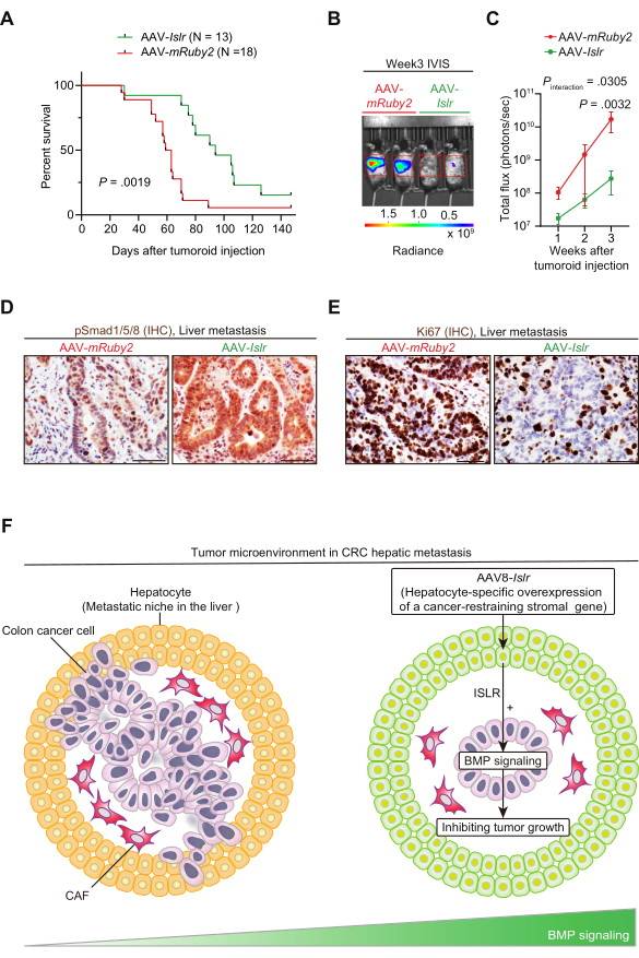
Figure 5: AAV8-mediated gene delivery of Islr to hepatocytes increases BMP signaling, reduces tumor proliferation, and improves mouse survival in a portal vein injection model of CRC liver metastasis. (A) Kaplan-Meier survival curve. Two weeks after tail vein injection of AAV-Islr or AAV-mRuby2, portal vein injection of CRC tumoroids was performed. (B and C) Tumoroid-derived luciferase signals were evaluated using IVIS. (B) Representative picture. (C) Quantification of IVIS signals. N = 5 (AAV-mRuby2) and 8 (AAV-Islr) mice. Mean ± S.E.M. (D) Representative pictures. Immunohistochemistry (IHC) for phosphorylated Smad1/5/8 (pSmad1/5/8). Smad1/5/8 is a downstream molecule of bone morphogenetic protein (BMP) signaling that is phosphorylated upon BMP signaling activation49. N = 4 mice each. (E) Representative pictures. Immunohistochemistry for Ki67. N = 4 mice each. (F) Graphical summary. AAV-mediated hepatocyte-directed gene delivery of a cancer-restraining stromal gene might serve as a therapeutic strategy to inhibit CRC metastagenesis. AAV8-mediated overexpression of Islr in hepatocytes remodeled the metastatic niche by augmenting BMP signaling and limited CRC metastasis progression. For detailed information, refer to Kobayashi et al., Gastroenterology, 202125. Log-rank test (A), and two-way repeated-measures ANOVA (analysis of variance) with post-hoc Sidak's multiple comparison test at Week 3 (C). Scale bars, 50 µm. Figure 5A–C has been reprinted from Gastroenterology, Vol 160(4), Kobayashi et al., The Balance of Stromal BMP Signaling Mediated by GREM1 and ISLR Drives Colorectal Carcinogenesis, Pages 1224-1239.e30, Copyright (2021), with permission from Elsevier. Please click here to view a larger version of this figure.
In this study, we have shown that portal vein injection of mouse CRC organoids reproducibly generates fibroblast-rich liver metastases that mimic histological features of human CRC hepatic metastases. Furthermore, when combined with stroma-directed therapeutics such as AAV8-mediated gene therapy, this preclinical model serves as a useful tool to assess therapeutic effects on mouse survival and tumor growth.
There are, at least, two critical steps in the protocol. Firstly, it is important to prepare a single-cell suspension of tumoroids by fully trypsinizing the organoids and using a mesh filter to remove cell clumps. Incomplete dissociation of tumoroids results in large cell aggregations, injection of which can occlude the portal vein. This causes infarction of the liver and animal death22. Secondly, during portal vein injection, it is imperative to minimize bleeding from the portal vein. The needle should be properly inserted into the portal vein. To avoid puncture of the other side of the portal vein, the angle of the inserted needle should be kept almost parallel to the portal vein. To prevent tearing of the portal vein during injection, the needle should not be moved once it is fully inserted into the portal vein. Immediately after removing the needle from the portal vein, it is vital to apply pressure to the injection site with a cotton bud for at least 5 min.
The cell number for injection needs to be modified according to the recipient mouse (e.g., immunocompetent vs. immunodeficient mouse) and tumorigenicity of the organoid line. In our experiments, injection of 5.0 x 10⁵ single cells in 100 µL suspension was sufficient to generate liver metastases in immunocompetent mice. Increasing the cell number could enhance the risk of embolism in the portal vein and death of animals14, and thus should be carefully considered. Given that previous papers used 5 x 10⁴ to 5 x 10⁵ cells in 100 µL for tumoroid injection into the portal or mesenteric vein25,26,27,28, we consider that this cell number range would be a good starting point for optimization.
One limitation of the portal vein injection approach is that it does not fully recapitulate the entire cascade of CRC metastagenesis. Hepatic metastasis of cancer requires five major steps: (1) cancer cell invasion at the primary site, (2) intravasation into the vessel, (3) cell survival in the portal circulation, (4) extravasation from the portal vein to the liver parenchyma, and (5) colonization in the metastatic niche50. The portal vein injection model only permits investigation of steps (3)-(5). To gain insights into the other metastatic processes, it is necessary to utilize other models such as in vitro models and/or a Notch1-mutant primary CRC mouse model that frequently metastasizes to the liver51.
The significance of the portal vein injection method with respect to the existing methods includes liver-specific growth and higher tumor burden. Peritoneal dissemination in other injection models and primary tumor growth in primary CRC models can confound analysis of liver metastasis progression. In contrast, our portal vein approach rapidly generates liver-specific tumors, simplifying survival and tumor growth analyses.
One major advantage of organoid transplantation over cell line injection is that portal vein injection of tumoroids generated the fibroblast-rich tumor microenvironment that phenocopies a desmoplastic feature of human metastatic CRC. Organoid culture conditions recapitulate features of the tumor microenvironment and support intestinal stem cell populations, including embedding cells in a rich and complex 3D ECM with a defined combination of growth factors24,52. This results in tumor cell propagation in a manner that preserves the genetic landscape of the source tumor52,53. In contrast, traditional 2D culture of tumor cell lines is associated with clonal selection and aberrant genomic/transcriptomic alterations that do not faithfully reflect characteristics of the original cancers54. We speculate that the relatively harsh culture conditions for 2D culture of cancer cell lines force cells to adapt to lack of ECM/growth factor signals that may then result in stroma-poor tumors in vivo, as the cells no longer require stroma-derived signals for survival.
By combining the organoid portal vein injection model with AAV8-mediated gene therapy, we found that delivery of a cancer-inhibitory payload to hepatocytes before cancer cell colonization in the liver could modify the (pre-)metastatic niche to inhibit CRC metastasis growth25. Encouragingly, clinical trials have demonstrated that AAV-mediated in vivo gene transfer to the liver induces long-term expression of a transgene and could be an effective and safe modality to treat non-neoplastic genetic diseases30,31,55. In the future, harnessing hepatocytes to prevent liver metastases through the AAV approach might have potential clinical value in cancer patients at high risk of metastases.
In summary, our paper shows that CRC organoid transplantation via the portal vein generates fibroblast-rich liver metastases, which can be exploited for the development of therapeutic strategies targeting the stroma. By coupling the portal vein injection with stroma-directed therapy such as AAV-mediated gene delivery, one may identify novel stromal targets to restrain CRC metastasis progression.
The authors have nothing to disclose.
This study was supported by grants from the National Health and Medical Research Council (APP1156391 to D.L.W., S.L.W.) (APP1081852 to D.L.W., APP1140236 to S.L.W., APP1099283 to D.L.W.,); Cancer Council SA Beat Cancer Project on behalf of its donors and the State Government of South Australia through the Department of Health (MCF0418 to S.L.W., D.L.W.); a Grant-in-Aid for Scientific Research (B) (20H03467 to M.T.) commissioned by the Ministry of Education, Culture, Sports, Science and Technology of Japan; AMED-CREST (Japan Agency for Medical Research and Development, Core Research for Evolutional Science and Technology (19gm0810007h0104 and 19gm1210008s0101 to A.E.); the Project for Cancer Research and Therapeutic Evolution (P-CREATE) from AMED (19cm0106332h0002 to A.E.); Japan Society for the Promotion of Science Overseas Challenge Program for Young Researchers (to H.K.), Takeda Science Foundation Fellowship (to H.K.), Greaton International Ph.D. Scholarship (to H.K.), Lions Medical Research Foundation Scholarship (to K.G.).
We thank Dr. Leszek Lisowski at Vector and Genome Engineering Facility (VGEF), Children's Medical Research Institute (CMRI) (NSW, AUSTRALIA) for producing recombinant AAV vectors.
10% Formalin | Sigma | HT501128 | |
15 mL centrifuge tube | Corning | 430791 | |
33-gauge needle | TSK | LDS-33013 | For portal vein injection |
4-0 vicryl suture | ETHICON | J494G | |
40-µm cell strainer | Corning | 431750 | |
5 mL Syringe | BD | 302130 | Used to apply saline to the intestine after portal vein injection |
50 mL centrifuge tube | Corning | 430829 | |
50 mL syringe | TERUMO | SS*50LE | Luer lock syringe for perfusion fixation |
70% Isopropyl alcohol wipe | Briemar | 5730 | |
Anaesthesia machine | Darvall | 9356 | |
αSMA antibody | DAKO | M0851 | Clone 1A4. 1/500 dilution for immunohistochemistry |
Buprenorphine | TROY | N/A | ilium Temvet Injection, 300 µg/ml Buprenorphine |
Cotton buds | Johnson & Johnson | N/A | Johnson's pure cotton bud applicators. Need to be autoclaved before use. |
D-luciferin | Biosynth | L-8220 | |
Electric shaver | Sold by multiple suppliers | ||
Forceps | Sold by multiple suppliers | ||
Hamilton syringe | HAMILTON | 81020 | For portal vein injection |
Heat box (animal warming chamber) | Datesand | MK3 | |
Heat lamp | Sold by multiple suppliers | ||
Hemostatic sponge | Pfizer | 09-0891-04-015 | Gelfoam absorbable gelatin sponge, USP, 12-7 mm |
India ink | Talens | 44727000 | |
Injection syringe and needle | BD | 326769 | For tail vein injection |
Islr probe (RNAscope) | ACD | 450041 | |
Isoflurane | Henry Schein | 988-3244 | |
IVIS Spectrum In Vivo Imaging System | Perkin Elmer | 124262 | |
Living Image Software | Perkin Elmer | 128113 | |
Matrigel | Corning | 356231 | |
MRI fibrosis tool | N/A | N/A | https://github.com/MontpellierRessourcesImagerie/imagej_macros_and_scripts/wiki/MRI_Fibrosis_Tool |
Phosphate-buffered saline (PBS) | Sigma | D8537 | |
RNAscope kit | ACD | 322300 | |
Rodent restrainer | Sold by multiple suppliers | ||
Rosa26-Cas9 mouse | The Jackson Laboratory | 024858 | |
Saline | Pfizer | PHA19042010 | |
Scissors | Sold by multiple suppliers | ||
Skin staplers | Able Scientific | AS59028 | 9 mm wound clips |
Stapler applicator | Able Scientific | AS59026 | 9 mm wound clip applicator |
Stapler remover | Able Scientific | AS59037 | Wound clip remover |
Surgical drape | Multigate | 29-220 | |
Surgical gauze | Sentry Medical | GS001 | |
Topical anesthesia cream | EMLA | N/A | EMLA 5% cream, 25 mg/g lignocaine and 25 mg/g prilocaine |
TrypLE Express | Gibco | 12605028 | Recombinant cell-dissociation enzyme mix |
Y-27632 | Tocris | 1254 |