With advancements in laparoscopic techniques, laparoscopic radical antegrade modular pancreatosplenectomy (L-RAMPS) has been widely recognized. However, owing to several technical difficulties in this procedure, the artery-first approach in L-RAMPS still remains uncommon. Here, we developed the dorsal-caudal artery approach for L-RAMPS, which might be safe and beneficial for pancreatic neck tumors.
Laparoscopic radical resection of the pancreatic neck is one of the most complicated radical operations for pancreatic cancer, especially for patients who have had neoadjuvant chemotherapy. Here, we present a technique to perform laparoscopic radical antegrade modular pancreatosplenectomy (L-RAMPS) using the dorsal-caudal artery approach by making full use of the high-definition vision and operation modes of the laparoscope.
The innovation and optimization of this operation are provided in the protocol. Priority should be given to the dorsal resection plane, including the dorsal side of the superior mesenteric artery (SMA), the dorsal side of the pancreatic head, the root of the celiac artery (CeA), the ventral side of the left renal vessels, and the renal hilum. On the condition that the operation for pancreatic neck-body cancer is feasible and safe, the second step is to perform tumor resection en bloc surrounding the SMA and CeA from the caudal to the cephalic side to increase the rate of R0 (radical zero) resection and further prognosis.
Radical antegrade modular pancreatosplenectomy (RAMPS) is an exquisite procedure for malignant tumors located in the pancreatic body or tail, first described by Strasberg in 2003. This operation strategy was designed based on the blood and lymph node drainage of the pancreas, in order to achieve tumor-free dissection planes and radical resection of regional lymph nodes1. RAMPS is becoming increasingly valued by surgeons as it could be conducive to obtain tumor-free margins and relatively favorable survival results2,3,4. With advancements in minimally invasive surgical instruments and techniques, laparoscopic RAMPS (L-RAMPS) has been gradually popularized on account of several advantages, including less intraoperative blood loss, decreased requirement for blood transfusions, and fewer incision events such as pain and infection1. For well-selected patients with distal pancreatic ductal adenocarcinoma (PDAC), recent studies have demonstrated that L-RAMPS could be an effective and safe approach1,5,6.
In pancreaticoduodenectomy (PD) for a malignant tumor around the pancreatic head region, the artery-first approach is a widely accepted strategy with several advantages. The core principle of this method is to explore the superior mesenteric artery (SMA) in the early stages of surgery in order to determine the feasibility of radical resection before pancreatic transection or the ligation of major vessels7,8,9. Recent studies have demonstrated that this artery-first approach may relieve the formation of venous congestion of the distal pancreas and spleen and contribute to the effective bleeding control of the operative regions; besides, it makes lymph node dissection around the SMA more adequate7,10,11. For these reasons, the artery-first strategy is becoming an important component for PD and provides an insight into left-sided pancreatic cancer.
Until now, only some artery-first approaches for L-RAMPS procedures have been reported5,6. The core concept of these approaches is that during pancreatic cancer surgery, tumor infiltration to the SMA should be detected before the performance of irreversible surgical steps, such as transection of the pancreas parenchyma or ligation and resection of the major vessels12,13.
Here, we developed the dorsal-caudal artery approach for L-RAMPS, which might be safe and beneficial for tumors in the pancreatic neck. Our procedure further optimized the routine artery-first approaches for L-RAMPS procedures which were reported by Yamamoto and Kawabata12,14. In other words, we explored and separated the SMA first via the dorsal-caudal approach, a method that has never been reported previously. The goal and advantages of this procedure are to ensure the feasibility and safety of the operation for pancreatic neck-body cancer, which might improve the rate of R0 resection and further prognosis.
Surgeons who intend to use this procedure, however, must have substantial experience in laparoscopic pancreatic surgery. Even if they have passed the learning curve, it is critical to evaluate the patient's condition, including tumor type, vascular condition, and other parameters, because this procedure necessitates sophisticated resection techniques.
In this article, we present a case of a 50-year-old male patient with PDAC, confirmed by endoscopic ultrasound-guided fine-needle aspiration (EUS-FNA) biopsy, who underwent L-RAMPS after preoperative neoadjuvant chemotherapy. Our aim is to demonstrate the clinical safety and feasibility of L-RAMPS using the dorsal-caudal artery approach, and its oncologic outcomes in patients with PDAC located in the pancreatic neck, body, or tail.
The present protocol follows the guidelines of the Ethics of the Second Affiliated Hospital of Guangzhou University of Chinese Medicine. Informed consent was obtained from the patient for this article and the video.
1. Patient Selection
2. Surgical technique
3. Postoperative follow-up
A 50-year-old man with upper abdomen discomfort and chronic malnutrition had a 3.2 cm x 2.5 cm tumor in the pancreatic neck identified. He had previously been healthy and had a normal BMI (19.9 kg/m2).
No distant metastasis, major vessels (besides the splenic artery and vein), or lymph node infiltration were detected on the preoperative imaging evaluation. Endoscopic ultrasound-guided fine-needle aspiration (EUS-FNA) was performed to confirm the pathological diagnosis of PDAC. The patient underwent four cycles of neoadjuvant chemotherapy using gemcitabine plus nab-paclitaxel program. The medication helped to ease clinical symptoms, and carbohydrate antigen 199 (CA-199) decreased from 4,666 U/mL to 1,350 U/mL, while the tumor's maximal diameter decreased from 3.2 cm to 2.5 cm (see Figure 3).
The outcome of the surgery is presented in Table 1. The total time for the procedure was 240 min, with a blood loss of 50 mL. The patient's recovery was uncomplicated, and he was discharged on the 9th day after the surgery. On postoperative day 3 (POD 3), the amylase level in the drainage fluid was 1,645 U/L. The drain was removed in POD 7 when the amylase level was 54 U/L. This was graded as grade A POPF. No peritoneal fluid was found on the postoperative CT examination on POD 7 (see Figure 4). The patient recovered well and was discharged on POD 9.
Histopathology revealed a moderately poorly differentiated ductal adenocarcinoma with interstitial fibrosis, thus confirming the preoperative diagnosis. The pancreatic interstitial fibrosis may be due to preoperative neoadjuvant chemotherapy (see Table 1). The resection margins of the pancreatic neck and posterior peritoneum were microscopically radical (R0). Only 15 lymph nodes were detected, and none of those were involved. The tumor was staged as T2N0M0 (AJCC 8th edition). The patient underwent six cycles of adjuvant chemotherapy using gemcitabine plus nab-paclitaxel program.
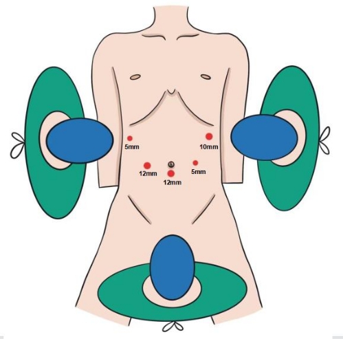
Figure 1: The position of the surgeons. The first surgeon is to the patient's right, the first assistant is to the left, and the second assistant, who is holding the laparoscope, is between the patient's legs. The procedure is performed using a five-port technique. Please click here to view a larger version of this figure.
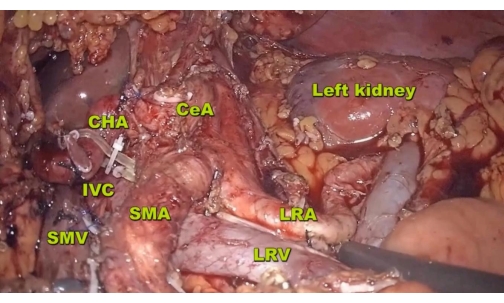
Figure 2: Resection. The resection range extends up to the diaphragmatic crus, down to the LRV, and to the posterior left lateral part of the aorta on the posterior side. Abbreviations: LRA = left renal artery, IVC = inferior vena cava. Please click here to view a larger version of this figure.
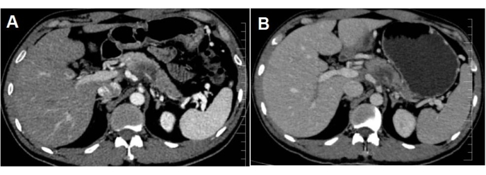
Figure 3: The images show the mass in the pancreatic neck. After neoadjuvant chemotherapy, the tumor maximum diameter was reduced from (A) 3.2 cm to (B) 2.5 cm. Please click here to view a larger version of this figure.
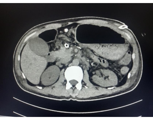
Figure 4: The CT image shows no peritoneal fluid was found on POD 7. Please click here to view a larger version of this figure.
Variable | Outcome |
Intraoperative | |
operative time,minutes | 240 |
Intraoperative blood loss,mL | 50 |
Postoperative | |
Postoperative pancreatic fistula(POPF) | Grade A |
Drain removal,postoperative day | 7 |
Postoperative hospital stay,days | 9 |
Pathological diagnosis | Radically(R0) resected ductal adenocarcinoma with interstitial fibrosis,2cm |
Table 1: Representative outcome of the surgery.
Despite the 5 year survival rates of PDAC, this is still unsatisfactory for patients who have the chance for radical resection; surgery has been the only curative therapeutic method till now22. Resectability status is mostly assessed by the conditions of the regional critical vessels, including the SMA, CeA, CHA, and SMV24,25. Although preoperative radiography can provide adequate information about vascular conditions, surgical exploration is the straightforward method for patients who have ambiguity evaluation, especially for the ones who previously had neoadjuvant chemotherapy. Hence, the early assessment of radical resection during surgery is of great importance in order to avoid unnecessary subsequent operation and improve R0 resection rate.
Laparoscopic radical resection of the pancreatic neck is one of the most complicated radical operations for pancreatic cancer. This study provides an L-RAMPS surgical strategy via the dorsal-caudal artery approach, which might develop to be a standardized, reproducible, and oncologically effective procedure in high-volume centers with experienced surgeons. It may offer a new vision of an artery-first aspect by minimally invasive approaches and could determine the feasibility of radical resection at the early stage of RAMPS.
The most important part of this technique is that priority should be given to confirm the dorsal resection plane, which contains the following parts: the dorsal side of the superior mesenteric artery (SMA), the dorsal side of the pancreatic head, the root of the celiac artery (CeA), the ventral side of the left renal vessels, and the renal hilum. On the condition that all the important parts are not infiltrated by the tumor, surgeons could ensure the feasibility and safety of the radical operation, and then resect the tumor en bloc around the SMA and CeA from the caudal side to the cephalic side. This surgical strategy is aimed at improving the rate of R0 resection and further long-term survival.
This complex procedure needs to be performed by highly experienced surgical teams with both open and laparoscopic pancreatic surgical skills. The learning curve is steep as the implementation of this surgical method has high requirements for the cooperation of the surgical team, anatomical cognition of this complicated area, and flexible response capability against unexpected variations. For these reasons, limited cases make the randomized clinical trials difficult to design and accomplish, so high-level evidence for the perioperative and survival results of this technique are difficult to establish.
The authors have nothing to disclose.
None.
3D Laparoscope | STORZ | TC200,TC302 | |
Echelon Flex Powered Plus Articulating Endoscopic Linear Cutter and Endopath Echelon Endoscopic Linear Cutter Reloads with Gripping Surface Technology | Ethicon Endo-Surgery | ECR60G/GST60G | |
HARMONIC ACE Ultrasonic Surgical Devices | Ethicon Endo-Surgery | HAR36 | |
Ligating Clips | Teleflex Medical | 5,44,22,05,44,23,05,44,000 | |
Ultrasonic Surgical & Electrosurgical Generator | Ethicon Endo-Surgery | GEN11CN |