Riboflavin-5'-phosphate (or flavin mononucleotide; FMN) is sensitive to visible light. Various compounds, including reactive oxygen species (ROS), can be generated from FMN photolysis upon irradiation with visible light. The ROS generated from FMN photolysis are harmful to microorganisms, including pathogenic bacteria such as Staphylococcus aureus (S. aureus). This article presents a protocol for deactivating S. aureus, as an example, via photochemical reactions involving FMN under visible light irradiation. The superoxide radical anion ( ) generated during the FMN photolysis is evaluated via nitro blue tetrazolium (NBT) reduction. The microbial viability of S. aureus that is attributed to reactive species was used to determine the effectiveness of the process. The bacterial inactivation rate is proportional to FMN concentration. Violet light is more efficient in inactivating S. aureus than blue light irradiation, while the red or green light does not drive FMN photolysis. The present article demonstrates FMN photolysis as a simple and safe method for sanitary processes.
Riboflavin-5′-phosphate (FMN) is formed by phosphorylation at the riboflavin 5′-position of the ribityl side-chain and is required by all flavoproteins for numerous cellular processes to generate energy. It also plays the role of vitamin for some functions in the human body1. FMN is approximately 200 times more soluble in water than riboflavin2.
The antibacterial photodynamic inactivation (aPDI) of bacteria is an efficient way to control resistance to bacteria3,4 because it does not depend on the mode of bacterial resistance. Clinically, aPDI is used to treat soft tissue infections in order to decrease infection of nosocomial skin due to multi-resistant bacteria5,6,7,8,9. aPDI also produces cell death by generating reactive oxygen species (ROS). ROS, such as superoxide radicals ( ), singlet oxygen, hydroxyl radicals (•OH), and peroxyl radicals, are free radicals or molecules that contain reactive oxygen10,11,12 and are normally reactive13. Similar to DNA damage that is caused by ROS, membrane peroxidation and destruction of endothelial cells are also adverse biochemical reactions that are attributed to ROS in cells12.
The use of aPDI for pathogenic bacteria involves a visible or UV light source to inactivate microorganisms in the presence of chemical compounds, such as methylthioninium chloride14, PEI-ce6 conjugate15, porphyrin16, titanium dioxide17, toluidine blue O18, and zinc oxide nanoparticles19. Toluidine blue O and methylene blue are phenothiazinium dyes and methylene blue has toxic properties. Zinc oxide nanoparticles and UV irradiation are linked to adverse health and environmental effects. As such, the exploitation of a reliable, secure, and simple photosensitizer via photolysis under visible irradiation deserves further study.
The micronutrient, riboflavin or FMN, is not toxic and is indeed used for food manufacturing or feeding20. Both FMN and riboflavin are highly sensitive to light irradiation2. Under UV1,2,21,22,23 and blue light irradiation10,24, these two compounds achieve an excited state. The activated riboflavin or FMN that is produced by photolysis is promoted to its triplet state and ROS are generated simultaneously2,25. Kumar et al. reported that riboflavin activated by UV light selectively causes increased injury to the guanine moiety of DNA in pathogenic microorganisms26. Under irradiation by UV light, photodynamically activated riboflavin is demonstrated to promote the generation of 8-OH-dG, which is a biomarker for oxidative stress, in double-stranded DNA27. It is reported that S. aureus and E. coli are deactivated by ROS during riboflavin or FMN photolysis10,24,28. A previous study by the authors showed that the photolytic reactions involving riboflavin and FMN reduce crystal violet, a triarylmethane dye and an antibacterial agent that generates , and eliminate most of the antimicrobial capability of crystal violet28. When flavin adenine dinucleotide or FMN is irradiated by blue light, the resulting ROS produce apoptosis in HeLa cells for their poisoning in vitro29. Using photochemical treatment in the presence of riboflavin, Cui et al. inactivated lymphocytes by inhibiting their proliferation and cytokine production30.
The photolysis of riboflavin is used for the inactivation of blood pathogen by UV10,24, but blood components can be impaired under UV light irradiation30. It is also reported that platelets exposed to UV progressively enhance the performance of the activation markers P-selectin and LIMP-CD63 on their membranes. The cytotoxicity of UV and high-intensity irradiation needs to be investigated and a photosensitizer that is uncomplicated and safe during an FMN photoreaction involving visible light would be of great use.
Light of shorter wavelengths has more energy and is much more likely to cause severe damage to cells. However, in the presence of a suitable photosensitizer, irradiation with low-intensity violet light can inhibit pathogenic microorganisms. The photosensitization and the generation of by FMN when irradiated with violet light is thus important to study, in order to determine the pathway by which ROS from FMN photolysis increases the inactivation of bacteria.
Antimicrobial control is a common issue and the development of new antibiotics frequently takes decades. After irradiation with violet light, photoinactivation that is intermediated by FMN can annihilate environmental pathogenic bacteria. This study presents an effective antimicrobial protocol in vitro using violet light for driving FMN photolysis and thus generating for aPDI. The microbial viability of S. aureus is used to determine the feasibility of FMN-induced aPDI.
1. Photolysis system setup
- Mount six light-emitting diodes (LED) (DC 12 V) on the inside of an opaque plastic cup (8 cm x 7 cm) as shown in Figure 1 to establish a photolysis system31.
- Add reactants (see below) into the glass test tubes (13 mm in diameter and 100 mm in height) and secure the tubes at the top of the cup as shown in Figure 1. Place the experimental setup in a room with a steady temperature of 25 ± 3 °C.
- Monitor and record the temperature of test units throughout the photolytic reactions by an infrared thermometer.
NOTE: Figure 2 shows the emission spectra for blue, green, red, and violet lights, as recorded using a calibrated UV-Vis optical spectrometer. The wavelengths corresponding to the absorbance maximum for blue, green, red, and violet LED lights used in the study are 465, 529, 632, and 405 nm, respectively. The LED chips can heat the apparatus during irradiation experiments. The whole photoreaction system in each experiment was, therefore, placed in a room where the temperature was kept constant at 25 ± 3 °C.
2. The effect of light wavelength on the photolysis of FMN (Figure 3)
- Prepare a 0.1 mM FMN solution in 100 mM potassium phosphate buffer (PB) (pH 7.8). Expose FMN samples (3 mL each) to blue, green, red, or violet light at an intensity of 10 W/m2 for 5 min. Keep 3 mL of FMN solution in dark as a control.
- Record the absorbance of irradiated FMN samples in the 250-750 nm range using a UV/Visible spectrophotometer.
3. Nitro blue tetrazolium (NBT) reduction method to detect (Figure 4).
NOTE: The NBT reduction method used here was slightly modified from the assay for riboflavin photoreaction. The NBT reduction method is used to evaluate the level of generated from FMN photolysis. The photochemically excited FMN is first reduced by L-methionine into a semiquinone, which then donates an electron to oxygen giving rise to 31. The as-generated reduces NBT to formazan, which has a characteristic absorbance at 560 nm32.
- Add 109.3 mg of L-methionine to 73.3 mL of PB (100 mM; pH 7.8). Vortex the solution to dissolve the L-methionine.
- After the L-methionine is fully dissolved, add 10 mg of NBT powder and 1.53 mL of 0.117 mM FMN to the solution. For each 1 mL of the reaction solution (i.e., a mixture of FMN, L-methionine, and NBT) applied, the final concentrations of FMN, methionine, and NBT are 2.4 x 10⁻⁶ M, 1.0 x 10⁻² M, and 1.6 x 10⁻⁴ M, respectively.
- Expose the reaction solution (1 mL) to blue or violet LED light irradiation at 10 W/m2 for 1-5 min.
- Detect the species (from the absorbance at 560 nm) produced by the photochemical reaction, which reduces NBT and produces formazan.
4. S. aureus viability after FMN photolysis (Figure 5)
- Transmit a colony of S. aureus(BCRC 10451) from a cultured plate into 10 mL of Lysogeny broth taken in a 15 mL screw-capped test tube. Culture in a shaker at 37 °C for 16 h.
- Transfer 0.5 mL of the culture to a 1.5 mL centrifuge tube. Add sterilized water into the centrifuge tube to dilute the culture to an optical density of 0.5 at 600 nm (OD600) (~6 x 10⁷ CFU/mL).
- Transfer 0.5 mL of the culture to a 1.5 mL centrifuge tube, centrifuge at 14,000 x g for 10 min and decant the supernatant to obtain a cell pellet.
- Add 1 mL of FMN-buffered solution (30, 60, and 120 µM FMN in PB) to the cell pellets obtained as in step 4.3, and vortex. For irradiated control, add 1 mL of PB alone.
- Transfer 1 mL each of viable bacterial cell solutions containing 30 µM FMN and PB alone into glass tubes, and irradiate them with violet light at 10 W/m2 for 30 min.
- Transfer 1 mL each of viable bacterial cell solutions containing 30, 60, and 120 µM FMN and PB alone into glass tubes and irradiate them with blue light at 20 W/m2 for 120 min. Set up another glass tube with 1 mL of viable bacterial cell solution containing 120 µM FMN and irradiate it with blue light at 20 W/m2 for 60 min.
- Set up test tubes as described in steps 4.5 and 4.6 and cover them with thick aluminum foils. These tubes serve as dark controls.
- Keep the irradiation chamber (as well as the dark controls) in a cold room at 9 ± 1 °C during the 30-120 min irradiation period.
NOTE: Heat released by LED lights cannot be ignored as the LED chips placed inside the cup can heat the photoreaction system during irradiation experiments. The experiments were, therefore, conducted in a cold room maintained at 9 ± 1 °C. - After irradiation, transfer 0.2 mL from each of the reaction solutions onto a Luria agar (LA) plate. Spread the bacteria over the plate by an L-shaped glass rod and incubate overnight at 37 °C.
- Calculate the viable plate count and the inactivation rates of S. aureus after overnight growth.
NOTE: The inactivation rate of S. aureus is calculated as the percentage reduction, which is equal to [1 –I / D] ×100%, where I and D denote, respectively, the number of CFUs in the irradiated sample and dark control. The percentage reduction is defined as a negative value of the inactivation rate.
5. Detection of in S. aureus (Figure 6)
- Prepare the S. aureus samples as described in step 4.
- Dilute the bacterial density of the samples with sterilized water to an optical density of 0.5 at 600 nm (OD600, ~6 x 10⁶ CFU/mL). Transfer 0.5 mL of the culture to a 1.5 mL centrifuge tube. Centrifuge at 14,000 x g for 10 min and decant the supernatant to obtain a cell pellet.
- Add 0.1093 g of L-methionine, 0.1 g of NBT, and 25 mL of FMN (400, 240, or 120 µM) to 75 mL of PB. For each 1 mL of the reactant applied, the final concentrations of FMN, methionine, and NBT are 100 (60 or 30) x 10-6 M, 7.3 x 10⁻³ M, and 1.2 x 10⁻³ M, respectively.
- Add 1 mL of each reactant solution (i.e., with varying FMN concentrations) to the cell pellets obtained as in step 5.2. Irradiate the solutions by violet light at 10 W/m2 for 10 min.
- Centrifuge the mixture at 14,000 x g for 10 min and decant the supernatant. Resuspend the pellet in 1 mL of dimethyl sulfoxide (DMSO) to extract the reduced NBT. Detect the produced species from the absorbance at 560 nm.
6. Statistical analysis
- Express data as mean ± standard deviation (SD) of three independent tests.
- Apply a homoscedastic two-sample t-test to evaluate whether two measurements are different. p-values < 0.05 are considered statistically significant.
Effect of light wavelength on FMN
The absorbance spectra of 0.1 mM FMN that is irradiated using blue, green, red, and violet LEDs are shown in Figure 3. There are two peaks for FMN (372 nm and 444 nm) for the dark control. Green and red light have no effect because changes in the spectra are insignificant. On the other hand, the respective absorbance of FMN at 444 nm is reduced by about 19% and 34%, respectively, after blue and violet light irradiation at 10 W/m2 for 5 min, suggesting that irradiation with blue/violet light increases FMN photolysis.
Detection of  from FMN that is irradiated using blue or violet light
NBT reduction was used to determine the formation of 32. FMN is highly sensitive to visible and UV light even for short periods of radiation. is formed from the intermediate photochemical reactions during the photolysis of FMN. Figure 4 shows that the photochemical effect of FMN on NBT reduction depends on the irradiation time, as is formed from FMN exposed to blue or violet light irradiation in a time-dependent manner. The average photolytic effect of blue light and violet light is, however, similar as shown in Figure 4.
Effect of FMN photolysis on S. aureus viability
The effect of FMN exposed to violet or blue light irradiation on S. aureus viability was determined. FMN photolysis using blue or violet light irradiation results in significant inactivation of S. aureus. The greater the concentration of FMN, the higher the inactivation rate of S. aureus that is exposed to blue light irradiation at 20 W/m2 for 120 min as shown in Figure 5A. Inactivation rates of 26%, 39%, 43%, and 97% were achieved for S. aureus using 0, 30, 60, and 120 µM FMN, respectively, under 20 W/m2 blue light irradiation for 120 min. As shown in Figure 5A, there is no significant difference (p-value = 0.20) in the inactivation rate for S. aureus using blue light irradiation with either 60 µM FMN at 20 W/m2 for 120 min (energy dose, 14.4 J/cm2) or 120 µM FMN at 20 W/m2 for 60 min (energy dose, 7.2 J/cm2). On the other hand, as shown in Figure 5B, inactivation rates of 53% and 96% were achieved for S. aureus without and with 30 µM FMN, respectively, under violet light irradiation at 10 W/m2 for 30 min (energy dose of 1.8 J/cm2). Therefore, FMN exposed to blue or violet light irradiation has a greater effect on the viability of S. aureus by increasing the bacterial inactivation. FMN treated with violet light irradiation is more efficient in S. aureus inactivation.
Detection of in FMN-treated S. aureus under violet light irradiation
The production of in FMN-treated S. aureus under violet light irradiation was determined using the NBT reduction method. The absorbance value at 560 nm was used to evaluate the extent of production. The photochemical effect of FMN on generation in S. aureus is proportional to the FMN concentration (Figure 6). The 560 nm absorbance values for S. aureus without FMN treatment are 0.31 and 0.07 under 10 W/m2 violet light irradiation for 10 min and in the dark, respectively (Figure 6). This suggests that little was produced in untreated S. aureus after violet light irradiation. The respective 560 nm absorbance values for S. aureus treated with 100 µM FMN are 1.36 and 0.08 under 10 W/m2 violet light irradiation for 10 min and in the dark, respectively. Therefore, the photochemical effect on NBT reduction in FMN-treated S. aureus increases under violet irradiation, as is produced abundantly.
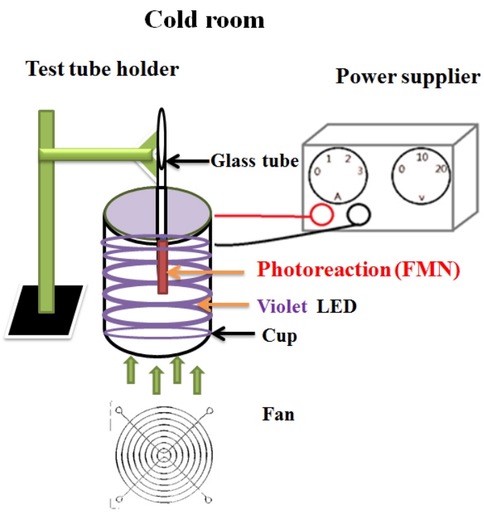
Figure 1: Schematic representation of the photoreaction system. Please click here to view a larger version of this figure.
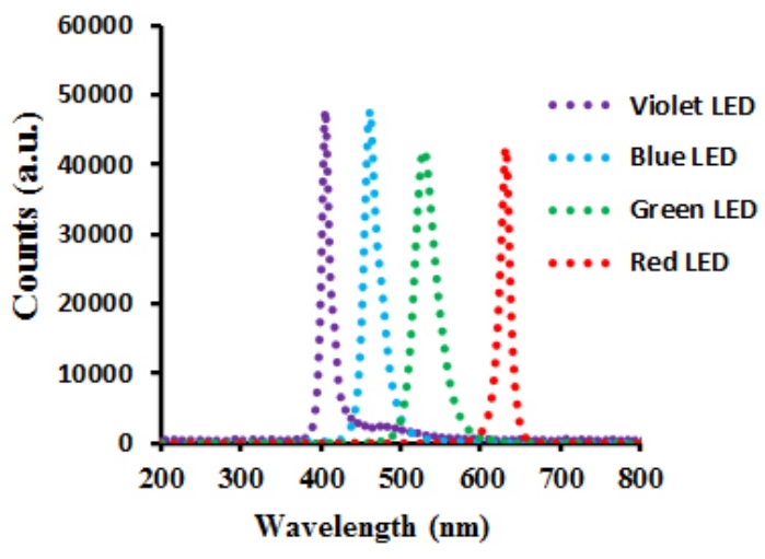
Figure 2: The emission spectra of blue, green, red, and violet LED lights in this study. The emission maxima for the blue, green, red, and violet lights are at 465, 529, 632, and 405 nm, respectively, and the spectral widths at half-height (W1/2) are 26, 31, 14, and 12 nm, respectively. Please click here to view a larger version of this figure.
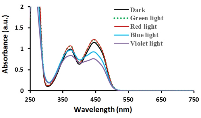
Figure 3: Absorbance spectra of FMN treated with LEDs of various wavelengths at 10 W/m2 for 5 min. Please click here to view a larger version of this figure.
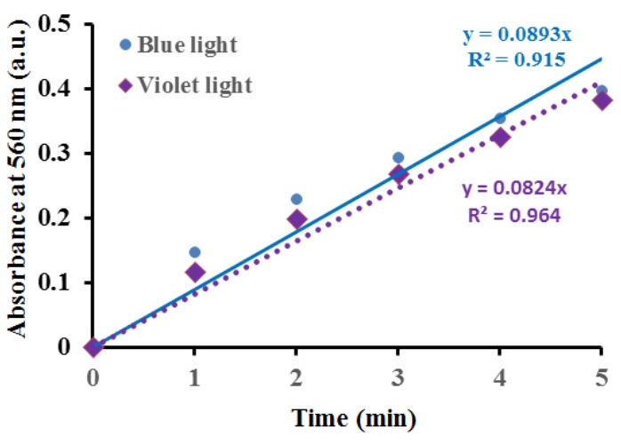
Figure 4: NBT reduction to determine the effect of blue and violet light irradiation at 10 W/m2 for 1-5 min on FMN photolysis. Please click here to view a larger version of this figure.
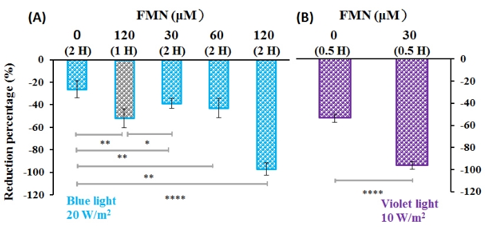
Figure 5: Effect of FMN photolysis on S. aureus viability. The percentage reduction is defined as a negative value of the inactivation rate for S. aureus treated with (A) FMN under blue light irradiation at 20 W/m2 for 120 min and (B) FMN under violet light irradiation at 10 W/m2 for 30 min. FMN concentrations and irradiation times are indicated at the top of the figure. Note that there is no significant difference (p = 0.20) in the inactivation rate for S. aureus using 60 µM FMN at 20 W/m2 blue light irradiation for 120 min or 120 µM FMN at 20 W/m2 blue light irradiation for 60 min (A). Results are expressed as mean ± SD, where n = 3. *p < 0.05; **p < 0.01; ****p < 0.0001. Please click here to view a larger version of this figure.
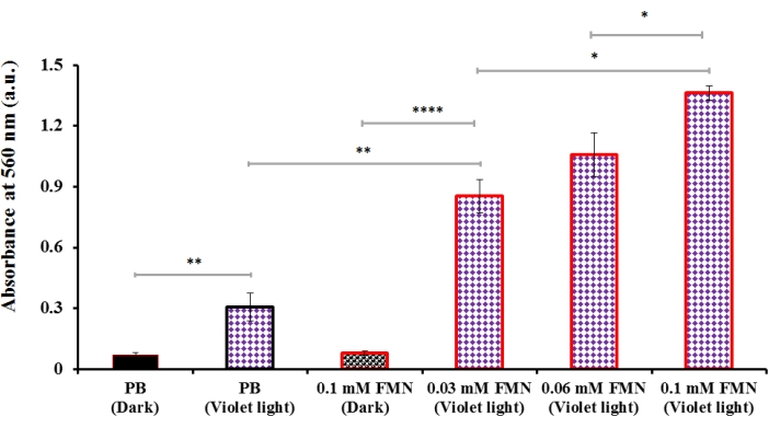
Figure 6: Effect of FMN concentration on generation in S. aureus subjected to violet light irradiation at 10 W/m2 for 10 min. Overall, little is produced in S. aureus in the absence of FMN although violet light irradiation slightly increases in the irradiated sample (PB (Violet light)) relative to dark control (PB (Dark)). Results are expressed as mean ± SD, where n = 3. * p < 0.05; ** p < 0.01; **** p < 0.0001. Please click here to view a larger version of this figure.
A photosensitizer increases the photochemical reaction of chemical compounds to generate ROS. Pathogenic microorganisms can be inactivated by light irradiation in the presence of photosensitizers. This study determines the aPDI of S. aureus due to ROS generated by violet light irradiation of an exogenous photosensitizer, FMN.
As shown in Figure 3, for FMN, the absorbance at 444 nm is reduced significantly after 5 min of irradiation using violet or blue light and there is no significant change in the FMN absorbance under irradiation with green or red light. NBT reduction is used to determine the formation of via a charge-transfer process10,33. A previous study by the authors used FMN under blue, green, or red-light irradiation to determine the formation of using NBT reduction. Blue light irradiation produces an enormous amount of whereas the photolytic efficiency of green and red light is less than 3% of that achieved by blue light10. These results show that a charge-transfer process is produced when FMN is exposed to blue or violet light irradiation and that there is no significant change upon irradiation by green or red light.
A previous study by the authors34 showed that the mechanism for FMN photolysis can be described with primary FMN photolytic reactions (Equation 1):
 (1),
(1),
where 1FMN* and 3FMN* are the singlet and triplet states of the excited FMN, respectively. The photolytic reaction mechanisms for these photoinduced FMN are possibly initiated by a triplet−triplet annihilation or a triplet-ground state quenching process35,36,37,38. The ground state of FMN (FMN0) is an electron donor that provides an electron to 3FMN* and generates the semi-reduced (FMN•-) and semi-oxidized (FMN•+) forms, as shown in Equation 2
 (2)
(2)
The photodegradation pathways for FMN result from photosensitization. FMN is promoted to the electronically excited state, FMN•, via a photolytic reaction and then, a neutral FMN is reduced when FMN• loses an electron, as shown in Equations 3–8 below:
 (3)
(3)
 (4)
(4)
 (5),
(5),
where FMN•+ is a radical cation species.
The interaction of FMN•- with O2 (3Σg–) produces the reactive superoxide radical, , followed by the hydroperoxyl radical, HOO•, which is generated in the reaction between and a proton and eventually leads to the degradation of FMN0:
 (6)
(6)
 (7)
(7)
 (8),
(8),
where FMN•- is a radical anion.
In the present study, in the absence of FMN, the inactivation rate in the case of S. aureus is 53% under violet light irradiation at 10 W/m2 for 30 min and 26% under blue light irradiation at 20 W/m2 for 120 min, suggesting that blue light irradiation deactivates fewer bacteria than violet light (Figure 5). It has been reported earlier that irradiation at wavelengths longer than 430 nm without an exogenous photosensitizer does not inactivate S. aureus cells, although porphyrins in S. aureus have a maximum absorbance at 405 ± 5 nm and cause bacterial inactivation after irradiation39.
A previous study by the present authors showed that E. coli and S. aureus are deactivated by DNA cleavage that is induced by the formation of through photolysis of riboflavin or FMN10,24,40. The respective inactivation rates for S. aureus are 97% and 96%, respectively, using 120 µM FMN under 20 W/m2 blue light irradiation (energy dose of 14.4 J/cm2) for 120 min and 30 µM FMN under 10 W/m2 violet light irradiation (energy dose of 1.8 J/cm2) for 30 min (Figure 5). Therefore, violet light irradiation is more effective in deactivating S. aureus as low-energy doses of violet light can deactivate S. aureus in the presence of lower FMN concentrations and shorter illumination periods as compared with blue light. Although the extent of FMN photolysis achieved by violet light is higher compared with blue light (Figure 3), the average photolytic effects of FMN on NBT reduction under blue and violet light irradiation are similar (Figure 4).
E. coli is a common Gram-negative bacterium. Riboflavin or FMN photolysis by blue light irradiation inactivates E. coli via ROS10,24,41 with an inactivation rate of 96% when using FMN10,24. S. aureus, on the other hand, is a Gram-positive bacterium, which is effectively inactivated by FMN photolysis under violet light irradiation, as demonstrated here (Figure 5). Therefore, FMN photolytic reaction under blue or violet light irradiation can produce ROS and inactivate both Gram-negative and Gram-positive bacteria.
In the absence of FMN, however, little is produced in S. aureus that is exposed to violet light irradiation (Figure 6); therefore, endogenous photosensitizers only slightly decrease the viability of S. aureus under violet light irradiation, compared to the effect in the presence of exogenous photosensitizer such as FMN. Under the same conditions, FMN that is exposed to violet light irradiation decreases the viability of S. aureus to result in a 96% inactivation rate, which is much higher than that for endogenous intracellular photosensitizers alone. The production of in FMN-treated S. aureus under violet light irradiation has significantly increased, as shown in Figure 6. These results show that FMN when exposed to violet light irradiation causes an elevated level of bacterial inactivation.
FMN can achieve a photo-excited state when irradiated using UV1,2,21,22,23, blue10,24, and violet light31; however, the cytotoxicity that is caused by UV radiation and high-intensity short-wavelength irradiation must be considered42. On the other hand, red and green radiations with longer wavelengths do not produce significant amounts of ROS during FMN photolysis10. Therefore, the ROS-induced aPDI of S. aureus requires irradiating FMN with violet light that has an intermediate wavelength. The spectral width at half height (W1/2) for violet light must be narrow to avoid any overlap with the UV absorption and inhibit risk due to UV radiation31. The micronutrient, FMN, is as such harmless, being an essential vitamin for the human body. Using an appropriate photosensitizer such as FMN, low-energy violet light irradiation can suppress pathogenic bacteria, providing a safe and effective means of the sanitary process. In addition to the photolytic reaction of FMN on S. aureus, other issues deserve further investigation, such as the phototoxicity of FMN on microbes, arising from the stress caused by , which can be studied using an RNA-seq technique along with qPCR.
FMN photolysis via violet light irradiation leads to photochemical oxidation by which ROS is generated and in turn enhances the inactivation of pathogenic bacteria. The use of violet light is considered a critical step in FMN photodecomposition. The limitation of the current technique is that the wavelength interval of violet light must be quite narrow to avoid the risk due to UV irradiation. The current method has the potential for future medical applications, such as infection treatment of surface skin along with deep subcutaneous tissues or muscles by inserting an optical fiber to direct the violet light to the infected areas. Photochemical treatment using FMN is therefore a safe and simple way to ensure effective hygiene practices.
