Synovial fluid analysis under transmitted, polarized, and compensated light microscopy is used to evaluate the inflammatory or non-inflammatory nature of a sample through simple steps. It is particularly useful in osteoarthritis to detect calcium crystals and identify a more severe subset of osteoarthritis.
Synovial fluid (SF) analysis is important in diagnosing osteoarthritis (OA). Macroscopic and microscopic features, including total and differential white blood cell (WBC) count, help define the non-inflammatory nature of SF, which is a hallmark of OA. In patients with OA, WBC in SF samples usually does not exceed 2000 cells per microliter, and the percentage of inflammatory cells, such as neutrophils, is very low or absent. Calcium crystals are frequent in SF collected from OA patients. Although their role in the pathogenesis of OA remains unclear, they have been associated with a mild inflammatory process and a more severe disease progression. Recently, calcium crystals have been described in both the early and late stages of OA, indicating that they may play a vital role in diagnosing different clinical subsets of OA and pharmacological treatment. The overall goal of SF analysis in OA is two-fold: to ascertain the non-inflammatory degree of SF and to highlight the presence of calcium crystals.
Osteoarthritis (OA) is a complex and multifactorial chronic joint disease, with an estimated pooled global prevalence of 16% in subjects aged 15 and over, and 23% in subjects aged 40 and over1. The incidence of OA is expected to increase due to an aging population and an increase in risk factors, such as obesity and metabolic syndrome2.
Among the major issues associated with OA are the difficulty in diagnosing the disease in its early stages and currently available treatments limited to pain management and symptomatic slow-acting drugs (SYSADOAs) such as glycosaminoglycans. The diagnosis of OA is based on clinical symptoms and imaging findings. However, the lack of correlation between the two assessments can cause years of delay in OA diagnosis and treatment initiation2. OA is characterized by degenerative processes and low-grade inflammation, which can be easily investigated via synovial fluid (SF) analysis. SF is a viscous plasmatic dialysate rich in hyaluronic acid, which lubricates the joint space, provides nutrients and oxygen to cartilage, and removes metabolic waste. SF also acts as a shock absorber, thus protecting the joints during stress and strain3.
SF analysis is a simple and reliable method that must always be performed during the initial evaluation of patients with musculoskeletal symptoms and joint effusions3. Recommendations from the American College of Rheumatology, the British Society for Rheumatology, and others include SF analysis among the diagnostic testing for rheumatic diseases that must be undertaken mainly in evaluating acute monoarthritis4,5. Total and differential leucocyte counts obtained from SF analysis provide a snapshot of the pathological process occurring in the joint, thus classifying the degree of inflammation. The identification of pathogenic crystals, such as monosodium urate (MSU) and calcium pyrophosphate (CPP) crystals, under polarized light, is vital in the diagnosis and treatment of crystal arthritis (e.g., gout and pseudogout). Furthermore, the presence of microorganisms suggests a diagnosis of septic arthritis.
Calcium crystals are frequent in samples collected from patients with OA6. Basic calcium phosphate (BCP) crystals and CPP have been reported in approximately 22% and 23% of SF samples, respectively, from patients with OA6. Although their role remains unclear, these crystals have been associated with more severe forms of OA7 and are considered an epiphenomenon of the pathological process itself. Calcium crystals have been detected in 100% of tissue samples from OA patients undergoing knee replacement8. Furthermore, it has been hypothesized that calcium crystals may be involved in the pathogenesis of OA owing to their inflammatory effects, demonstrated by several studies9 and mediated, at least in part, by the NLRP3 inflammasome10.
More recently, calcium crystals have been described in both early and late stages6 of OA, indicating that they may play a vital role in diagnosing different clinical subsets of OA and pharmacological treatment.
The overall goal of SF analysis is twofold: to determine the inflammatory degree of SF and to diagnose crystal or septic arthritis by identifying specific crystals or microorganisms. It is a particularly useful, simple, and reliable tool in diagnosing OA, owing to the typical non-inflammatory pattern with a very low percentage or absence of neutrophils.
Advantages over alternative techniques
SF analysis consists of simple procedures that include total and differential leucocyte counts and crystal search. Manual cell counting performed by expert laboratory technicians3,11 remains the gold standard for the cytological analysis of synovial and other body fluids. However, due to the time-consuming limitation and the inter- and intra-observer variability of this method, automated cell counters have gradually replaced manual counting in large routine clinical laboratories where blood and urine analyzers have been adapted to enable SF analysis11,12. Nevertheless, manual counting presents some advantages in specific settings: (1) in the ambulatory, to obtain a total and differential WBC value on time; (2) to identify cell types such as cytophagocytic mononuclear (Reiter) cells or non-hematopoietic cells such as synoviocytes, that automated counters cannot recognize; (3) when the sample is too small to be handled by the instrument; (4) to create local laboratory registries that are readily accessible for research purposes. Another advantage of manual counting is seen insupravital staining, a method that allows the differentiation of cells very quickly and immediately after glass slide preparation. By contrast, traditional stainings, such as the Wright and the May-Grünwald-Giemsa procedures, require air-dried SF smears and time to stain the cells13. Although not suitable for time-sensitive routine analyses, these staining methods reveal more detailed cell populations in the sample, including erythrocytes, basophils, eosinophils, polymorphonuclear leukocytes, lymphocytes, and platelets.
Finally, routine SF analysis for crystals is performed using a simple technique based on polarized light microscopy, which provides fast results14. Alternative methods, such as scanning and electron microscopy, yield more accurate and sensitive results, but their use in everyday clinical practice is not feasible due to high costs and time-consuming sample preparation and analysis.
The present protocol complies with the guidelines of the Ethics committee of Padova University Hospital. SF was collected with patient consent from the knee joints of patients receiving therapeutic arthrocentesis for joint effusion at their initial presentation to the clinic or in response to an arthritic flare. Contraindications to the procedure were: coagulopathy, anticoagulant medications, skin lesions, dermatitis, or cellulitis overlying the joint. All SF samples were deidentified.
1. Synovial fluid preparation
2. Macroscopic examination
NOTE: Macroscopic evaluation of SF samples consists of subjective, qualitative, and semiquantitative assessments. There are no reference standard scales, and no control samples are used.
3. Microscopic examination
NOTE: The SF microscopic analysis consists of cytological examination, including total and differential white blood cell (WBC) counts and the search for crystals.
4. Crystal detection
5. Alizarin red staining
Large joints affected by OA are often swollen and produce significant amounts of SF, which are drained via arthrocentesis15. The macroscopic characteristics of SF evaluated immediately after arthrocentesis essentially include the quantity, color, clarity, and viscosity17. Despite their low specificity, they provide preliminary data on the degree of inflammation. The color depends on SF cellularity and the degree of fragmentation of extracellular matrix macromolecules. The SF collected from patients with OA is pale yellow, a shade generally associated with non-inflammatory samples, whereas dark yellow is associated with inflammatory effusions (Figure 2). The presence of small amounts of blood in the sample, for instance, due to capillary ruptures, results in slightly orange color. Bloody effusions must be carefully considered as they might indicate a potential hemarthrosis. Clarity is a hallmark of SF collected from patients with OA. In fact, non-inflammatory SF is poor in cells, debris, hyaluronic acid, and fibrin fragments, unlike the turbid appearance of inflammatory SF (Figure 2).
Viscosity represents a good inflammatory index as it is associated with the integrity of matrix macromolecules, mainly hyaluronic acid (Figure 2). These molecules depolymerize during inflammation, resulting in a less viscous SF. Depending on the disease stage and severity, the viscosity may be preserved or slightly reduced in OA.
The number of leukocytes never exceeds 500-1,000 cells/mm3 in OA, and they are mostly monocytes (Supplementary Figure 5A) and other mononuclear cells like synoviocytes (Supplementary Figure 5B). One of the most relevant and frequent findings in OA SF concerns CPP and BCP crystals. CPP crystals are easily detected under compensated polarized light microscopy (Figure 5), whereas the identification of BCP crystals is rendered more difficult due to their submicroscopic size (Figure 6). In fact, the length of a single BCP crystal is less than 1 µm, and although these crystals form aggregates, the clumps appear as non-birefringent amorphous-looking globules. Although nonspecific, positive alizarin red staining generally reveals the presence of BCP crystals.
Contrary to pseudogout, wherein CPP crystals are numerous and associated with high leukocyte counts and inflammatory clinical features, in OA, the number of CPP crystals in SF is very low and not linked to any altered humoral response. Nevertheless, SF samples with CPP or BCP crystals display higher levels of inflammatory cytokines when compared with OA without crystals21. Regardless of WBC count, SFs with crystals are also associated with increased percentages of PMN cells5. From a clinical point of view, the relationship between calcium crystals and inflammation may help define a particular subset of patients at an increased risk of developing more severe disease.
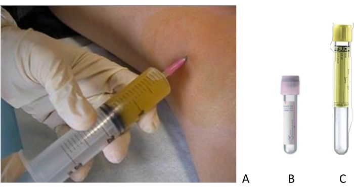
Figure 1: Joint aspiration of a swollen knee. (A) Arthrocentesis is performed after accurate disinfection of the skin with sterile materials. The SF is collected in (B) EDTA tubes for total cell count and (C) no-additive tubes for differential cell count and crystal search. Please click here to view a larger version of this figure.
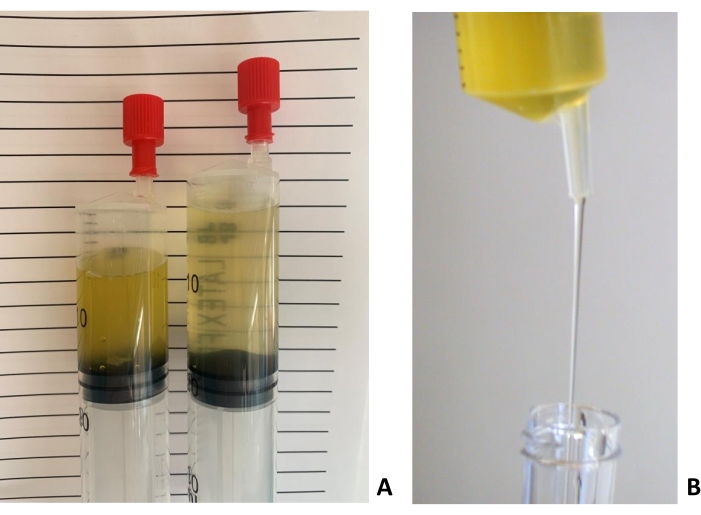
Figure 2: Characteristics of inflammatory (left) and non-inflammatory (right) SF samples. (A) Color and clarity of inflammatory (left) and non-inflammatory (right) SF samples. (B) The viscosity of a non-inflammatory SF evaluated through the "string" test. The laboratorist evaluates the length of the "string" formed by a falling drop of SF from a syringe or a pipette. The sample in the picture produces a long string. Please click here to view a larger version of this figure.
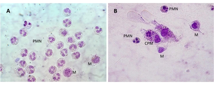
Figure 3: SF cells observed in a pre-stained slide. (A) A group of polymorphonuclear (PMN) cells with multilobed nuclei and two monocytes with their bulky nuclei in the lower right side of the image. (B) PMN cells and monocytes (M) with a cytophagocytic mononuclear (CPM) cell in the center of the image. Bright field; 1,000x magnification. Please click here to view a larger version of this figure.
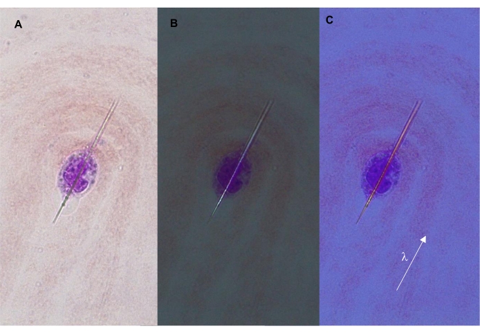
Figure 4: Intracellular MSU crystal. The crystal forms a typical needle shape shown under (A) transmitted, (B) polarized, and (C) compensated polarized light (400x). The arrow indicates the orientation of the λ filter (compensator), which is parallel to the long axis of the crystal. In this configuration, the crystal appears yellow/orange. Please click here to view a larger version of this figure.
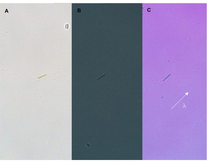
Figure 5: Intracellular CPP crystal. The crystal is shown under (A) transmitted, (B) polarized, and (C) compensated light (400x). The arrow indicates the orientation of the λ filter (compensator), which is parallel to the long axis of the crystal. In this configuration, the crystal appears blue. Please click here to view a larger version of this figure.
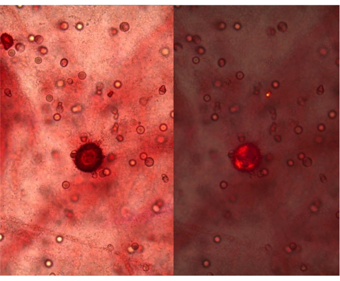
Figure 6: Positivity to the alizarin red test of an SF from OA, 400x. Red precipitates are visible under transmitted light (left panel) and polarized light (right panel), 400x magnification. Please click here to view a larger version of this figure.
Table 1: Degree of inflammation of SF according to the total number of leucocytes. Please click here to download this Table.
Table 2: Colors exhibited by MSU and CPP crystals under compensated polarized light and signs of birefringence. Please click here to download this Table.
Supplementary Figure 1: Blood diluting pipettes according to Malassez-Potain for leucocytes. Please click here to download this File.
Supplementary Figure 2: The layout of the chamber grid for WBC count. Please click here to download this File.
Supplementary Figure 3: Pre-stained, ready-to-use slides for a quick and easy differential WBC morphology evaluation. Please click here to download this File.
Supplementary Figure 4: May-Grünwald-Giemsa staining of an SF smear. Eosinophilic granules are clearly visible (arrow), 1,000x magnification. Please click here to download this File.
Supplementary Figure 5: Monocyte and synoviocytes collected from an SF sample. (A) Monocyte from an SF sample collected from a patient with osteoarthritis (1,000x). (B) Synoviocytes from an SF sample collected from a patient with osteoarthritis (1,000x). Please click here to download this File.
In OA, SF analysis helps define disease characteristics through simple steps: total and differential leukocyte counts and searching for crystals, including CPP and BCP. Furthermore, the detection of MSU crystals may highlight important comorbidities.
Despite low costs and simple execution, the sensibility of the tests and reliability of the results may be affected due to inexperienced analysts-mainly as it pertains to crystal identification. Training and experience of the analyst are crucial in distinguishing the shape and birefringence of CPP and MSU crystals and matching their shape to their degree of birefringence. Some studies have reported discrepancies in crystal identification between different laboratories; thus, training laboratory technicians is essential to obtain reliable and consistent results14. Crystal identification is also affected by misinterpretation of birefringent particles that may be confused for pathogenic crystals. These may derive from external sources (i.e., dust or fibers) or may be present inside the joint cavity (i.e., corticosteroids or lipids). A thorough cleaning of the slide can resolve the former, but the latter can only be overcome through training and experience14.
Another limitation of SF analysis regards the use of alizarin red staining for BCP identification. As mentioned earlier, this is not a specific test, and thus caution must be exercised when interpreting results. The analyst must be able to recognize red precipitates with a peculiar morphology (Figure 11). More sensitive techniques, such as scanning electron microscopy (SEM), can be used to identify BCP crystals, but they are not used in routine examinations of SF in everyday clinical practice. Importantly, it has been shown that results obtained with alizarin red staining show a good, albeit not optimal, concordance with those obtained by SEM22, thus enhancing the value of the former.
Another salient issue to consider regards what to do when arthrocentesis yields very small amounts of SF and how to store the sample. When only a few drops of SF are collected from small joints, such as inter- and metacarpophalangeal joints and the wrist, keeping the material in the syringe and spreading it directly onto a clean glass slide is recommended. Using a micropipette and some tricks, it is possible to prepare more than one slide and perform a complete analysis. In detail, the analyst draws the SF with the counting pipette from the slide prepared for crystal searching and prepares the hemacytometer chamber, whereas some microliters can be aspirated for the supravital staining.
As far as the storage is concerned, SF samples degrade over time, and their analysis requires fresh preparations to obtain reliable results. Alternatively, SF can be preserved at 4 °C and used within 24 h, and no later than 48 h, for cytological examination. To avoid cell clumping and SF coagulation, attention must be paid to collecting the specimen in at least one EDTA tube. Frozen SF samples stored at -20 °C for a longer period of time can be used only for crystal search.
SF analysis is an irreplaceable pathognomonic test in rheumatology, owing to its utility, simplicity of execution, and affordability. The main challenge for future applications of SF analysis is a more accurate diagnosis of rheumatic diseases. This will be possible by integrating this method with more advanced and innovative technologies23, making it possible to characterize SF composition and identify specific molecular biomarkers and proteomics at the site of inflammation.
The authors have nothing to disclose.
The Authors wish to acknowledge Professor Leonardo Punzi for his precious mentorship in the field of synovial fluid analysis and Padova University Hospital for its support.
Alizarin red S | Merck | A5533 | For BCP crystal search |
Burker chamber | Merck | BR718905 | For total white blood cell count |
Cover glasses | Merck | C7931 | For microscopic examination |
EDTA tubes | BD | 368861 | For SF collection |
Glass slides | Merck | S8902 | For crystal search |
Lambda filter (compensator) | Any | Refer to microscope company | For crystal identification |
Malassez-Potain pipette | Artiglass | 54830000 | For dilution of synovial fluid |
Methylene blue solution | Merck | 3978 | For total white blood cell count |
Polarized microscope | Leica, Nikon, others | Depending on the model and company | For complete synovial fluid analysis |
Polarizing lens | Any | Refer to microscope company | For crystal identification |
Testsimplet | Waldeck | 14386 | Supravital staining for cell differentiation |