Methods are described for the generation of large amounts of recombinant adenoviruses, which can then be used to transduce the native rodent urothelium allowing for expression of transgenes or downregulation of endogenous gene products.
In addition to forming a high-resistance barrier, the urothelium lining the renal pelvis, ureters, bladder, and proximal urethra is hypothesized to sense and transmit information about its environment to the underlying tissues, promoting voiding function and behavior. Disruption of the urothelial barrier, or its sensory/transducer function, can lead to disease. Studying these complex events is hampered by lack of simple strategies to alter gene and protein expression in the urothelium. Methods are described here that allow investigators to generate large amounts of high-titer adenovirus, which can then be used to transduce rodent urothelium with high efficiency, and in a relatively straightforward manner. Both cDNAs and small interfering RNAs can be expressed using adenoviral transduction, and the impact of transgene expression on urothelial function can be assessed 12 h to several days later. These methods have broad applicability to studies of normal and abnormal urothelial biology using mouse or rat animal models.
The urothelium is the specialized epithelium that lines the renal pelvis, ureters, bladder, and proximal urethra1. It comprises three strata: a layer of highly differentiated and polarized often bi-nucleate umbrella cells, whose apical surfaces are bathed in urine; an intermediate cell layer with a population of bi-nucleate transit-amplifying cells that can give rise to superficial umbrella cells in response to their acute loss; and a single layer of basal cells, a subset of which function as stem cells that can regenerate the entirety of the urothelium in response to chronic injury. Umbrella cells are chiefly responsible for forming the high-resistance urothelial barrier, components of which include an apical membrane (rich in cholesterol and cerebrosides) with low permeability to water and solutes, and a high-resistance apical junctional complex (comprised of tight junctions, adherens junctions, desmosomes, and an associated actomyosin ring)1. Both the apical surface of the umbrella cell and its junctional ring expand during bladder filling and return to their pre-filled state rapidly after voiding1,2,3,4,5. In addition to its role in barrier function, the urothelium is also hypothesized to have sensory and transducer functions that allow it to sense changes in the extracellular milieu (e.g., stretch) and transmit this information via release of mediators (including ATP, adenosine, and acetylcholine) to underlying tissues, including suburothelial afferent nerve processes6,7,8. Recent evidence of this role is found in mice lacking urothelial expression of both Piezo1 and Piezo2, which results in altered voiding function9. Additionally, rats overexpressing the tight-junction pore-forming protein CLDN2 in the umbrella cell layer develop inflammation and pain analogous to that seen in patients with interstitial cystitis10. It is hypothesized that disruption of urothelial sensory/transducer or barrier function may contribute to several bladder disorders6,11.
A better understanding of the biology of the urothelium in normal and disease states depends on the availability of tools that will allow investigators to readily downregulate endogenous gene expression or allow for the expression of transgenes in the native tissue. While one approach to downregulate gene expression is to generate conditional urothelial knockout mice, this approach depends on the availability of mice with floxed alleles, is labor intensive, and can take months to years to complete12. Not surprisingly then, investigators have developed techniques to transfect or transduce the urothelium, which can lead to results on a shorter time scale. Published methods for transfection include the use of cationic lipids13, anti-sense phosphorothioated oligodeoxynucleotides14, or antisense nucleic acids tethered to the HIV TAT protein penetrating 11-mer peptide15. However, the focus of this protocol is on the use of adenoviral-mediated transduction, a well-studied methodology that is efficient at gene delivery to a broad range of cells, has been tested in numerous clinical trials, and most recently was used to deliver the cDNA encoding the COVID-19 capsid protein to recipients of one variant of the COVID-19 vaccine16,17. For a more thorough description of the adenovirus life cycle, adenoviral vectors, and clinical applications of adenoviruses, the reader is directed to reference17.
An important milestone in the use of adenoviruses to transduce the urothelium, was a report by Ramesh et al. that showed short pretreatments with detergents, including N-dodecyl-β-D-maltoside (DDM) dramatically enhanced transduction of the urothelium by an adenovirus encoding β-galactosidase18. Using this proof-of-principle study as a guide, adenoviral-mediated transduction of the urothelium has now been used to express a variety of proteins, including Rab-family GTPases, guanine-nucleotide exchange factors, myosin motor fragments, pore-forming tight junction-associated claudins, and ADAM1710,19,20,21,22. The same approach was adapted to express small interfering RNAs (siRNA), the effects of which were rescued by co-expressing siRNA-resistant variants of the transgene22. The protocol described here includes general methods to generate large amounts of highly concentrated adenovirus, a requirement for these techniques, as well adaptations of the methods of Ramesh et al.18 to express transgenes in the urothelium with high efficiency.
Experiments involving the generation of adenoviruses, which requires BSL2 certification, were performed under approval from the University of Pittsburgh Environmental Health and Safety offices and the Institutional Biosafety Committee. All animal experiments performed, including adenoviral transduction (which requires ABSL2 certification), were done in accordance with relevant guidelines/regulations of the Public Health Service Policy on Humane Care and Use of Laboratory Animals and the Animal Welfare Act, and under the approval of the University of Pittsburgh Institutional Animal Care and Use Committee. Gloves, eye protection, and appropriate garb are worn for all procedures involving recombinant viruses. Any liquid or solid waste should be disposed of as described below. The bedding of the animals post transduction, and any resulting animal carcasses, should be treated as biohazardous materials and disposed of according to institutional policies.
1. Preparation of high-titer adenovirus stocks
NOTE: Effective transduction of rodent bladders depends on the use of purified and concentrated viral stocks, typically 1 x 10⁷ to 1 x 10⁸ infectious viral particles (IVP) per µL. This portion of the protocol is focused on generating high-titer adenovirus stocks from existing virus preparations. All steps should be performed in a cell culture hood using sterile reagents and tools. While the available strains of adenovirus used today are replication defective, most institutions require approval to use adenoviruses and recombinant DNA. This often includes designation of a cell culture room as a BSL2 approved facility to produce and amplify adenoviruses. Some general considerations include use of masks, eye protection, gloves, and appropriate garb at all stages of virus production and purification. When performing centrifugation, safety caps are recommended if the centrifuge tubes lack tight-fitting caps. All non-disposable materials, including potentially contaminated centrifuge safety caps, bottles, and rotors are treated with an antiviral solution (see Table of Materials), and then rinsed with water or 70% ethanol. Liquid wastes are treated by adding bleach to a final concentration of 10% (v/v). Disposal of these liquid wastes will depend on institutional policies. Solid wastes are typically disposed of in biohazardous waste.
2. Transduction of rodent bladder
NOTE: If new to this technique, it is recommended that the number of animals transduced at one time be limited to 2-4. This can be accomplished by staggering the start times for each animal, particularly during the detergent treatment in step 2.2, and then the virus incubation in step 2.3. Experienced investigators can transduce up to six animals at a time.
Virus preparation
An example of virus purification by density gradient centrifugation is shown in Figure 1A. The light pink band, found at the interface of the loaded cellular material and the 1.25 g/mL CsCl layer, is primarily composed of disrupted cells and their debris (see magenta arrow in Figure 1A). It derives its pinkish color from the small amount of culture medium that is carried over from step 1.5 in the protocol. The virus particles of interest, which appear as a milky white band, are found at the interface of the 1.25 g/mL CsCl and 1.40 g/mL CsCl solutions (see yellow arrow in Figure 1A). One may also observe a band of material that floats 2-3 mm above the enriched virus particles (see thin black arrow in Figure 1A). It comprises unassembled viruses and debris, contains few IVP, and should be avoided when collecting the virus sample.
Further purification of the virus and buffer exchange using a PD10 column is depicted in Figure 1B, and the OD260 readings of the resulting fractions after elution are shown in Figure 1C. The void volume of these columns is approximately 3 mL, and thus the virus begins to appear in fraction 6 and peaks in fraction 9. In this experiment, fractions 6-9 were pooled. While fraction 6 and 7 are relatively low in virus particles, they contain sufficient virus to merit recovery, and they serve to dilute the very high titer fractions to a more reasonable concentration. Fractions 10-12 were not included as the increase in OD260 in fraction 11 indicates the possible presence of a second contaminating peak, which is variably observed in these preparations. The pooled fraction will typically have 1 x 10⁷ to 1 x 10⁸ IVP/µL, and the expected yield would be in the order of 1 x 10¹⁰ to 2 x 10¹¹ total IVP. This is sufficient amount of virus to perform hundreds of transductions. While it is possible to titer the virus by plaque assays or by counting colonies of cells expressing fluorescent proteins22, the 1% rule is sufficient in most cases. This rule states that 1% of purified virus particles, estimated by measuring the OD260 of the sample, are IVP. Aliquots of virus stored at -80 ˚C have a shelf-life of 2-5 years, although infectivity decreases over the long term. Thawed aliquots of virus can be refrozen at -80 °C one time without significant loss of infectivity. However, repeated thawing and freezing negatively impacts viral infectivity.
Bladder transduction
An important first step when assessing the impact of transduction is to confirm transgene expression. This can be assessed using several techniques, including tools that detect mRNA (e.g., RNAScope), western blot analysis, or use of immunofluorescence9,10,23. Figure 2 is an example of mouse urothelium that was transduced with an adenovirus that encodes V5-epitope tagged human growth hormone (V5-hGH)23. This protein is packaged into discoidal/fusiform vesicles and can be exocytosed during bladder filling19,23. Western blot analysis of urothelial lysates revealed V5-hGH expression in the urothelium of transduced bladders, but not in untransduced ones (Figure 2A). Expression was also confirmed by immunofluorescence, in this case using antibodies that recognized hGH or the V5 epitope tag (untransduced bladders lacked signal, not shown) (Figure 2B).
An additional example is transduction of the rat bladder with a virus encoding CLDN2, a pore-forming tight junction-associated protein24,25. CLDN2 increases paracellular flux of cations (including K+) and its overexpression results in inflammation and development of visceral pain10. Western blot analysis confirmed expression of CLND2 in rat bladders transduced with adenovirus encoding Cldn2, but not those transduced with a control GFP-encoding virus (Figure 2C). In general, GFP is not considered to be toxic when expressed in cells and it thus serves as a useful control. The use of GFP also allows the investigator to confirm that transduction is working. Immunofluorescence analysis further confirmed exogenous CLDN2 expression in urothelial umbrella cells transduced with virus encoding Cldn2 cDNA (red signal in Figure 2D). Furthermore, similar to endogenous CLDN2 (not shown), the expressed CLDN2 is localized to TJP1-labeled tight junctions, as well as the basolateral surfaces of the umbrella cells (CLDN2 is labeled green in Figure 2E)10. In the case of CLDN2 expression, it was highest one day after transduction, but then trailed off, and was barely detectable after 15 days.
A second consideration is, which cell types will be targeted by adenoviral transduction. While in rats it is possible to mostly transduce the umbrella cell layer19, in mouse, all the layers of the urothelium can be transduced, although transduction of the intermediate and basal cell layers can be variable. Importantly, in the entirety of the bladder wall, only the urothelium is transduced and no other tissue is targeted by the instilled adenovirus (Figure 3).
While analyses of transduced cells can be performed at the single cell level, when exploring an overall bladder phenotype, one must transduce the majority of the urothelial cells. Thus, it is important to define the efficiency of transduction (i.e., what fraction of urothelial cells are transduced). For example, in the case of the Cldn2-expressing adenovirus, >95% of umbrella cells were transduced (see Figure 2D). An additional example is the image field shown in Figure 3, where counting the number of transduced umbrella cells (in this case, expressing the Ca2+ sensor GCAMP5G), reveals an efficiency that approaches 95%. However, one must examine cells in random fields taken throughout the bladder wall to achieve an accurate and unbiased estimate of the transduction efficiency.
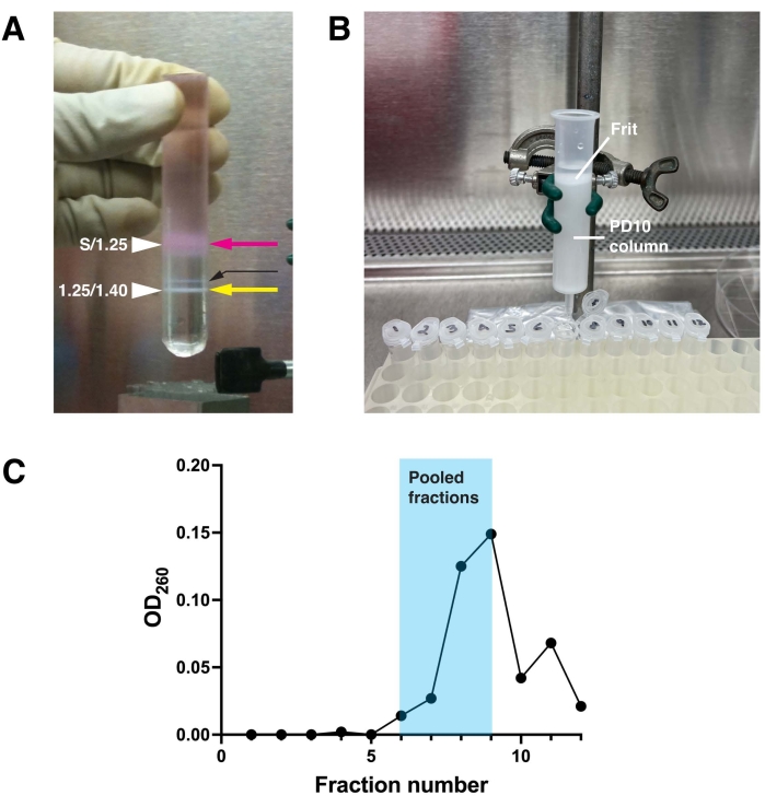
Figure 1: Purification of adenovirus. (A) Adenovirus particles produced by infected HEK293T cells were purified by centrifugation on a discontinuous CsCl gradient made up of a layer of 1.4 g/mL CsCl, a layer of 1.25 g/mL CsCl, and a layer of sample (S) diluted in Tris-EDTA solution. The cellular material (pink arrow) accumulates at the S/1.25 interface, while the purified adenovirus floats at the 1.25/1.4 interface (yellow arrow). A small band of improperly assembled virus floats above the latter (thin black arrow). (B) The adenovirus-rich band in the gradient is recovered using a needle, and the CsCl is removed from the adenovirus by gel filtration using a G25 Sephadex-filled PD10 column equilibrated with PBS containing 10.0% (v/v) glycerol. The surface of the Sephadex is protected by a porous, plastic frit. (C) Fractions, 0.5 mL, were collected from the PD10 column. The OD260 of the fractions were measured in a spectrophotometer and the values plotted. Virus-rich fractions elute in the void volume, which begins at fraction 6 and extends to fraction 9. Pooled fractions for this representative experiment are shaded in blue. Please click here to view a larger version of this figure.
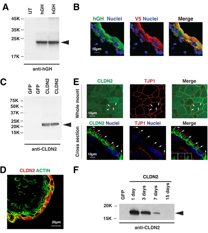
Figure 2: Transduction of rodent bladder urothelium with adenoviruses encoding V5-hGH or CLDN2. (A) Mouse urothelium was left untransduced (UT) or transduced with adenoviruses encoding V5 epitope tagged human growth hormone (hGH). After 24 h, the bladders were recovered, urothelial lysates were prepared and subjected to sodium dodecyl sulfate polyacrylamide gel electrophoresis, and V5-hGH expression confirmed using western blots probed with an antibody against hGH. (B) Detection of V5-hGH in mouse urothelium sectioned and stained using antibodies to hGH (green) or the V5 epitope (red) and fluorophore-tagged secondary antibodies. Immunofluorescence was captured using confocal microscopy. Samples were counterstained with TO-PRO3 to label nuclei. (C–F) Rat urothelium was transduced with a virus encoding rat CLDN2 (common name claudin-2) or GFP (common name green fluorescent protein) as a control. (C) Detection of exogenous CLDN2 by western blotting using an antibody again CLDN2. Note that the endogenous CLDN2 is expressed at low levels and is not detected in this experiment. (D) Transduction of the umbrella cell layer is revealed by immunofluorescence and confocal microscopy. The borders of the cell are revealed by co-staining the tissue with FITC-phalloidin, which labels the cortical actin cytoskeleton. (E) Detection of exogenous CLDN2 and TJP1 (common name ZO1) by immunofluorescence and confocal microscopy of whole-mounted or cross-sectioned urothelium. Small white arrows indicate the location of the tight junction and small magenta arrowheads mark the location of intracellular accumulations of CLDN2 in the cell cytoplasm. These were previously revealed to be Golgi-associated CLDN210. (F) Following transduction, the animals were euthanized on the indicated days post infection. Exogenous CLDN2 expression was detected using western blotting. Animals expressing GFP were euthanized after day 1. The data in Figure 2C–E have been modified from Montalbetti et al.10, and are reproduced with permission of the American Physiological Society. Please click here to view a larger version of this figure.
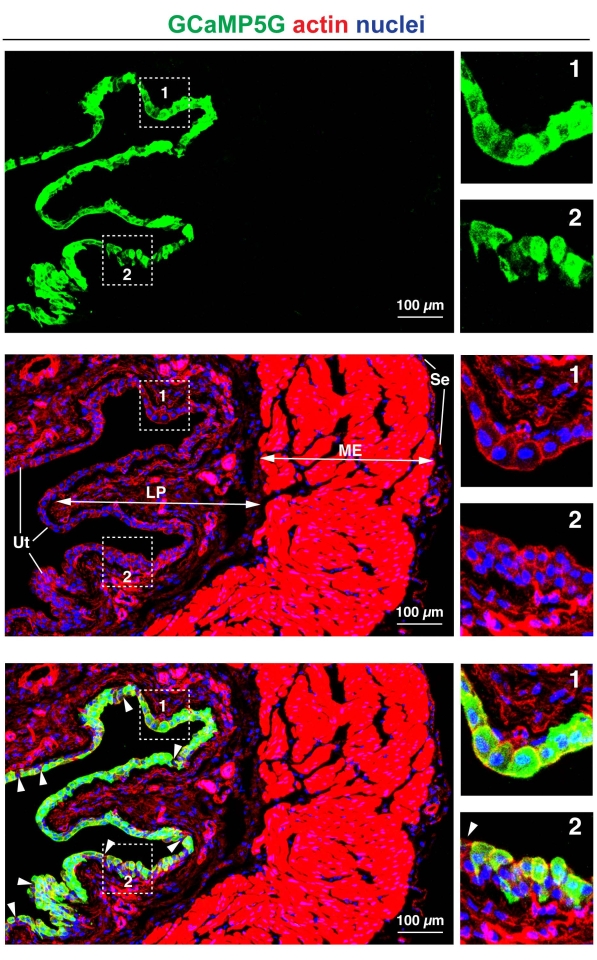
Figure 3: Efficiency of adenoviral transduction. Mouse bladder urothelium was transduced with an adenovirus encoding the calcium sensor GCaMP5G. The upper panels show staining for GCaMP5G (detected using an antibody to green fluorescent protein; GFP, green), the middle panels show the distribution of DAPI-stained nuclei (blue) and rhodamine-phalloidin-labeled actin (red), and the bottom panels are a merger of the three signals. The white arrowheads in the lower panel are the rare umbrella cells that are not transduced. The overall efficiency of transduction of umbrella cells in this image is ~95%. Note that only the urothelium is transduced. The boxed regions are magnified in the panels to the right. The region in box 1 primarily comprises transduced umbrella cells. The region in box 2 is urothelium showing efficient transduction of umbrella cells, but less efficient transduction of the underlying cell layers. LP = lamina propria; ME = muscularis externa; Se = serosa; Ut = urothelium. Images were acquired using a confocal microscope (see Table of Materials). Please click here to view a larger version of this figure.
While Ramesh et al. were focused on developing strategies to use adenoviral transduction in the treatment of bladder cancer18, more recent reports have demonstrated the utility of these techniques in studying normal urothelial biology/physiology and pathophysiology10,18,19,20,21. The important features of this approach include the following: (i) In the entirety of the rodent bladder wall, only the urothelium is transduced10,19,20,21,22. In the case of rats, transduction is mostly limited to the umbrella cell layer, while in mice, the entirety of the urothelium can be targeted; (ii) This approach can be used to express proteins or siRNAs10,19,20,21,22; (iii) The efficiency of transduction can approach 70%-95% (e.g., see Figure 3)18,20; (iv) One day after transduction, the ultrastructure of the urothelium, the integrity of the tissue, presence of a high-resistance barrier (assessed by measuring trans-epithelial resistance), and the expression of urothelial differentiation markers is "normal"10,19. The only morphological effect noted thus far is a decrease in umbrella cell diameter (when viewed from above); however, they revert to their normal size after 3-4 days10,19; (v) The urothelium can be transduced with up to three different adenoviruses simultaneously18; (vi) Transgene expression can be altered by titrating the number of IVP, which affects the amount of protein expression, lengthening or shortening the time of incubation before the animal is sacrificed, or use of different promoters such a tet-regulated system (tet-off)19. In the latter case, one can co-express a virus encoding the transactivator/tet-repressor, and then modulate the expression of the transgene by altering the concentration of doxycycline in the animal's diet.
While the overall protocol is relatively simple, there are some critical steps that must be considered when performing these experiments. One of these is the availability of high-titer virus stocks that are of reasonable purity. Adenoviruses can be obtained from commercial vendors, from institutional virus production cores, from other investigators, or they can be produced in house. In the case of the latter, the Bert Vogelstein laboratory AdEasy system is recommended because its components can be purchased from Addgene, and adenoviruses generated using this approach work well in urothelial transduction4. This technique allows one to generate adenoviruses encoding cDNAs as large as 8 Kb using the pAdEasy-1 packaging plasmid (the insert replaces viral genes E1 and E3), the pAdEasier-1 bacterial cells (which encode the adenoviral genes necessary to produce virus), and production of replication-defective adenoviruses in HEK293T cells (which express the required viral E1 protein). However, substituting the pAdEasy-1 packaging plasmid with the pAdEasy-2 packaging plasmid increases the packaging size by an additional 2.7 kB, but necessitates using a different cell line (E1-transformed human embryonic retinal 911 cells) as the viruses lack early gene E4 in addition to E1 and E34. Expression of siRNAs can be accomplished using several systems, including pAdloss, which is described in detail in Kasahara et al.26. While it may be possible to use virus-rich cell culture medium to transduce the urothelium, this method can be unreliable. Instead, large-scale virus amplification, CsCl gradient purification, followed by gel filtration provides the most reliable way to generate large amounts of high-titer virus. The relative stability of virus at -80 °C, coupled with relatively large yields of virus, makes this an ideal way to have a consistent stock of virus that can be used over a large number of experiments.
Additional critical steps in this protocol include those associated with the transduction protocol. These include use of PBS (without divalent cations) as a washing agent and diluent, and the use of DDM detergent. As the junctional complex is dependent on Ca2+ for proper function, PBS likely promotes viral entry by disrupting the junction-associated barrier. DDM is also critical; as Ramesh et al. reported, the efficiency of adenovirus transduction is exceedingly low in the absence of detergent treatment18. However, the mode of action of the detergent is not clear. A 10 min incubation with DDM is ideal, but this can be shortened to 5 min; however, it is not recommended that the incubation extend beyond 10 min as the detergent may cause unexpected damage to underlying tissues. The amount of virus used is a critical parameter, with higher concentrations generally leading to greater amounts of transgene expression and higher transduction efficiencies. However, the optimal virus concentration that gives the best combination of expression, efficiency, and phenotype must be empirically determined. As a starting concentration, a range of 5.0 x 10⁶ to 2.0 x 10⁷ IVP per animal is recommended. Depending on the protein being expressed, this results in efficiencies in the 70%-95% range10,19,20,21,22; however, some dominant-negative GTPase constructs are less efficient (30%-50%) and require a single-cell analysis approach4,20. These differences in efficiency may reflect the turnover of the transgene, its toxicity, or the purity and yield of virus used for transduction. The latter can be ruled out by performing plaque assays. Although not hyper-critical, the time the virus remains in the bladder must be long enough for the virus to attach to its CXADR receptor. Times less than 30 min can lower the efficiency of transduction, while extending the incubation period to 45 min can increase efficiency; however, this can be transgene-dependent. The final critical step in this protocol is determining how long the animal will be held before a phenotype is assessed. Transgenes exhibit the highest expression after 1-2 days19, but the expression can decrease after that. In the case of siRNAs, it depends on the turnover rate of the protein itself. For example, when expressing siRNAs that target expression of Rab11a, a Rab-family GTPase, efficient downregulation is only observed 72 h after transduction23. Thus, long-lived proteins (with half-lives measured in days) may not be suitable targets using this approach.
The investigator should also be aware of the caveats associated with this approach. First, its utility may be limited to female rodents because of difficulties in catheterizing male rodents through their penis. However, it may be technically possible to perform this protocol in males by introducing a catheter into the dome of the bladder, similar to the preparation employed when performing cystometry9. This would allow one to introduce a detergent or a virus, and perform washes as needed. A second caveat is that overexpression of proteins can exhaust cell pathways and resources, which can lead to events such as protein aggregation, activation of cell stress pathways, and death27. Thus, it is generally wise to limit transgene expression to non-toxic levels (unless of course that is the intention of transgene expression), as assessed using markers of cell stress and cell death. A third caveat is that in some settings, long-term adenovirus expression can trigger immune responses28. While there is no evidence that adenoviral transduction of the urothelium per se stimulates immune responses10, this is transgene-dependent and as noted above CLDN2 overexpression leads to cystitis.
Currently, investigators have a variety of methods that they can use to modulate gene and protein expression in the urothelium, including use of transgenic or conditional urothelial knockout mice, use of transfection reagents, or use of adenoviral transduction. The latter method, the subject of this protocol, is relatively easy, efficient, and reproducible. Other than the initial expense of cell culture reagents needed to generate the virus stocks, the very large amount of virus produced (enough to perform hundreds of transductions), results in a relatively low cost on a per animal basis. Adenoviral transduction can be exploited to study urothelial biology. For example, adenoviral transduction was used to define the importance of Rho-family and Rab-family GTPases, guanine-nucleotide exchange factors, myosin motor fragments, and ADAM17 in exocytosis and endocytosis10,19,20,21,22, and adenoviral transduction was recently used to study urothelial mechanotransduction9. Likewise, adenoviral transduction may be relevant when exploring and treating human disease. For example, Ramesh et al. initially proposed adenoviral transduction may be a useful way to target tumor cells18. While their studies were more a proof-of-principle approach, one can imagine that expression of toxins in cancer cells or expression of epitopes that could be recognized by the immune system would be useful strategies. Adenoviral transduction may also be useful in understanding diseases where overexpression or underexpression of gene products leads to disease. As one example, biopsies from the bladders of patients with interstitial cystitis exhibit a 90-fold increase in CLDN2 expression29. Interestingly, over-expressing CLDN2 using adenoviral transduction replicates many of the symptoms of this disease in rats10. Thus, CLDN2 could be one target in the treatment of patients with this disorder.
The authors have nothing to disclose.
This work was supported by a pilot project award through P30DK079307 (to M.G.D.), NIH grant R01DK119183 (to G.A. and M.D.C.), NIH grant R01DK129473 (to G.A.), an American Urology Association Career Development award and a Winters Foundation grant (to N.M.), by the Cell Physiology and Model Organisms Kidney Imaging Cores of the Pittsburgh Center for Kidney Research (P30DK079307), and by S10OD028596 (to G.A.), which funded the purchase of the confocal system used to capture some of the images presented in this manuscript.
10 mL pipette | Corning Costar (Millipore Sigma) | CLS4488 | sterile, serological pipette, individually wrapped |
12 mL ultracentrifuge tube | ThermoFisher | 06-752 | PET thinwall ultracentrifuge tube |
15 mL conical centrifuge tube | Falcon (Corning) | 352097 | sterile |
18 G needle | BD | 305196 | 18 G x 1.5 in needle |
20 mL pipette | Corning Costar (Millipore Sigma) | CLS4489 | sterile, serological pipette, individually wrapped |
50 mL conical centrifuge tube | Falcon (Corning) | 352098 | sterile |
5 mL pipette | Corning Costar (Millipore Sigma) | CLS4487 | sterile, serological pipette, individually wrapped |
Cavicide | Henry Schein | 6400012 | Anti-viral solution |
Cell culture dish – 15 cm | Falcon (Corning) | 353025 | sterile, tissue-culture treated (150 mm x 25 mm dish) |
Cell scraper | Sarstedt | 893.1832 | handle length 24 cm, blade length 1.7 cm |
CsCl | Millipore Sigma | C-4306 | Molecular Biology grade ≥ 98% |
DMEM culture medium (high glucose) | Gibco (ThermoFisher) | 11965092 | with 4.5 g/L glucose + L-glutamine + phenol red |
EDTA | Millipore Sigma | EDS | Bioiultra grade ≥ 99% |
Fetal bovine serum | Hyclone (Cytiva) | SH30070.03 | defined serum |
Glass pipette | Fisher Scientific | 13-678-20A | 5.75 in glass pipette, autoclaved |
Glycerol | Millipore Sigma | G-5516 | Molecular Biology grade ≥ 99% |
HEK293 cells | ATCC | CRL-3216 | HEK293T cells are a variant of HEK293 cells that express the SV40 large T-antigen |
Isoflurane | Covetrus | 29405 | |
IV catheter – mouse | Smith Medical Jelco | 3063 | 24 G x 3/4 in Safety IV catheter radiopaque |
IV catheter – rat | Smith Medical Jelco | 3060 | 22 G x 1 in Safety IV catheter radiopaque |
KCl | Millipore Sigma | P-9541 | Molecular Biology grade ≥ 99% |
KH2PO4 | Millipore Sigma | P5655 | Cell culture grade ≥ 99% |
Na2HPO4•7 H2O | Millipore Sigma | 431478 | ≥ 99.99% |
NaCl | Millipore Sigma | S3014 | Molecular Biology grade ≥ 99% |
N-dodecyl-β-D-maltoside | Millipore Sigma | D4641 | ≥ 98% |
Nose cone for multiple animals | custom designed | commercial options include one from Parkland Scientific (RES3200) | |
PD-10 column | GE Healthcare | 17-085-01 | Prepacked columns filled ith Sephadex G-25M |
Penicillin/streptomycin antibiotic (100x) | Gibco (ThermoFisher) | 15070063 | 100x concentrated solution |
Spectrophotometer | Eppendorf | BioPhotometer | |
Stand and clamp | Fisher Scientific | 14-679Q and 05-769-8FQ | available from numerous suppliers |
Sterile filter unit | Fisher Scientific (Nalgene) | 09-740-65B | 0.2 µm rapid-flow filter unit (150 mL) |
Sterile filter unit 0.2 µm (syringe) | Fisher Scientific | SLGV004SL | Millipore Sigma Milex 0.22 µm filter unit that attaches to syringe |
Super speed centrifuge | Eppendorf | 5810R | with Eppendorf F34-6-38 fixed angle rotor (12,000 rpm) |
Syringe (1 mL) | BD | 309628 | 1-mL syringe Luer-lok tip – sterile |
Syringe (3 mL) | BD | 309656 | 3-mL syringe slip tip – sterile |
Table-top centrifuge (low speed) | Eppendorf | 5702 | with swinging bucket rotor |
Transfer pipettes | Fisher Scientific | 13-711-9AM | polyethylene 3.4 mL transfer pipette |
Tris-base | Millipore Sigma | 648310-M | Molecular Biology grade |
TrypLE select protease solution | Gibco (ThermoFisher) | 12604013 | TrypLE express enzyme (1x), no phenol red |
Ultracentrifuge | Beckman Coulter | Optima L-80 XP | with Beckman SW41 rotor (41,000 rpm) |
Vaporizer | General Anesthetic Services, Inc. | Tec 3 | Isoflurane vaporizer |
Vortex Mixer | VWR | 10153-838 | analog vortex mixer |