A point-of-care ultrasound (POCUS) is a simple, non-invasive, and portable tool that enables dynamic airway assessment. Several studies have attempted to determine the role of ultrasound parameters as an adjunct to clinical examination in predicting difficult laryngoscopies.
Airway management remains a crucial part of perioperative care. The conventional approach to assessing potentially difficult airways emphasizes the LEMON method, which looks for and evaluates the Mallampati classification, signs of obstruction, and neck mobility. Clinical findings help predict a higher likelihood of difficult tracheal intubation, but no clinical result reliably excludes difficult intubation. Ultrasound as an adjunct to clinical examination can provide the clinician with a dynamic anatomical airway assessment, which is impossible with clinical examination alone. In the hands of anesthesiologists, ultrasound is becoming more popular in the perioperative period. This method is particularly applicable for identifying proper endotracheal tube positioning in specific patient populations, such as those who are morbidly obese and patients with head and neck cancer or trauma. The focus is on identifying the normal anatomy, correctly positioning the endotracheal tube, and refining the parameters that predict difficult intubation. Several ultrasound measurements are clinical indicators of difficult direct laryngoscopy in the literature. A meta-analysis revealed that the distance from the skin to the epiglottis (DSE) is most associated with a difficult laryngoscopy. An ultrasound of the airway could be applied in routine practice as an adjunct to the clinical examination. A full stomach, rapid sequence intubation, gross visual anatomical abnormalities, and restricted neck flexibility prevent using ultrasound to assess the airway. The airway evaluation is performed with a linear array transducer of 12-4 MHz, with the patient in the supine position, with no pillow, and with the head and neck in a neutral position. The central axis of the neck is where the ultrasound parameters are measured. These image acquisitions guide the standard ultrasound examination of the airway.
Airway management is a crucial part of a patient's perioperative care and is an essential skill for an anesthesiologist. Failure to secure a proper airway can result in unplanned intensive care admissions and complications, prolonged hospital stays, and an increased risk of brain damage and death. The American Society of Anesthesiologists (ASA) 2022 difficult airway task force updated the definition of a difficult airway to include the following: difficult mask ventilation, a difficult laryngoscopy view, a high number of intubation attempts, the use of advanced airway adjuncts, and difficult extubation or ventilation1. The visual assessment of the airway before intubation includes looking for, evaluating, and allocating a Mallampati score, observing for signs of obstruction, and assessing the neck mobility. This is commonly known as the LEMON method. Additional assessments include radiographic, oropharyngeal, or external anatomic airway structure assessments and the upper lip bite test2. No method is without limitations as a predictor of significant intubation difficulty. These many quality assessments may explain why the incidence of difficult airways varies from 5% to 22% and the positive predictive value (PPV) is low. A recent meta-analysis showed a low prevalence of difficult intubation in patients with a Mallampati score of III or IV, making the Mallampatti scoring system less sensitive and specific than measured ultrasound parameters3. Images of the airway provided on ultrasound are comparable to radiography, rendering it an appealing alternative. Ultrasound of the airway has been gaining momentum as an adjunct in airway management since point-of-care ultrasound protocols were introduced and shown to be supported by clinical data based on identifying endotracheal tube placement in trauma patients4. Ultrasound provides the clinician with a dynamic anatomical assessment, which is impossible with clinical examination alone.
Studies indicate the added value of specific ultrasound parameters in determining a difficult laryngoscopy visualization. The feasibility of point-of-care ultrasound (POCUS) for airway management in the perioperative setting is still an area of great interest. Ultrasound reliably images all the structures visualized by CT, and infrahyoid airway structures agree well with the parameters measured by CT5. Various ultrasound measurements at different levels of the neck have been studied. The following measurements correlate with difficult direct laryngoscopy: (1) the hyomental distance (HMD); (2) the thyrohyoid membrane (THM); (3) the distance from the skin to the epiglottis (DSE); (4) the distance from the skin to the hyoid bone (SHB); and (5) the distance from the skin to the vocal cords (SVC). This method is suitable for general populations and specific populations, such as those with obesity. A full stomach, rapid sequence intubation, gross visual anatomical abnormalities, and restricted neck mobility from different causes preclude using ultrasound to assess the airway.
This narrative review discusses the significant ultrasound parameters in the POCUS of the airway and supplies training suggestions that can be used in everyday practice. Ultrasound is simple, portable, easy, and has a short learning curve.
Sound above a frequency of 20 MHz is called ultrasound, and medical imaging uses 2-15 MHz. Ultrasound waves are transmitted and received by an ultrasound transducer, commonly called an ultrasound probe. The resistance of the ultrasound wave traveling through tissue is called the acoustic impedance. Ultrasound waves reflect from the tissue-air interface back to the transducer, and different tissues have different acoustic impedances. Bone gives a strong echo, meaning it is referred to as being hyperechoic and appears white. In addition, bone absorbs the ultrasound waves, and nothing passes beyond it. This phenomenon is described as acoustic shadowing. Airway structures that contain cartilage create a small echo; they are described as hypoechoic structures and appear dark on the ultrasound image. As calcifications develop with aging, these structures appear more echogenic5. A more heterogenic appearance is seen with muscle and connective tissue. Glandular tissue appears brighter, meaning this tissue is hyperechoic. It is essential to understand the air-tissue border concept. The ultrasound waves do not travel through the air but return to the transducer, creating a strong reflection. The returning echo signal is a dispersion artifact – a reverberation causing multiple white lines. The ultrasound beam at the air-mucosa interface creates a bright white line. Denser tissue appears brighter on the screen, and the structures beyond cannot be observed. Clinically, only the tissue from the skin to the anterior luminal surface of solid tissue is visualized. The posterior wall of the pharynx and larynx cannot be visualized. Acoustic shadowing reflects the ultrasound beams returning to the probe6.
The ultrasound transducers include a curved low-frequency (C5-1 MHz) transducer, a high-frequency linear array (L12-4 MHz), (L12-5) MHz, or (L13-6 MHz) transducer. The airway structures are superficial within 2-3 cm from the skin but are deeper in obese patients due to the increased anterior neck fat tissue. The curved low-frequency C5-1 MHz transducer displays a broader field of view for a better submandibular view. If only one transducer is available, then the high-frequency linear array performs all ultrasound examinations relevant to the airway assessment. The transducer must have complete contact with the skin. A generous amount of conductive gel is needed to maintain the skin contact. In males, it is challenging to prevent air from being trapped between the skin and the transducer due to the prominent thyroid cartilage. In this instance, minimal caudal and cranial adjustments can be used to optimize the image.
This scanning protocol is for clinical training and has not been published elsewhere. The ultrasound images were obtained from a volunteer and de-identified. As per the institutional guidelines, this protocol is beyond the Common Rule and FDA definition of the human research subject, and formal IRB approval is not required.
1. Transducer and image optimization
2. Patient position
3. Transducer technique for image optimization
4. Hyomental distance (HMD, Figure 1)
5. Thyrohyoid membrane (THM, Figure 2)
6. Distance from the skin to the epiglottis (DSE, Figure 3)
7. Distance from the skin to the hyoid bone (SHB, Figure 4)
8. Distance from the skin to the vocal cords (SVC, Figure 5)
This paper aims to provide significant ultrasound parameters that are predictive of a difficult laryngoscopy. To date, 30 studies have analyzed several different ultrasound parameters. Two meta-analyses have identified the five most studied parameters that significantly differ between easy and difficult direct laryngoscopy views and have higher sensitivity and specificity than the classic Mallampatti classification12. This narrative review follows the scanning protocols from the studies shown in Table 1 and Table 2.
Distance from the skin to the epiglottis (DSE)
To obtain the DSE at the level of the thyroid membrane, the patient is placed supine, with the head and neck in a neutral position and without a pillow. The transducer is placed transversely along the anterior surface of the neck and is moved from the floor of the mouth to the sternal notch level. The epiglottis is a hypoechoic (dark) curvilinear structure visualized via the thyroid membrane anteriorly and the bright air-mucosa interface posteriorly. The transducer tail is slightly angled cephalad or/and caudal for optimal visualization. Swallowing enables a mobile view of the epiglottis. The measurements are from the skin to the posterior border of the epiglottis along the central axis and 1 cm to the left and right and are averaged.
A recent meta-analysis by Carsetti et al. of 15 eligible studies found the distance from the skin to the epiglottis (DSE) was the parameter that correlated the most with a difficult direct laryngoscopy12. The DSE was higher in patients with a higher Cormack-Lehane laryngoscopy grade. The average DSE ultrasound measurement was >2-2.5 cm, with a positive predictive value (PPV) of 30%-49.4%, indicating a 30%-50% probability of difficult intubation. The negative predictive value (NPV) ranged from 95%-97%, meaning the likelihood of easy intubation with the above ultrasound parameter would be 95%-97%. In clinical practice, a positive result prompts caution in the intubation method12.
Hyomental distance (HMD) and hyomental distance ratio (HMDR)
The HMD is determined by obtaining a submandibular image, which involves placing the transducer in the sagittal plane – longitudinally – in the submental space along the long central axis of the body. The image of the floor of the mouth shows a fine tissue echogenicity between the acoustic shadows of the mentum and the hyoid bone. The hard palate projects a hyperechoic white line. The HMD is measured from the upper border of the hyoid bone to the lower edge of the mentum of the mandible. The HMDR is the ratio of the hyomental distances in the neutral head position and the extended head position. The HMDR reflects the ability to estimate the submandibular space, which is essential during laryngoscopy. The hyoid bone moves with the extension of the neck, increasing the submandibular area. The inability to visualize the hyoid bone on ultrasound increases the likelihood of a difficult direct laryngoscopy. The below parameters are associated with a difficult direct laryngoscopy and are predictive in both the obese and general populations13,14:
1. HMD in the neutral position in the range of 3.43-4.55 cm (sensitivity: 100%, specificity: 71.4%)
2. HMD in the extended head position less than 5.50 cm (sensitivity: 100%, specificity: 71.4%)
3.HMDR less than 1.20 cm (sensitivity: 75%, specificity: 76.2%)
Distance from the skin to the vocal cords (SVC)
Placing the ultrasound transducer on the thyroid cartilage in the transverse position allows the vocal cords to be visualized within a large upside-down V-shaped structure. The vocal cords display fine tissue echogenicity. With age, the thyroid cartilage calcifies at the level of the vocal cords. The vocal cords move with respiration. They are hypoechoic and triangular in shape, overlie the vocal cord muscles, and are medially attached to the hyperechoic ligaments; with phonation, the vocal cords close at the midline. The false vocal cords are hyperechoic because they contain fat, are parallel and cephalad, and do not move during phonation. Fine movements of the transducer cephalad and caudad distinguish the true vocal cords from the false vocal cords. The false vocal cords are hyperechoic, more prominent, and circular to oval. The true vocal cords are often only distinguished by the hyperechoic vocal cord ligaments.
A study by Ezri reported an overall 0.27 cm higher SVC value in difficult direct laryngoscopy and SVC measurements from 1.10-2.80 cm. The sensitivity and specificity were 53% and 66%, respectively10. A second study noted a distance between 0.92-1.30 cm with greater than 0.38 cm difference and a sensitivity and specificity of 75% and 80.6%, respectively, correlating with a difficult laryngoscopy11,15.
Distance from the skin to the hyoid bone (SHB)
The placement of the probe transversally over the hyoid bone optimizes the view. The hyoid bone is a bright echogenic line that is curved upside. Below it, there is a hypoechoic shadow.
A distance of greater than 1.28 cm from the skin to the hyoid bone correlates with difficult direct laryngoscopy. The sensitivity is 85.7%, and the specificity 85.1%. In addition, a difference of 0.2 cm differentiates an easy airway from a difficult airway. In contrast, the Mallampatti airway classification is inconsistent, less sensitive, and less specific12. The ability to visualize the hyoid bone is associated with a lower Cormack-Lehane laryngoscopy grade and easy intubation13.
Thyrohyoid membrane (THM)
The thyrohyoid membrane expands from the caudal border of the hyoid bone to the cephalad border of the thyroid cartilage. The view is optimized with the transducer in the transverse position between these two structures. The epiglottis is a hypoechoic (dark) curvilinear structure at this level. The thyrohyoid membrane distance is measured from the skin to the anterior border of the epiglottic space.
Adhikari et al. and Pinto et al. found that the anterior neck soft tissue thickness at the thyrohyoid membrane level is an independent predictor of difficult laryngoscopy8,16. Compared to an easy direct laryngoscopy, a 0.24 cm lower THM value was statistically significant for a difficult direct laryngoscopy. A value of more than 2.8 cm was predictive of a difficult laryngoscopy. Adhikari et al. did not report the sensitivity or specificity8. In the study from Pinto et al.16, the sensitivity was 64.7%, and the specificity was 77.1%. These two studies did not find an association between ultrasound measurements and clinical evaluation. Still, they concluded that the ultrasound measurement at the level of the THM was a better predictor than the SVC measurements.
Two other parameters are often mentioned in the ultrasound evaluation of the airway: the distance from the skin to the anterior surface of the first tracheal cartilage and the thickness of the tongue. However, these parameters were identified by small studies with inconsistent results, and a larger sample size is needed to supply substantial evidence17.
Table 1: Ultrasound parameters associated with difficult direct laryngoscopy. Please click here to download this Table.
Table 2: Ultrasound parameters in a difficult airway. Please click here to download this Table.
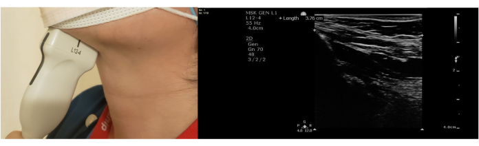
Figure 1: Hyomental distance (HMD). Please click here to view a larger version of this figure.
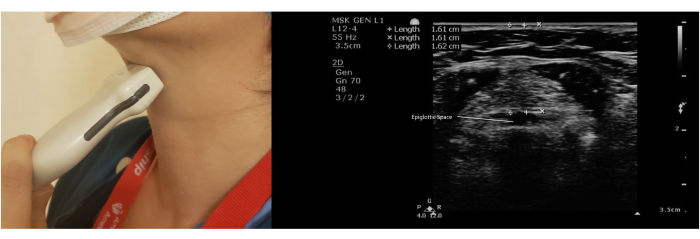
Figure 2: Thyrohyoid membrane (THM). Please click here to view a larger version of this figure.
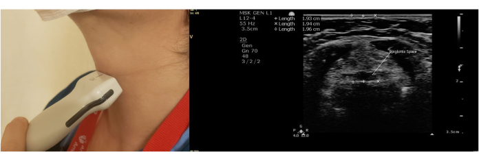
Figure 3: Distance from the skin to the epiglottis (DSE). Please click here to view a larger version of this figure.
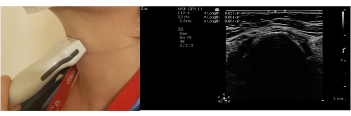
Figure 4: Distance from the skin to the hyoid bone (SHB). Please click here to view a larger version of this figure.
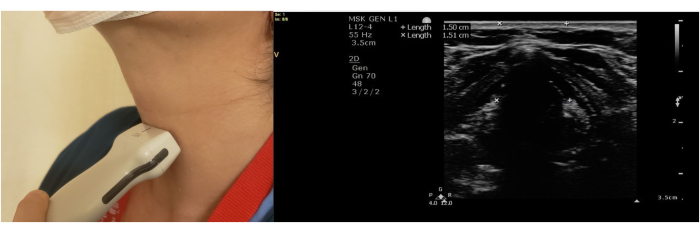
Figure 5: Distance from the skin to the vocal cords (SVC). Please click here to view a larger version of this figure.
Ultrasound of the airway is an effective methodology to examine the airway. The goal is to incorporate airway examination into daily practice to give additive value to the standard pre-anesthetic assessment of the airway before the induction of anesthesia.
It is best to start the scanning protocol from the submandibular space with the transducer positioned along the long axis of the body – the sagittal plane. From there, the transducer is turned in the transverse position along the midline and slowly moved caudally as each parameter comes into view. All the steps should be performed consistently and systemically, and focused training and practice are critical to maintaining good images for future studies.
Alternatively, the scanning protocol starts at the sternal notch, and the tracheal rings will come into view; at this point, one should slowly move the transducer cephalad as the ultrasound parameters focus into view. The scanning sequence can be organized and the order of the views can be changed depending on the sonographer's experience. The submandibular space is best seen with the curvilinear C5-1 MHz transducer, which gives a broad view. A high-frequency linear array 12-4 MHz transducer can obtain all the airway images if only one transducer is available.
The most important aspect of this technique is the patient position. The patient should be supine, with a neutral head position and without a pillow. Small cephalad and caudal movements of the transducer are often necessary to obtain the best images. If specific parameters are difficult to visualize, the sonographer can start over from the most cephalad position with the transducer positioned transversely in the midline and slowly move the transducer caudally.
The linear array L12-4 MHz transducer is a high-frequency transducer that reaches 8 cm in depth. The lines on the right of the ultrasound screen depict the depth to which the ultrasound waves will reach. The depth knob adjusts between shallow or deep depth. A good depth is 3.5-4 cm. Changing the gain knob up or down alters the overall gain, thus making the picture brighter or darker. The gain should be adjusted for the optimal visualization of all the structures. The near/far field and time gain compensation (TGC) fine-tune and adjust the gain at a specific depth in the grayscale ultrasound images. The TGC was middle negative to middle positive from the top to the bottom rows of the narrative review images. The focus knob adjusts the area of interest of the ultrasound image.
The limitations of this technique include the availability of ultrasound and the required training in basic airway ultrasound. Chalumeau-Lemoine et al. concluded that a comprehensive training of 8.5 h, with 2.5 h of didactic sessions and three 2 h hands-on sessions, allowed individuals to achieve competency in essential ultrasound examination, even without prior knowledge of the ultrasound technique18. Additionally, interpretation improved with experience18. A consensus or guidelines about the parameters' cutoff values do not exist. The different populations studied can explain this incongruity, and the results cannot be generalized to other groups. Ultrasound measurements are in centimeters (cm), and the pressure applied to the anterior neck may alter the measured values. A minimal pressure to the anterior neck that allows for the maintenance of skin contact should be applied. A higher risk of difficult laryngoscopy in obese or pregnant patients precluded their inclusion in these study groups.
The thickness of the anterior neck measured by ultrasound has superior sensitivity and specificity than traditional airway assessment in predicting difficult laryngoscopy. Combined with standard bedside clinical assessments, an ultrasound examination of the anterior neck can significantly improve the prediction of difficult laryngoscopy. To date, the studies are small, and there is no common use of ultrasound in airway management other than to confirm the placement of an endotracheal tube or to localize the cricothyroid membrane in case of an emergent surgical airway.
Despite the uncertainty, in the future, portable and handheld ultrasound devices are likely to be accepted as an adjunct to clinical examinations for immediate bedside assessment and management, just as the stethoscope, mobile airway, and other management devices were established previously. This acceptance involves setting up standard protocols and incorporating ultrasound into airway management guidelines. Quality assessment and improved patient safety require training and regular simulation education. Three-dimensional and handheld ultrasound devices are likely to push the boundaries for quality images and the widespread accessibility of point-of-care devices.
Clinical airway examination by the LEMON method is an external assessment of the airway above the hyoid bone. Ultrasound examination is the internal assessment of the structures below the hyoid bone. The study results show that ultrasound-based airway management could be a valuable adjunct to traditional bedside assessment and a helpful tool in predicting difficult airways. The integration of POCUS is increasingly becoming the mainstay for difficult airway management. Its novelty and portability mean that it is feasible to integrate POCUS in the perioperative setting.
The authors have nothing to disclose.
This study was supported, in part, by the National Institutes of Health/National Cancer Institute (Bethesda, Maryland) Cancer Support Grant P30 CA008748.
Gel-Lubricant jelly | MediChoice | 13143 gram, LUB Sterile | Bacteriostatic,water soluble-alcohol free. |
Philips SPARQ Point of Care System | Philips | Transducer L12-4 MHz | Broadband linear. 128elements. 38.4 mm. |