The clinical microfluidic chip is an important biomedical analysis technique that simplifies clinical patient blood sample preprocessing and immunofluorescently stains circulating tumor cells (CTCs) in situ on the chip, allowing the rapid detection and identification of a single CTC.
Circulating tumor cells (CTCs) are significant in cancer prognosis, diagnosis, and anti-cancer therapy. CTC enumeration is vital in determining patient disease since CTCs are rare and heterogeneous. CTCs are detached from the primary tumor, enter the blood circulation system, and potentially grow at distant sites, thus metastasizing the tumor. Since CTCs carry similar information to the primary tumor, CTC isolation and subsequent characterization can be critical in monitoring and diagnosing cancer. The enumeration, affinity modification, and clinical immunofluorescence staining of rare CTCs are powerful methods for CTC isolation because they provide the necessary elements with high sensitivity. Microfluidic chips offer a liquid biopsy method that is free of any pain for the patients. In this work, we present a list of protocols for clinical microfluidic chips, a versatile CTC isolating platform, that incorporate a set of functionalities and services required for CTC separation, analysis, and early diagnosis, thus facilitating biomolecular analysis and cancer treatment. The program includes rare tumor cell counting, clinical patient blood preprocessing, which includes red blood cell lysis, and the isolation and recognition of CTCs in situ on microfluidic chips. The program allows the precise enumeration of tumor cells or CTCs. Additionally, the program includes a tool that incorporates CTC isolation with versatile microfluidic chips and immunofluorescence identification in situ on the chips, followed by biomolecular analysis.
Circulating tumor cells (CTCs) are significant in cancer prognosis, diagnosis, and anti-cancer therapy. CTC enumeration is vital since CTCs are rare and heterogeneous. The enumeration, affinity modification, and clinical immunofluorescence staining of rare CTCs are powerful techniques for CTC isolation because they offer the necessary elements with high sensitivity1. Rare number of tumor cells mixed with normal blood closely mimics real patient blood since 2-3 mL of real patient blood only contains 1-10 CTCs. To solve a critical experimental problem, instead of using a large number of tumor cells introduced in PBS or mixed with normal blood, the use of rare number of tumor cells provides us with a low number of blood cells, which is closer to reality when performing an experiment.
Cancer is the leading cause of death in the world2. CTCs are tumor cells shed from the original tumor that circulate in the blood and lymphatic circulation systems3. When CTCs move to a new survivable environment, they grow as a second tumor. This is called metastasis and is responsible for 90% of deaths in cancer patients4. CTCs are vital for prognosis, early diagnosis, and for understanding the mechanisms of cancer. However, CTCs are extremely rare and heterogeneous in patient blood5,6.
Microfluidic chips offer a liquid biopsy that does not invade the tumor. They have the advantage of being portable, low cost, and having a cell-matched scale. The isolation of CTCs with microfluidic chips is classified mainly into two types: affinity-based, which relies on antigen-antibody binding7,8,9 and is the original and most widely used method of CTC isolation; and physical-based chips, which utilize size and deformability differences between tumor cells and blood cells10,11,12,13,14,15, are label-free, and are easy to operate. The advantage of microfluidic chips over alternative techniques is that the physical-based approach of big-ellipse microfilters firmly captures CTCs with high capture efficiency. The reason for this is that ellipse microposts are organized into slim tunnels of line-line gaps. The line-line gaps are different from the traditional point-point gaps formed by microposts such as rhombus microposts. Wave chip-based capturing of CTCs combines both physical property-based and affinity-based isolation. Wave chip-based capture involves 30 wave-shaped arrays with the antibody of anti-EpCAM coated on circular microposts. The CTCs are captured by the small gaps, and the big gaps are used to accelerate the flow rate. The missed CTCs have to pass the small gaps in the next array and are captured by the affinity-based isolation integrated inside the chip16.
The goal of the protocol is to demonstrate the counting of rare numbers of tumor cells and the clinical analysis of CTCs with microfluidic chips. The protocol describes the CTC isolation steps, how to obtain a low number of tumor cells, the clinical physical separation of small-ellipse filters, big-ellipse filters, and trapezoid filters, affinity modification, and enrichment17.
Patient blood samples were supplied by Longhua Hospital Affiliated to Shanghai Medical University.The protocol follows the guidelines of Peking University Third Hospital's human research ethics committee. Informed consent was obtained from the patients for using the samples for research purposes.
1. Pre-experiment to check the capture efficiency with cultured tumor cells
2. Clinical experiment on the chip to enumerate the circulating tumor cells (CTCs)
The whole setup includes a syringe pump, a syringe, and a microfluidic chip. The cell suspension in the syringe is connected to the syringe pump, and the cell suspension is introduced into the microfluidic chip to capture the cells. The capture efficiency for all the microfluidic chips utilized was around 90% or above. For the wave chip, we designed microstructures with varied gaps. The small gaps are used to capture the CTCs, and the big gaps are used to accelerate the flow rate. The cell suspension flows quickly in the big gap areas. The missed CTCs tend to be captured by the small gaps in the subsequent array16. For ellipse chips, we designed line-line gaps instead of point-point gaps to form a slim tunnel to enhance the capture. Therefore, high capture efficiency was achieved1. We designed an ellipse structure to avoid edges and corners to maintain viability. The trapezoid filters have two circular spiral channels with embedded trapezoid and circular micropost barriers. For trapezoid filters, the capture efficiencies for MCF-7, MDA-MB-231, and HeLa were 94%, 95%, and 93%, respectively18.
Figure 1 shows that all the tumor cells were captured by the wave chip. Since all the tumor cells were concentrated around the wave micropost array, this indicates high capture efficiency for this microfluidic chip, as demonstrated by the number of tumor cells that were captured. Therefore, this setup makes it much easier to capture rare tumor cells; indeed, the chip is fabricated to capture a large number of tumor cells as well as rare number of tumor cells. For example, if the chip is solid or reproducible enough to capture 10,000 tumor cells, it is easy for the chip to capture 10-100 cells. Video 1 shows how rare number of tumor cells were obtained for the pre-experiment. A hollow needle made using a micropipette puller was used to aspirate 1, 2, 3, 4, 5, 6, 7, 8, 9, and 10 tumor cells from a culture dish with diluted tumor cells stained with calcein AM in PBS. The cells were absorbed by the silica gel tube connected with the hollow needle. A tumor cell with green immunofluorescence was suctioned into the hollow needle. Blowing into the hollow tube led to the tumor cell inside the hollow needle being discharged into a microcentrifuge tube1. This is the procedure to obtain rare tumor cells. Figure 2 shows CTCs from a gastric cancer patient captured using small-ellipse microfilters. Figure 3 shows the CTCs from a colorectal cancer patient captured using trapezoid microfilters and emitting both blue and green fluorescence. Figure 4 shows tumor cells grown on the chip after capture, which are ready to be treated with anti-cancer medicine. These findings illustrate that big-ellipse microfilters do not have any negative effects on cell viability.
Figure 5 shows the clinical immunofluorescence analysis of colorectal CTCs captured on the microfluidic chip. These are seen in brightfield and stained with Hoechst, CK-FITC, and CD45-PE staining. The CTCs were recognized as DAPI+/CK+/CD45−, and the WBCs were identified as DAPI+/CK−/CD45+. Figure 6 shows colorectal tumor cells cultured on the big-ellipse chip after capture. Figure 7 shows colorectal tumor cells captured on the big-ellipse microfilters. Clinically, CTCs in patient blood were captured by wave chips, trapezoid microfilters, and big-ellipse microfilters, indicating that these three chips are successful in capturing CTCs. Potentially, they could be applied in CTC products, such as CTC isolation products, with high efficiency.
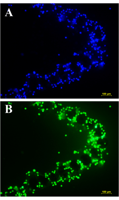
Figure 1: Wave chip capture. All the tumor cells of MCF-7 were captured around the array of the wave chip without any cells missing. Since there were not any tumor cells in any other area besides the array, this indicates the high capture efficiency of the chip. A large number of tumor cells were captured. Therefore, rare tumor cells can also be easily captured. Tumor cells of MCF-7 were captured by wave chip and (A) stained with Hoechst and (B) stained with calcein AM. Scale bar: 100 µm. Please click here to view a larger version of this figure.
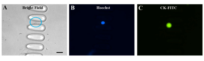
Figure 2: Clinical sample for CTCs captured by small-ellipse microfilters. Clinical CTCs of a gastric cancer patient captured by small-ellipse microfilters. The CTCs were identified in brightfield and with both blue and green fluorescence. The blue circle in (A) indicates a CTC of a gastric cancer patient captured inside the chip. From the images taken, it can be seen that there were no other cells in any other areas, indicating that the capture purity was high for this chip. Tumor cells of MCF-7 were captured by small-ellipse microfilters and seen in (A) brightfield, (B) stained with Hoechst, and (C) stained with CK-FITC. Scale bar: 20 µm. Please click here to view a larger version of this figure.
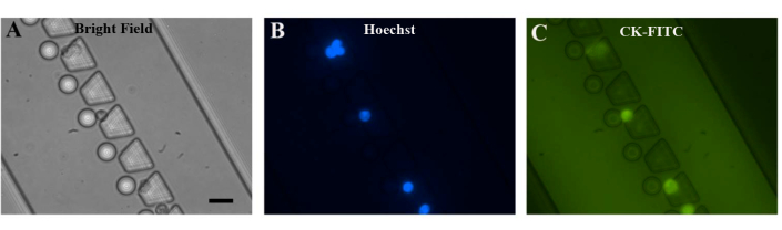
Figure 3: Clinical sample of CTCs captured by trapezoid microfilters. Clinical CTCs from a colorectal cancer patient were captured by trapezoid microfilters. In these images, it can be seen that six CTCs were captured, indicating that the capture efficiency was high in the small field. Additionally, no other cells appeared, indicating that the capture purity was extremely high for this chip. Tumor cells of MCF-7 were captured by trapezoid microfilters and seen in (A) brightfield, (B) stained with Hoechst, and (C) stained with CK-FITC. Scale bar: 20 µm Please click here to view a larger version of this figure.
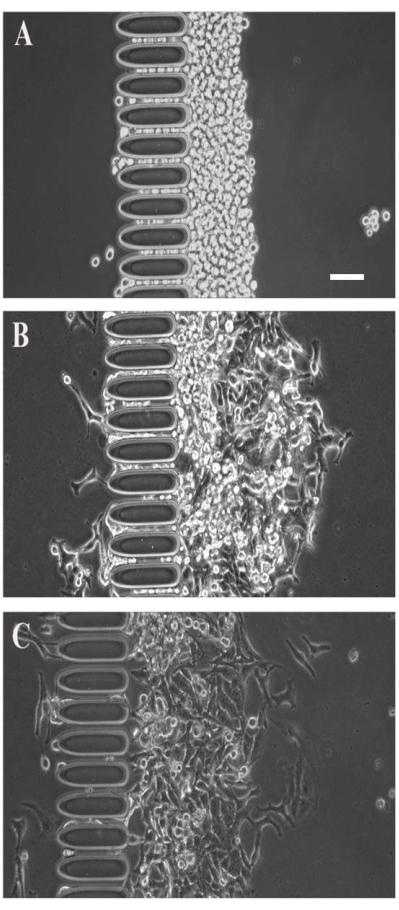
Figure 4: Culture of CTCs captured by big-ellipse microfilters. Tumor cells of MCF-7 were captured by big-ellipse microfilters in front of a big-ellipse micropost array. No tumor cells passed through the array, indicating high capture efficiency for this chip. After capture, the tumor cells grew for 24-48 h. This indicates that both the capture efficiency and viability were very high for the big-ellipse microfilters. The cultured tumor cells of MCF-7 were captured by big-ellipse microfilters and seen at (A) 0 h after capture, (B) 24 h after capture, and (C) 48 h after capture. Scale bar: 50 µm. Please click here to view a larger version of this figure.
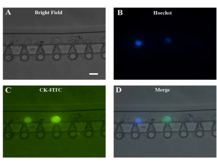
Figure 5: Example of a clinical sample used for CTC capture by trapezoid microfilters. Clinical CTCs from a colorectal cancer patient were captured by trapezoid microfilters. In these images, it can be seen that two CTCs were captured, indicating high capture efficiency. There was no other cell disturbance except residues of RBCs in the chip. Thus, for the clinical pre-processing of CTC capture, it is better not to use red blood cell lysis. CTCs of colorectal cancer were captured by trapezoid microfilters and seen in (A) brightfield, (B) stained with Hoechst, (C) stained with CK-FITC, and (D) seen in merged images. Scale bar: 20 µm. Please click here to view a larger version of this figure.
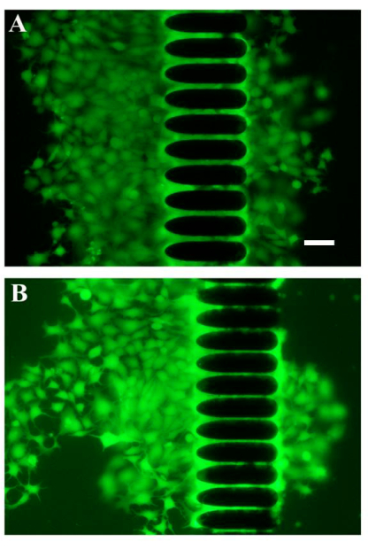
Figure 6: Example of cultured CTCs captured by a big-ellipse chip. Tumor cell cultures of MCF-7 cells in front of the big-ellipse micropost array and behind the big-ellipse microfilters. The tumor cells stained with calcein AM emitting green fluorescence grew as desired, indicating that the cell viability for this chip was very high. In total, there were 15 arrays, with varied gaps for each array organized by big-ellipse microposts. MCF-7 tumor cells were cultured on the big-ellipse chip after capture (A) in one array and (B) in another array. Scale bar: 50 µm. Please click here to view a larger version of this figure.
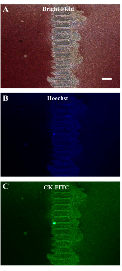
Figure 7: Example of a clinical sample used for CTC capture by big-ellipse microfilters. Clinical CTCs from a colorectal cancer patient were captured by big-ellipse microfilters. From the images, it can be seen that there was RBC contamination. This indicates that the whole patient blood sample must be diluted and that the chip needs to be flushed after capture. CTCs of clinical colorectal cancer patient blood samples were captured on the big-ellipse chip and seen in (A) brightfield, (B) stained with Hoechst, and (C) stained with CK-FITC. Scale bar: 100 µm. Please click here to view a larger version of this figure.
The prognosis and early diagnosis of cancer have a significant effect on cancer treatment1. CTC isolation with microfluidic chips offers a liquid biopsy with no invasion. However, CTCs are extremely rare and heterogeneous in the blood1, which makes it challenging to isolate CTCs. CTCs have similar properties to the original tumor sources from which they originate. Thus, CTCs play a vital role in cancer metastasis1.
The proposed protocol allows a complete analysis of CTC isolation with the microfluidic chip. The protocol includes all the key procedures related to CTC isolation. For example, this protocol includes RBCL analyses, which are recorded meticulously and organized into different packages, rare tumor cell acquisition, optimal flow rate determination, capture efficiency, clinical characterization, and enumeration. The careful management of the operations facilitates the clinical patient sample processing. The protocol allows for the pre-processing and processing of clinical patient samples and offers vital services for clinicians.
For CTC isolation, the pre-processing of rare tumor cells is extremely difficult to perform. This difficulty was solved by using a hollow needle pulled through a needle puller to suck 1, 2, 3, 4, 5, 6, 7, 8, 9, and 10 tumor cells as desired. Then, a close-to-real clinical patient blood sample was prepared with the desired number of tumor cells mixed into normal blood, mimicking a patient blood sample. With these artificial patient blood samples, a close-to-real experiment was carried out. This method has solved a difficult problem usually met in CTC isolation for pre-experiments. This approach is not easy to perform and has never been reported elsewhere; however, it is a useful approach before performing a real clinical experiment.
CTC isolation with microfluidic chips is classified into three types: affinity-based, physical-based, and immunomagnetic-based. For affinity-based isolation, the microfluidic chip needs to be modified. The specific modification procedures have been included for modification with anti-EpCAM. The CTCs were identified through immunofluorescence staining with Hoechst, CK-FITC, and CD45-PE. Since many tumor cells express EpCAM, anti-EpCAM is an important antibody to capture CTCs in patient blood. However, anti-EpCAM is very expensive; thus, many aptamers have been developed to solve this problem. We mainly utilize physical-based microfluidic chips such as small-ellipse microfilters and trapezoid microfilters to capture CTCs since they are simple, easy to operate, and effective.
The advantage of this technique over alternative techniques is that the physical-based chip of big-ellipse microfilters firmly captures CTCs with high capture efficiency. The reason for this is that there are slim tunnels of line-line gaps. Wave chips capture CTCs by combining both physical property-based and affinity-based isolation. The CTCs are captured by the small gaps, and the big gaps can be used to accelerate the flow rate. The missed CTCs have to pass through the small gaps in the following arrays and are also captured by the affinity-based isolation.
The protocol has resolved several major problems in CTC isolation, especially for clinical experiments. However, it is impossible to include every detailed step in CTC isolation, such as regarding the role of nanoparticles and nanostructures and aptamer modification. These aspects also play an essential role in CTC isolation. However, they are not critical in solving problems such as improving capture efficiency. Instead, these aspects enrich the CTC isolation with new contents.
This work concentrates on the clinical isolation of CTCs with the designed microfluidic chip. Most microfluidic chips used are mainly physical-based, such as big-ellipse filters and small-ellipse filters. Wave chips are made by combining both affinity-based and physical-based chip properties1. The microposts of big-ellipse filters were designed to be shorter to enhance the capture purity in this work. The small-ellipse filters can also be improved by designing them with varied micropost sizes for different arrays. Wave chips can be better designed by adding more arrays to enhance the capture.
Big-ellipse filters are organized with long elliptical microposts to form line-line tunnels that achieve high capture efficiency. However, the limitation of this capture is that the capture purity is not high enough or high capture purity is not easily obtained. For wave chip, the small gaps are used to capture the CTCs, and the missed CTCs are captured by the following arrays. The big gaps are used to accelerate the flow rate and eliminate disturbance from RBCs, thus improving the capture purity; however, the capture efficiency is solid at above 90% and is reproducible.
Clinical validation is significant in CTC isolation with microfluidic chips. This work presents the clinical isolation of CTCs from colorectal patients with big-ellipse filters, small-ellipse filters, and wave chips. The aim of the designed microfluidic chips is to clinically enumerate CTCs. For the existing methods of some other systems or platforms, the capture efficiency is low, or the capture efficiency is not high enough for the method to be effectively used in clinical applications.
Based on the clinical performances of these three microfluidic chips, which have high capture efficiency, they can potentially be applied in CTC products, especially after modification. The strength of our protocol is that it demonstrates the enumeration of rare number of tumor cells to mimic real clinical samples. In addition, the clinical experimental procedure is feasible and practical. This method can separate CTCs from whole patient blood. Therefore, the method is appropriate for clinical applications.
The authors have nothing to disclose.
This research work was supported by the Anhui Natural Science Foundation of China (1908085MF197, 1908085QB66), the National Natural Science Foundation of China (21904003), the Scientific Research Project of Tianjin Education Commission (2018KJ154), the Provincial Natural Science Research Program of Higher Education Institutions of Anhui Province (KJ2020A0239), and the Shanghai Key Laboratory of Multidimensional Information Processing, East China Key Laboratory of Multidimensional Information Processing, East China Normal University (MIP20221).
Calcein AM | BIOTIUM | 80011 | |
calibrated microcapillary pipettes | Sigma- Aldrich | P0799 | |
CD45-PE | BD Biosciences | 560975 | |
CK-FITC | BD Biosciences | 347653 | cytokeratin monoclonal antibody |
DMEM | HyClone | SH30081.05 | |
fetal bovine serum (FBS) | GIBCO,USA | 26140 | |
Hoechst 33342 | Molecular Probes, Solarbio Corp., China | C0031 | |
penicillin-streptomycin | Ying Reliable biotechnology, China | ||
Red blood cells lysis (RBCL) | Solarbio, Beijing | R1010 |