Point-of-care ultrasound (POCUS) of the optic nerve sheath diameter (ONSD) has been shown to be useful in identifying patients with increased intracranial pressure (ICP). However, the non-standardized technique for this POCUS has hampered its use. We present a standardized image acquisition protocol for use in the acute care setting.
The goal of this protocol is to develop a standardized method for acquiring images of the optic nerve sheath and measuring the optic nerve sheath diameter (ONSD). Diagnostic ultrasound of the ONSD to detect intracranial hypertension has traditionally faced many problems because of methodologic discrepancies. Due to inconsistencies in the measuring techniques, the potential for ONSD to become a non-invasive bedside monitoring tool for ICP has been hampered. However, establishing a transparent, consistent methodology for measuring the ONSD would support its use as a valid and reliable method of identifying intracranial hypertension. This is important as it has both high sensitivity and specificity in acute care settings. This narrative review describes ONSD POCUS image acquisition, including patient positioning, transducer selection, probe placement, the acquisition sequence, and image optimization. Further, visual aids are provided to assist in real-time during image acquisition. This method should be considered for patients for whom there are concerns regarding intracranial hypertension but who do not have an intracranial monitor in place.
The goal of this protocol is to allow bedside providers to rapidly evaluate patients for intracranial hypertension in a non-invasive fashion using point-of-care ultrasound. In recent years, bedside decision-making and treatment have been augmented by the emergence of point-of-care ultrasound (POCUS). POCUS involves the use of ultrasound for diagnostic or procedural guidance by a patient's primary treatment provider. This article focuses on the diagnostic POCUS of the optic nerve sheath.
The rationale behind this technique is that the optic nerve and sheath communicate with the central nervous system. Specifically, the subarachnoid space extends from inside the skull around the optic nerve. Thus, when the intracranial pressure increases, the optic nerve sheath increases in size. It has been shown in studies with magnetic resonance imaging (MRI) and computed tomography (CT) that the optic nerve does not change in size with increased ICP, but the optic nerve sheath does. The vitreous of the eye provides excellent transmission of sound waves, resulting in the optic nerve being clearly visible as a hypoechoic structure inserted on the posterior eye, with the sheath visible around it. For these reasons, ultrasound of the optic nerve sheath has been used to detect elevated ICP, thus allowing the diagnosis of potentially life-threatening instances of increased ICP1.
However, despite its clinical significance, the proficiency of physicians in using ONSD POCUS is variable2,3, which limits the appropriate use of this modality4. This study aims to describe a time-efficient yet thorough image acquisition protocol for diagnostic ONSD POCUS and to illustrate the abnormal findings commonly found in clinical practice. Multiple imaging protocols have been described in the literature, and they present variations in the structural interpretation, ultrasound settings, marker placement, and scan technique5. Different marker points have varying sensitivity to changes in ICP, and the placement affects the ability to distinguish between patients with normal and high ICP. For these reasons, in this paper, we outline a standard technique to obtain optic nerve sheath images and measure them consistently.
Differences in the measurement location and technique have resulted in widely different thresholds of what is considered to be an abnormal optic nerve sheath5,6. In a recent meta-analysis, the average ONSD for patients without intracranial hypertension was 4.1 mm, and the average ONSD for those with increased ICP was 5.6 mm. A generally accepted threshold for a dilated ONSD is 5.5 mm, but a change in ONSD from a normal baseline is much more predictive if a baseline is available. In the context of severe TBI, the ONSD of both eyes seems to change together7. There are some individual cases of increased ONSD on one side, but this is rare8.
There are additional challenges in the measurement of ONSD in the severe traumatic brain injury (TBI) population. Patients with severe TBI (defined as having a Glasgow coma score of <8) are not alert enough to follow commands. This means a different approach is required to measure the nerve in this population compared to in patients being evaluated in an ophthalmology office in an elective fashion for neuritis. An A-scan can be used to measure the optic nerve in a cooperative patient that can hold their eye still for a length of time, but this is not a useful technique in TBI patients, so a B-scan is the standard technique (see step 2.7 below).
The use of this method and protocol should be considered for patients for whom there are concerns regarding intracranial hypertension but who do not have an intracranial monitor in place. Specific patient populations that may benefit are trauma patients with a clinical concern for traumatic brain injury in the prehospital or emergency room setting. Additionally, patients in the ICU with a sudden change in neurologic status are good candidates for ONSD POCUS.
This procedure is in accordance with the ethical standards of the institutional committee on human experimentation and with the Helsinki Declaration of 1975. Ultrasound is considered minimal risk procedure, so written consent from the patient is not required.
1. Probe selection
NOTE: ONSD POCUS can be performed with a multitude of different transducers. To evaluate abnormalities in suspected cases of increased ICP, choose a linear high-frequency (5-14 MHz) probe to evaluate ONSD. The probe nomenclature varies by manufacturer; however, the linear probe will be flat and usually have an L in the probe name. Choose a probe 37 mm long or smaller. Almost any probe size can be used to obtain transverse images of the optic nerve. However, sagittal views are difficult to obtain with larger probe sizes. Using a probe 37 mm long consistently allows both transverse and sagittal images in adults, while a 55 mm probe can be used in some adults but not all. Some representative probe heads are shown in Figure 1.
2. Ultrasound setup
3. Patient selection
4. Patient positioning
5. Scanning technique
There are several findings that can be seen on the POCUS of the optic nerve sheath and several pitfalls that can occur in the measurements. As can be seen in Figure 5, the nerve typically heads back from the eye at an angle when the eye is in a neutral position. In patients undergoing elective ultrasound of the eye for optic neuritis, for example, the patient is typically asked to move the eye and straighten the nerve to facilitate the measurement of the diameter along the axis of the nerve. Patients with concern for TBI will rarely be able to follow commands, so the transverse diameter of the nerve sheath will need to be measured at an angle.
Another potential pitfall that can occur is a measurement of the optic nerve instead of the sheath. If appropriate settings are not used, or a low-resolution probe is used, such as those driven on a USB cable, it can be difficult to see the sheath, as is demonstrated in Figure 6. In this figure, two images are shown of the same patient, one demonstrating the nerve sheath and another, which was performed with a low-resolution probe, in which only the nerve is visible. It is important that the nerve sheath can be seen and measured for accuracy. The optic nerve itself does not change substantially in size with elevated ICP, so measuring the nerve instead of its sheath may give a false low measurement.
There are also several artifacts that can interfere with measuring the ONSD. A blooming artifact can confound the measurements. This artifact is hypothesized to be an acoustic shadow caused by the lamina cribrosa, which can be larger than the ONSD10. An example can be seen in Figure 7, where two images from the same patient are seen, one showing the optic nerve sheath and one showing a blooming artifact that is much larger in size. It is important that the head of the nerve be visualized as a rounded-off hypoechoic structure as it inserts into the posterior eye. The blooming artifact will extend from the optic disk and will not have a rounded-off head. Some authors11 recommend the use of color Doppler to clarify the course of the nerve and differentiate it from the blooming artifact. However, we do not think this is necessary for providers experienced in B-mode interpretation.
Normal findings vary, but in normal patients, the mean size of ONSD is 4.1 mm, and the ONSD cut-off value of 5.3 mm strongly predicts increased ICP (sensitivity: 94.7%; specificity: 84.3%)7. The average ONSD in patients with increased ICP is over 6 mm.
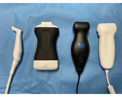
Figure 1: A series of representative ultrasound probes that can be used for ocular exams. From left to right: a 5-14 MHz probe with a 37 mm footprint (commonly referred to as a hockey stick probe) that can easily obtain transverse and sagittal views; a battery-powered handheld probe; an inexpensive USB probe with a 55 mm footprint that can acquire transverse images consistently and sagittal images on some patients; and a linear probe that cannot obtain sagittal views on most patients. Please click here to view a larger version of this figure.
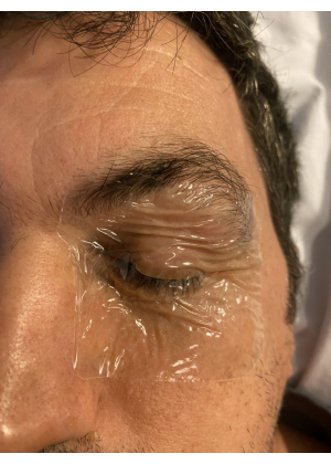
Figure 2: A transparent adhesive dressing is used to keep the eyelid closed during scanning to protect the cornea. Please click here to view a larger version of this figure.
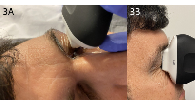
Figure 3: Transverse and sagittal views. (A) The transverse view is performed from the anteriormost aspect of the eye. The probe may need to be oriented slightly toward the midline to bring the nerve into the plane. (B) The sagittal view is performed in a similar fashion from the anteriormost aspect of the eye. Please click here to view a larger version of this figure.
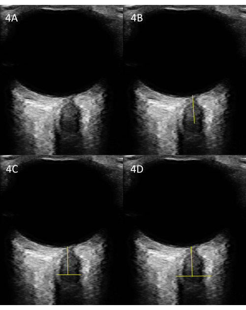
Figure 4: Measurement of the ONSD. (A) An unlabeled US image. (B) The same image with the appropriate place to measure the ONSD 3 mm posterior to the optic disk. (C) Transverse measurement of the optic nerve, not the sheath. (D) Measurement of the optic nerve sheath; this is the most relevant measure. Please click here to view a larger version of this figure.
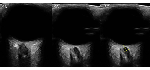
Figure 5: Imaging in patients with cooperative eye movements versus those who cannot cooperate. (A) If the patient is cooperative, have them move the eye so that the nerve is headed straight back, as in this example. (B) However, for severe TBI, the patient will not be able to cooperate, and the nerve will head back at an angle. (C) We recommend measuring along the axis of the nerve if it does not head straight back. Please click here to view a larger version of this figure.
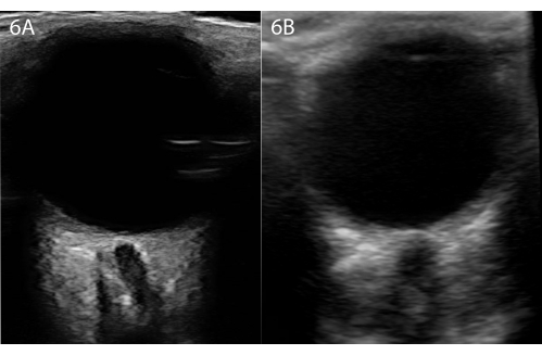
Figure 6: Two ultrasounds from the same patient at the same point in time. (A) The optic nerve sheath is visible. (B) US performed with an inexpensive USB-powered ultrasound; the nerve is visible, but not the sheath, even though this is the same patient. Please click here to view a larger version of this figure.
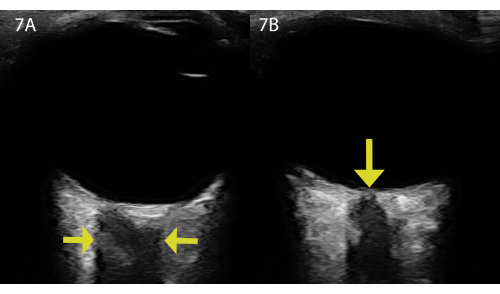
Figure 7: Two ultrasounds from the same patient and the same eye demonstrating a blooming artifact. (A) Demonstration of a blooming artifact from structures on the posterior eye. Note that there is no rounded nerve head, no sheath, and the shadow appears very rectangular. The arrows point to the sides of the blooming artifact. (B) The same patient with the optic nerve and sheath visible. Note that the rounded head of the nerve is demonstrated by the yellow arrow, and the sheath is visible. Please click here to view a larger version of this figure.
The most critical steps of this protocol are ensuring the correct settings are used, such as ophthalmic or small parts, to minimize the energy transmitted to the eye. Additionally, ensuring that the optic nerve and not a blooming artifact is being measured is important.
Within diagnostic POCUS, the evaluation of the ONSD is well-suited to investigating patients with traumatic brain injury to evaluate if there is increased ICP. Although invasive intracranial monitoring remains the gold standard for the accurate measurement of ICP, ONSD POCUS serves as an excellent non-invasive alternative for assessing the presence and magnitude of increased ICP. In a previous study, in terms of the diagnostic performance, ONSD demonstrated a superior correlation with invasive measurements in comparison to other ultrasound-based techniques, such as arterial and venous transcranial Doppler12. While most patients with severe TBI will have a monitor placed, POCUS can be especially useful in emergent decision-making when there is not an intracranial monitor in place, such as in the prehospital setting, emergency room, resource-limited institutions, or during an unexpected change in mental status. However, ONSD POCUS has some limitations. First, the inability to follow specific commands (e.g., adjusting their gaze to assist in sheath identification) in patients suffering from a severe TBI may limit the acquisition of an adequate image.
Another limitation of the technique is the variation in ONSD cutoff values, which greatly affects the diagnosis of increased ICP. The cut-off values vary greatly9,10,11,12and can inhibit the diagnostic accuracy of ONSD POCUS measurements. Different cut-off values may exist due to body habitus, age, sex, and, in some instances, ethnicity13. The lack of data for these individual groups hampers the tool's overall effectiveness but is not enough to totally impair its usability.
Finally, a surmountable limitation of ONSD POCUS is the lack of proficiency and inter-examination variability. Like any ultrasound technique, POCUS is very operator-dependent. One must know how to acquire clinically valuable images to make appropriate decisions. A thorough understanding of the potential artifacts visualized on ocular ultrasound will improve the accuracy. The standardization of ONSD POCUS practice and the establishment of educational curriculums have been highlighted as possible solutions to this limitation. This is a much-needed step to guide academic standards at the national and international levels and to continue to develop this essential diagnostic/procedural tool.
The authors have nothing to disclose.
This effort was partially sponsored by the US Government under Other Transactions (W81XWH-15-9-0001/W81XWH-19-9-0015) and the Medical Technology Enterprise Consortium (MTEC) under 19-08-MuLTI-0079.
Butterfly iQ+ with USB-C 2.0 | Butterfly | n/a | Used to obtain one of the images |
Clarius L7HD Portable Ultrasound Machine | Clarius | n/a | Used to obtain one of the images |
Ultrasound Gel | Parker | n/a | Used to obtain all images |
Transparent dressing | 3M | 9534HP | Used to protect eye |