The present protocol describes how to perform 64Cu PET/CT and PET/MRI imaging in humans to study copper-related disorders, such as Wilson disease, and the treatment effect on copper metabolism.
Copper is an essential trace element, functioning in catalysis and signaling in biological systems. Radiolabeled copper has been used for decades in studying basic human and animal copper metabolism and copper-related disorders, such as Wilson disease (WD) and Menke's disease. A recent addition to this toolkit is 64-copper (64Cu) positron emission tomography (PET), combining the accurate anatomical imaging of modern computed tomography (CT) or magnetic resonance imaging (MRI) scanners with the biodistribution of the 64Cu PET tracer signal. This allows the in vivo tracking of copper fluxes and kinetics, thereby directly visualizing human and animal copper organ traffic and metabolism. Consequently, 64Cu PET is well-suited for evaluating clinical and preclinical treatment effects and has already demonstrated the ability to diagnose WD accurately. Furthermore, 64Cu PET/CT studies have proven valuable in other scientific areas like cancer and stroke research. The present article shows how to perform 64Cu PET/CT or PET/MR in humans. Procedures for 64Cu handling, patient preparation, and scanner setup are demonstrated here.
Copper is a vital catalytic cofactor that drives multiple important biochemical processes essential for life, and defects in copper homeostasis are directly responsible for human diseases. Mutations in the ATP7A or ATP7B genes, encoding copper-transporting ATPases, cause Menke's and Wilson diseases, respectively. Menke's disease (ATP7A) is a rare lethal disorder of intestinal copper hyperaccumulation with severe copper deficiency in peripheral tissues and deficits in copper-dependent enzymes1. Wilson disease (WD) (ATP7B) is a rare disease characterized by the inability to excrete excess copper to bile, resulting in copper overload and subsequent organ damage, most severely affecting the liver and brain2.
Studies on copper metabolism have utilized radiolabeled copper (usually 64-copper [64Cu] or 67-copper) for decades, and these studies have proven invaluable for our understanding of mammalian copper metabolism, including absorption site and excretion pathways3,4,5,6. Previously, gamma counters were used to detect the radioactive signal with a limited anatomical resolution, but recently, 64Cu positron emission tomography (PET) combined with computed tomography (CT) or magnetic resonance imaging (MRI) has been introduced in both human and animal studies. Today, PET scanners have such a high sensitivity that it is possible to track 64Cu for up to 70 h after injection. The long half-life of 12.7 h for 64Cu allows for the long-term assessment of copper fluxes. This improvement in resolution has just recently entered the field of copper studies, and studies on normal and pathological copper metabolism, as well as studies evaluating the impact of specific treatments, are starting to emerge. Additionally, the introduction of whole-body PET scanners with an extended field-of-view will further enhance the sensitivity of these examinations.
This methodological paper aims to enable clinicians and scientists to add 64Cu PET CT/MRI to the existing repertoire of tools as a robust and easy-to-use method for assessing copper metabolism in a manner comparable between nuclear medicine departments. The production of 64Cu copper can be carried out using different methods and is usually performed at special facilities. Among the nuclear reactions, the 64Ni (p, n) 64Cu method is widely used, since a high production yield of 64Cu can be obtained with low energy protons in this route7,8. A detailed description of the production methods is out of the scope of this work, and availability will differ by country and region.
In this article, we first describe the preparation of the necessary radiochemistry and the tracer. Then, the principles for preparing the PET/CT or PET/MRI scanners are demonstrated.
A few clinical trials using this 64Cu PET/CT or PET/MRI protocol have been approved by the Regional Ethics Committee of Region Midt, Denmark [1-10-72-196-16 (EudraCT 2016-001975-59), 1-10-72-41-19 (EudraCT 2019-000905-57), 1-10-72-343-20 (EudraCT 2020-005832-31), 1-10-72-25-21 (EudraCT 2021-000102-25), and 1-10-72-15-22 (EudraCT 2021-005464-21)]. Written informed consent was obtained from the participants at enrollment. The inclusion criteria for all participants were age >18, and for females the use of safe contraception. The exclusion criteria for Wilson disease patients were decompensated cirrhosis, a Model for End-stage Liver Disease (MELD) score >11, or a modified Nazer score >6. The exclusion criteria for all participants were a known hypersensitivity to 64Cu or other ingredients in the tracer formula, pregnancy, breastfeeding, or a desire to become pregnant before the end of the trial.
1. Preparation of 64CuCl2
2. Preparation of PET scanner
3. Drawing of tracer for intravenous (IV) injection and per oral (PO) administration
4. Application of the tracer
5. PET scans
6. Image reconstruction
7. Data analysis
NOTE: The present study describes a simple method to quantify 64Cu content in the liver. The PET signal is measured as standard uptake value (SUV), the tissue radioactivity concentration adjusted for participant weight injected activity and/or kilobecquerel (kBq) per mL of tissue.
Dose calculation
Based on dosimetry calculations, the effective radioactivity dose for IV administration is 62 ± 5 µSv/MBq tracer10. Thus, a 50 MBq dose is recommended depending on the time frame. Up to 75-80 MBq is applicable for longer examinations and provides good-quality images without exceeding an ethically approved dose. The effective dose for oral administration is 113 ± 1 µSv/MBq tracer, due to intestinal accumulation of the tracer. Thus, a lower dose needs to be considered, and for up to 24 h post-injection, 30 MBq is sufficient to yield high-quality images. Fertile female participants should always be asked for a negative pregnancy test before tracer application.
Scan
For very long examinations, performed to follow the 64Cu biodistribution and kinetics for hours or days, the PET examination is performed as multiple separate static PET scans. This allows the patient to rest between the PET examinations. The duration of each PET examination is adjusted to achieve the best image quality (i.e., the scan time is prolonged as the injected tracer decays). An example of scan times providing good-quality images is 4.5 min/bed position for up to 20 h after tracer administration and 10 min/bed position for up to 68 h after tracer administration. Longer scan times may provide even better image quality, but too-long scans are unfeasible and uncomfortable for the patient. Thus, the length of the scans is limited by practicalities.
Data analysis
SUV is an excellent measure to compare individuals (because of the weight adjustment) and to compare the same individuals before and after an intervention. A standard deviation of the SUV in the VOI is available from the data analysis program (e.g., PMOD). This standard deviation increases with time after injection because the noise increases.
Figure 1 shows 64Cu in the body 6 h and 20 h after IV injection of ~70 MBq tracers in a healthy subject and a subject with WD10. The images are qualitatively easy to interpret as the 64Cu is quickly visible in the gallbladder (difficult to see in the figure), small intestine, and later in the colon, while it accumulates in the liver in the patient. The gut is also visible on the patient's scan, however this is not from 64Cu in the gut lumen but rather from intestinal blood vessels. The gut is seen by the 64Cu being more homogenously distributed along the entire gut segment, whereas in healthy subjects, the 64Cu is visible in segments with higher signals. The 64Cu content in the liver was further quantified by placing five spherical VOIs with a diameter of 10 mm in different planes in the right liver lobe, yielding a mean SUV in the organ for each participant, then calculating the group's mean SUV for comparison between groups.
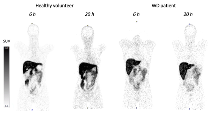
Figure 1: PET scan showing 64Cu distribution in healthy and WD subjects after IV administration. This figure shows 64Cu in the body 6 h and 20 h after IV injection of ~70 MBq tracers. Please click here to view a larger version of this figure.
Figure 2 shows the results of 64Cu scans with the orally administered tracer in two individuals. Both are WD patients, but the bottom individual is under zinc treatment, demonstrating that zinc treatment reduces copper uptake in the intestines and thus to the liver; this is a well-known effect of zinc treatment13. While orally administered tracer is the physiological way of ingesting copper, it may be difficult to use for diagnostics, as only 50% of the 64Cu is taken up from the intestines to systemic circulation (most of the tracer goes to the liver). However, to demonstrate the effects of pharmacologic drugs on copper uptake, which may be of great interest in WD, the method has shown to be valuable11. This can be seen in Figure 3, in which the same individual has been scanned using oral 64Cu before and after 4 weeks of treatment with zinc11. The study hypothesis was to quantify zinc's effect on blocking intestinal copper uptake by estimating the copper content in the liver. The study was performed with different zinc salts and dose regimens and demonstrates the method's qualities in testing treatment effects. The method's ability to quantify other treatment effects in animals and humans is being tested.
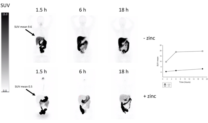
Figure 2: PET scan showing 64Cu distribution in two WD patients after per oral administration. The patient in the upper panel is without zinc treatment, and the patient in the lower panel is on zinc treatment. Note the signal difference in the liver. Graph depicting liver SUV. Please click here to view a larger version of this figure.
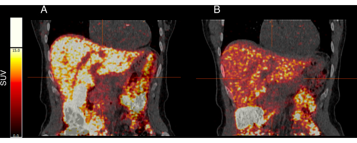
Figure 3: The effects of pharmacologic drugs on copper uptake. PET/CT scan using orally administered 64Cu before (A) and after (B) 4 weeks of zinc treatment. The participant is a healthy individual (notice the 64Cu in the gallbladder, which would not be seen in a WD patient). Zinc treatment reduced 64Cu content in the liver to around 50% of the pre-treatment content in the group (10 participants). Please click here to view a larger version of this figure.
The method is like any other PET method, but the long half-life of 12.7 h offers the opportunity to investigate long-term copper fluxes (we have good results from up to 68 h after IV tracer injection). All steps in the protocol must be handled by personnel familiar with PET, although they are no more critical than any other PET examination.
Troubleshooting
Because we often use 64Cu for long-term investigations, the PET signal will be noisier than usual. This is important to remember when quantifying PET signals, particularly in smaller organs such as the gallbladder. The signal in the gallbladder will be difficult to distinguish from spill-over from the liver and colon. In this case, smaller VOIs centrally in the organ are the most reliable.
The amount of 64Cu in the liver, from our experience, tends to vary between individuals despite IV injection (a fairly large variance in tracer uptake from the gut must be expected with an orally administered tracer). This limits the comparisons between individuals and calls for the use of ratios instead of definite numbers. If per oral tracer administration is preferred, keeping trial subjects on a standardized diet for a minimum of 24 h before the tracer intake is recommended to limit intra-individual differences, as different foodstuffs may interfere with copper and thus with 64Cu uptake11.
Limitations
When the 64Cu PET method is used, it is assumed that the "hot" copper (64Cu) acts like the "cold" copper in the body. However, this is not certain, and we thus cannot determine if the "hot" copper is treated differently in the body. From the current results, however, we believe that "hot" copper acts like "cold" copper. An increase in blood radioactivity after 20 h is observed in healthy individuals, indicating that the 64Cu is built into ceruloplasmin. This increase is not seen in WD patients, who cannot build copper into the copper-carrying protein because of their disorder. This and the lack of tracer excretion in patients point toward 64Cu acting as "cold" copper.
Although 68 h is a long time to follow a radioactive tracer, it should still be considered a temporary image of what happens to copper in the body. An example is that even though stalled 64Cu excretion is seen in individuals who are heterozygotous for the WD gene, and thus more 64Cu in the liver after 20 h, they do not have liver disease because, in the long term, they do not accumulate copper.
So far, it is not known if there is a correlation between short-term copper accumulation (up to 68 h) and long-term copper accumulation in the liver and other organs. Thus, the method cannot be used to determine disease severity or the long-term effects of pharmacological agents. However, the method is very useful in determining the short-term effects of treatment. It may be used to test whether treatment increases biliary or urinary excretion up to 68 h after copper intake, or if a treatment decreases intestinal copper uptake.
Significance
Experiments with 64Cu in WD is not a new technique. In fact, IV administration of the tracer and blood measurements of radioactivity goes back to the 1950s14. Today, high-resolution PET scanners and combination with CT or MR provide a unique opportunity to investigate 64Cu distribution in the whole body. With dynamic PET, the kinetic properties of the tracer can further be elucidated. So far, due to the limited field-of-view of PET scanners, conducting kinetic analyses of copper's biodistribution throughout the body has not been feasible. Currently, dynamic uptake has been restricted to the liver and upper abdomen, but the advent of whole-body scanners will enable the simultaneous investigation of larger areas. This will facilitate examination of the initial period after the injection of 64Cu in multiple organs, but given that late time points after injection are more relevant for copper-related disorders, whole-body scanners are expected to be more significant due to their heightened sensitivity. This allows for high-quality imaging even at low radioactivity levels, surpassing current scanners' capabilities.
Future applications
In humans, the technique has shown potential in diagnosing WD10 and quantifying the effect of different treatments on copper uptake11. In animals, the method has proven to be able to show the effect of gene therapy of WD by quantifying the hepatic retention of 64Cu as well as fecal excretion and changes in blood kinetics15. In the future, it is expected that 64Cu PET/CT or PET/MR will be seen in a clinical setting for both diagnosis and treatment evaluation in WD. The method is also highly likely to be part of many clinical trials involving new therapies for WD, especially gene therapy, in which fecal excretion of the IV injected tracer could be a surrogate marker of effect15. There is currently no good data for brain 64Cu uptake available, but this would be highly relevant for clinical studies in WD.
The technique has not been explored in Menke's disease yet, but could potentially show copper uptake from the gut and copper uptake into the brain as a treatment effect. The technique may also have potential in neurodegenerative diseases such as Alzheimer's disease, where copper metabolism may be altered16.
It is worth noting that 64Cu is becoming widely available in the US with the increasing use of 64Cu-Dotatate in neuroendocrine tumor (NET) diagnostics. Furthermore, 67Cu is showing potential in cancer theranostics; thus, this tracer may also become more available.
The authors have nothing to disclose.
Supported by a grant from The Memorial Foundation of Manufacturer Vilhelm Pedersen & Wife. The foundation played no role in the planning or any other phase of the study.
0.22 micrometer sterilizing filter | Merck Life Science | ||
Cannula 21 G 50 mm | BD Microlance | 301155 | |
Cannula 25 G 16 mm | BD Microlance | 300600 | |
Dose calibrator | Capintec CRC-PC calibrator | ||
PET/CT scanner | Siemens: Biograph | ||
PET/MR scanner | GE Signa | ||
PMOD version 4.0 | PMOD Technologies LLC | ||
Saline solution 0.9% NaCl | Fresenius Kabi | ||
Sodium acetate trihydrate BioUltra | Sigma Aldrich | 71188 | |
Solid 64CuCl2 | Danish Technical University Risø | ||
Sterile water | Fresenius Kabi | ||
Venflon 22 G 25 mm | BD Venflon Pro Safety | 393280 |