Here, we present a protocol for efficient and rapid electroacupuncture (EA) in mice or young rats using a three-dimensional (3D) printed holder. This technique allows simultaneous operation on multiple animals, saving time and increasing experimental efficiency.
Electroacupuncture (EA) is widely used to treat various health conditions. However, the underlying mechanism of EA treatment remains unclear, hindering its promotion. The mechanistic study requires mouse or rat models to address this issue. However, these animals are not obedient to the experimental process, which is time-consuming. To solve these problems, we designed a 3D-printed small animal body bulk fixator to improve the efficiency of EA’s animal experiments. This video shows in detail how to use the fixator to perform bulk EA on mice or young rats. For the selection of acupoints, the anterior oblique line of vertex temporal (MS6 head) and Tianshu point (ST25 belly) were chosen to verify the effect of the fixation device with prone positioning and supine positioning. Using the 3D-printed small animal holder allows multiple rodents to be immobilized and treated simultaneously, reducing the time and resources required for the experiment. This technique could be applied to other animal models by 3D printing different sizes and could potentially be used for various fixing conditions. The device is beneficial for the promotion of experimental scientific research in EA.
Electroacupuncture (EA) therapy is a unique method in which acupuncture needles are inserted into the scalp and connected to an electro-machine to stimulate specific points1. Unlike manual stimulation, EA allows better control of stimulation by stabilizing specific frequencies and waveforms to achieve optimal therapeutic effects2. According to a survey, 81.2% of medical institutions in China use EA or manual acupuncture to treat cerebral palsy, neuralgia, facial palsy, and other conditions. Despite its popularity, the specific mechanism of efficacy of EA is still unknown, which has hindered its promotion and application in the rehabilitation treatment of neurological diseases1,2,3. Further research is needed to fully understand the mechanism of EA's effectiveness and to promote its use in the rehabilitation treatment of neurological diseases.
As the influence of acupuncture expands worldwide, many studies have already investigated the mechanisms of EA performed on rodent models, such as rat or mouse models1,2,3. Several issues are often encountered in EA's rodent experiments. The first is how to immobilize rodents without anesthesia, as acupuncture performed on awake subjects is more reflective of clinical practice. In addition, some experiments require animals to be awake to observe treatment effects2,3. Another challenge is accurately locating the acupoints in mice or rats that correspond to those in humans. The precise localization of acupoints in experimental rodents is currently being studied by many scholars4,5. In this protocol, MS6 and ST25 were selected, which have been clearly defined in rodents by the transformation of human anatomical localization. MS6 is commonly used for the treatment of some brain diseases, such as Parkinson's disease6. ST25 is usually used to treat gastrointestinal problems, such as diarrhea7. These two acupoints were chosen primarily to demonstrate how rodents can be effectively immobilized in both supine and prone positions. Moreover, these acupoints have been extensively studied and offer significant insights for research purposes6,7.
The previous method of immobilizing a single rat for experiments is not only time-consuming but also difficult to handle by a single person8. Additionally, due to the non-cooperation of animals, the success rate is relatively low in practice. Therefore, there is a critical need to create an easily established animal model with stable characteristics to improve experiment efficiency. In this article, a 3D printed holder for small animals was introduced that could easily immobilize multiple rodents, leading to motor restriction. The aim of this paper is to administer EA treatments to a batch of young rats or mice, focusing on the strategies for bulk restriction of mice, identification, and stimulation of MS6 and ST25 acupoints.
The experiments in this study comply with the "3Rs" principle of animal ethics and have been approved by the Laboratory Animal Welfare and Ethnic Committee of the Army Medical University (AMUWEC20234543).
1. Preparation of rodent body fixation device
2. Preparation of rodents
3. Fixing rodents to the fixator (prone position)
4. Performing EA in mice or young rats (MS6)
5. Fixing rodents to the fixator (supine position)
6. Performing EA (ST25)
The floor plan of the animal body fixator we designed is shown in Figure 1. In addition, a 3D graphic model of this fixator was submitted to provide a comprehensive view of the design (Supplementary File 1). This is a device that allows 3 rodents to be immobilized and perform EA simultaneously. Rats and mice were restricted to the fixator in the awake state, and both prone and supine positions could be firmly fixed without causing injury to the rodents (Figure 2 and Figure 3). With the help of the fixator, the MS6 and ST25 acupoints were successfully located and needled in rodents (Figure 4 and Figure 5).
To control variables and validate the effectiveness of this fixation approach, one experimenter immobilized 3 rats (Video 1) or 3 mice for three consecutive trials in a comparative study (Table 1). The finding demonstrated that the average time for immobilization of 3 rats in prone and supine positions using this tool was 77.1 s ± 7.8 s and 74.9 s ± 8.6 s, respectively (mean ± SD). As for 3 mice, the average immobilization time in prone and supine positions using this fixator was 72.0 s ± 10.5 s and 62.3 ± 4.2 s, respectively (mean ± SD). Both groups of rats and mice were successfully immobilized on the device and did not escape during the 5-min EA treatment (Figure 2 and Figure 3). At 1-2 mA EA intensity, mice and rats felt comfortable and did not struggle violently. Respiration in mice and rats was stable, and no sudden deaths occurred in animals. After treatment, the mice and rats remained healthy and survived.
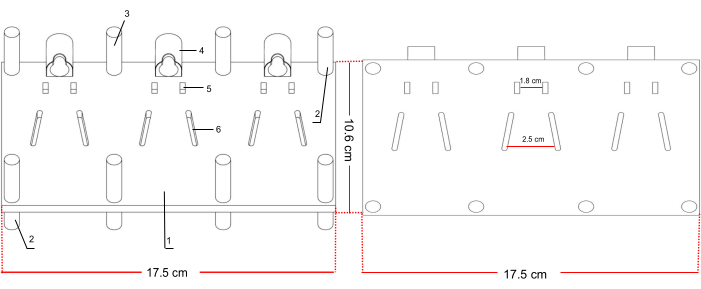
Figure 1. Batch rodent fixator floor plan. (1) Rodents holder device, divided into three parts. (2,3) Pillars: used to fix the limbs of mice or young rats. (4) Cave: used to fix the head. (5) Hole: used to fix the neck of the young rats. (6) Hole: used to fix the body. Please click here to view a larger version of this figure.
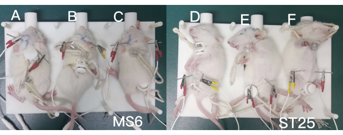
Figure 2: Fixed young rats in prone and supine positions. (A–C) EA treatment on MS6 acupoint.(D–F) EA treatment on ST25 acupoint. Please click here to view a larger version of this figure.
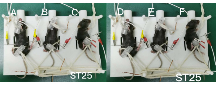
Figure 3: Fixed mice in prone and supine positions. (A–C) EA treatment on MS6 acupoint.(D–F) EA treatment on ST25 acupoint. Please click here to view a larger version of this figure.
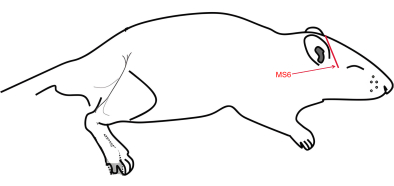
Figure 4: MS6 in the cerebral region. MS6 is located on the lateral side of the head and is positioned at the connection point between Shencong (Ex-hn1) and Xuanli (GB6), which is also known as the anterior parietal (Du21) region. Please click here to view a larger version of this figure.
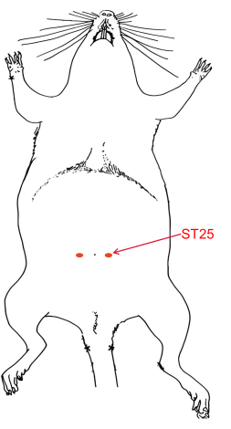
Figure 5: ST25 bilateral point in red. ST25 is located at the level of the umbilicus. Positioning this point in rodent requires first measuring the length of the rodent's paw. ST25 is located at 2/3 the length of the paw next to the navel. For example, if the measure paw of the rodent is 0.6cm, then the ST25 is 0.4 to the right and left of the umbilicus. Please click here to view a larger version of this figure.
Trials | Rats | Mice | ||
Prone position | Supine position | Prone position | Supine position | |
(time in seconds) | (time in seconds) | (time in seconds) | (time in seconds) | |
Trial 1 | 75.5 | 65.1 | 60.2 | 62.3 |
Trial 2 | 70.2 | 80.3 | 80.1 | 70.6 |
Trial 3 | 85.5 | 79.5 | 75.7 | 65.9 |
Average time (Mean ± SD) | 77.1 ± 7.8 | 74.9 ± 8.6 | 72.0 ± 10.5 | 62.3 ± 4.2 |
Table 1: Time spent on fixing rats and mice with prone and supine positions. The data (time in seconds) was obtained by one experimenter who immobilized 3 rats and 3 mice for 3 consecutive trials, respectively. The mean time required was determined by calculating the average and standard deviation of the time taken for the 3 trials.
Supplementary File 1: 3D printed bath rodent fixator design file. This 3D model can be dragged to achieve a full 360° view of the design. Please click here to download this File.
Video 1: Movie of prone and supine position fixation of young SD rats, as described in detail in the Figure 2 legend. This video demonstrates an experimenter restraining 3 rats in both supine and prone positions. Please click here to download this Video.
Electroacupuncture (EA) is a form of acupuncture that involves the use of electrical stimulation on acupuncture needles11. This technique involves the use of micro-pulse currents of specific intensity and frequency to stimulate acupuncture points and achieve enhanced therapeutic effects3. However, the mechanism of EA for healing is limited and requires extensive basic research to prove1,2,3. Rodent models are commonly used in basic EA studies8,12,13, and we aim to provide a fast and efficient method for conducting these experiments in rats and mice.
Currently, the EA experiment confronts many challenges. The primary challenge is to effectively immobilize rodents without anesthesia. Stable immobilization of animals is crucial and has a significant impact on the outcome of the animal experiment. Otherwise, the physiological functions of anesthetized animals are different from those of awake animals, and clinical acupuncture treatment is always performed on awake patients. Therefore, experimental studies with awake animals are more practical for clinical purposes. Previously, immobilization with a fixation plate was the preferred method, but it was time-consuming and only allowed for the operation of a single rodent14. Traditional immobilizers also required tying up the rodent's limbs, which was a time-consuming process and often resulted in pain due to the tight fastening. Several studies have implemented adhesive tape for mice, but their ability to easily escape poses significant challenges, particularly during acupuncture procedures8. When there are more experimental subjects, batch operation is often required to save time and effort2. Therefore, we have developed a 3D printed small animal body batch fixator that offers several advantages over traditional fixation methods15: 1) it shortens the fixation time, greatly improving the experimental efficiency16; 2) it is simple and easy to use and is not affected by subjective factors15,16; 3) It led less harmful to rodents; 4) Once fixed, the EA operation can be performed on several young rats at the same time, saving time and allowing simultaneous observation of treatment results.
The use of 3D printing technology for experimental studies of EA is a valuable expansion in the field of acupuncture research. The device can be adjusted to any size when 3D printed, which helps experimental acupuncture to be carried out more effectively. Fused deposition modeling (FDM), also known as fused filament fabrication (FFF), an additive manufacturing method10, is used for 3D printing the fixator. This device is suitable not only for this experiment but also for performing experiments like CT or MRI on rodents17,18. In addition, it can also be applied to any situation where bulk fixation is needed, except for EA studies. Due to the prevalence of 3D printing technology, this device is easily available. It can be made through a local 3D printing manufacturer. Furthermore, this fixing device can be modified and customized based on the design drawing. In this study, the fixing device is comprised of three sections, each capable of fixing a single rodent. If additional rodents or other animals need to be included, the device can be scaled proportionally to accommodate the larger number. For example, it can be designed as a device that can fix 5 mice or even as a fixing device for other animals such as rabbits.
Another challenge is to find acupoints in the rodents which correspond to humans. MS6 and ST25 acupoints were chosen to examine the effects of prone and supine fixation. Moreover, those two acupoints have been studied by many scholars6,7,19. MS6 is most often used in brain disease research, such as Parkinson's disease, dysfunction after cerebral stroke, etc6,13,19. MS6 could enhance the neurological function of rats suffering from ischemic stroke via modulation of the JAK/STAT signaling pathway that is mediated by IL-12 in a previous study13. ST25 is usually used for the treatment of the gastrointestinal tract, such as diarrhea and irritable bowel syndrome (IBS)7,12. A previous study has reported that EA applied solely at the ST25 point showed the potential to alleviate visceral hypersensitivity while also restoring normal slow-wave frequency and colon rhythm in IBS rats12. These two acupoints are also able to appropriately demonstrate the effects of supine and prone positioning in rodents. Also, these two acupoints have the significance of display6,7,19. Since these two acupoints have been studied by many scholars, we provide a protocol on how to locate these two acupoints in rodents. It is helpful for animal experiments where MS6 and ST25 studies are being conducted. However, the current study has some limitations. For instance, the position and insert depth of the needle cannot be objectively measured and may vary based on the operator's level of experience.
Our experiments comply with the "3Rs" principle of animal ethics and have been approved by the Laboratory Animal Welfare and Ethnic Committee of the Army Medical University. The most challenging and crucial step in this experiment is to find a reliable and efficient method to securely immobilize rodents. This is because any attempt to escape or death of the rodents during the 5-min EA stimulation will result in the failure of the experiment. The design of the front cave takes advantage of the rodents' preference for digging holes. Placing the rodents' head inside the cave is intended to provide psychological comfort. Furthermore, the ropes used to fasten the head and body of the rodents should be moderately secure, remaining neither excessively slack nor overtight to avoid the escape of the rodent and prevent respiratory distress. Following electrical stimulation, the rodents' behaviors and responses were carefully monitored. Ideally, when applying a current of 1-2 mA, rodents would not exhibit aggressive struggling or vocalizations in response to electrical shock20. The fixator can also be used to fix rodents in supine, prone positions and other types of fixations as needed. It is suitable for a variety of experimental needs, and the fixation operation is easy to perform21. However, this has led to a limitation that animals of varying sizes necessitate tailored immobilization equipment of varying sizes. Only animals with slight size discrepancies can be restrained using the same device. The batch immobilization fixator used in this protocol efficiently and reliably immobilizes rodents within the weight range of 10-40 g. For animals outside this weight range, customized batch immobilization fixators are needed to suit their specific requirements.
In addition, the 3D-printed batch rodent fixation used for bulk EA operations on rodents offers enhanced safety as it is resistant to rodent scratches and bites. Studies of EA on rodents have provided valuable insights into the potential benefits of this therapeutic modality in humans1,2,3,6,7,11,14. The device is beneficial for the promotion of experimental scientific research in EA. This technique allows researchers to conduct animal experiments that require the immobilization of animals. Moreover, it presents the exact location of ST25 and MS6, which can serve as a reference for those studying these two acupoints in the future. Ultimately, this methodology is expected to lead to valuable animal models for EA research.
The 3D-printed batch mice fixator described in this paper offers a simple and efficient method for conducting EA experiments in rats or mice. Its design considers the comfort and safety of the animals, as well as the need for quick and efficient fixation. This device has the potential to save time and effort in conducting animal experiments, promoting the advancement of electroacupuncture research and ultimately leading to better treatments for various diseases.
The authors have nothing to disclose.
Thanks to the Department of Neurosurgery of the Second Affiliated Hospital of Army Medical University for site support. Funding: This work was supported by the Natural Science Foundation of China (82104696).
3D printing batch mice fixator | MESH INVENT | Custom made | The fixator produced by 3D printer. The printing method is called fused deposition modeling (FDM), also known as fused filament fabrication (FFF). |
Electroacupuncture instrument | Hwato, Suzhou Medical Appliance Factory | SDZ-III | www.Hwato-med.com |
Disposable sterile acupuncture needle | Suzhou Medical Appliance Factory | N/A | 0.25 mm x 13 mm |