Here, we describe the measurement of cough using a noninvasive and real-time whole-body plethysmography (WBP) system and the normative procedures for harvesting tissue samples of mice and introduce some methods to assess airway inflammation.
Chronic cough, which lasts for more than 8 weeks, is one of the most common complaints requiring medical attention, and patients suffer from a huge socioeconomic burden and a marked decrement in quality of life. Animal models can mimic the complex pathophysiology of the cough and are important tools for cough research. The detection of cough sensitivity and airway inflammation is of great significance for studying the complex pathological mechanism of cough. This article describes the measurement of cough using a noninvasive and real-time whole-body plethysmography (WBP) system and the normative procedures for harvesting tissue samples (including blood, lung, spleen, and trachea) of mice. It introduces some methods to assess airway inflammation, including pathological changes in hematoxylin and eosin (HE)-stained lung and trachea sections, the total protein concentration, the uric acid concentration, and the lactate dehydrogenase (LDH) activity in the supernatant of bronchoalveolar lavage fluid (BALF), and the leukocytes and differential cell counts of BALF. These methods are reproducible and serve as valuable tools to study the complex pathophysiology of cough.
Cough is an important defense behavior to maintain airway patency and protect the lungs from potentially harmful substances. However, when dysregulated, cough becomes a pathological condition1. Chronic cough, usually defined as lasting eight or more weeks, is one of the most frequent symptoms requiring medical attention2. Because chronic cough frequently persists for years, patients suffer from a huge socioeconomic burden and a marked decrement in quality of life3,4,5. Chronic cough is widely considered a cough hypersensitivity syndrome and is characterized by troublesome coughing often triggered by low levels of thermal, mechanical, or chemical exposure6. The occurrence of cough hypersensitivity is closely related to airway inflammation7. However, the pathophysiological mechanisms underlying the modulation of cough sensitivity need to be further elucidated.
Animal models can mimic the complex pathophysiology of the cough and are important tools for cough research8,9. Previous studies have found that viral infection, intrapulmonary Interferon-γ (IFN-γ) instillation, esophageal perfusion of hydrochloric acid, pollutant exposure, cigarette smoke, and citric acid can induce cough in animals10,11,12,13,14,15,16,17. In order to better evaluate cough and airway inflammation, a mouse model of cough was established using a nonlethal dose of the H1N1 virus in this study. For the detection of cough, some cough measurement tools have been established clinically to measure cough, including subjective and objective methods18. The subjective evaluation tools to assess the cough severity primarily include a visual analog scale, the cough score, and quality of life questionnaires, etc19,20. However, they are unlikely to be used to assess cough in animals. In addition, cough can be objectively assessed through a cough challenge test and cough frequency monitoring. The cough challenge test using a whole-body plethysmography (WBP) system is an objective method widely used in animal studies to measure cough sensitivity and reveal the underlying mechanisms of cough13,16. Based on the neuroanatomical characteristics of the cough reflex, citric acid, capsaicin, adenosine 5'-triphosphate (ATP), allyl isothiocyanate (AITC), and inflammatory mediator bradykinin are commonly used as the tussive agents to induce cough21,22. Citric acid is one of the earliest and the most widely used tussive agents triggering cough reflexes, which has been validated for measuring cough sensitivity. Besides, the citric acid challenge has good safety, feasibility, and tolerability and is suggested to assess cough reflex sensitivity in response to cough therapies23. Therefore, this article will describe the method for measuring cough sensitivity in response to citric acid in mice using a noninvasive and real-time WBP system.
The studies of the pathophysiology of cough require test samples, including samples from the blood, bronchoalveolar lavage fluid (BALF), and lung and trachea tissues to verify changes in the levels of key factors24. Currently, there is a lack of normative procedures for harvesting tissue samples of mice, and related studies employ different approaches that complicate the assessment of airway inflammation. Bronchoalveolar lavage is an important method to evaluate airway inflammation in respiratory diseases25. Different methods of bronchoalveolar lavage will lead to a lack of comparability between related studies. Moreover, different bronchoalveolar lavage methods have an impact on inflammatory cells and inflammatory cytokines in BALF. Therefore, this article will describe the establishment of a mouse model of cough with a nonlethal dose of the H1N1 virus, the measurement of cough using a WBP system, and a reliable, safe, and highly successful bronchoalveolar lavage method of mice.
All the procedures were approved by the Animal Care and Use Committee of Guangzhou Medical University (20240248) and were performed in strict accordance with approved guidelines. Male specific pathogen-free C57BL/6 mice weighing 20-25 g were used in this study. All mice were housed under controlled temperature (22 ± 2 °C), humidity (50% ± 20%), and lighting (6:30 AM to 6:30 PM) in solid bottom cages with food and water available ad libitum. The time line of the protocol is shown in Figure 1.
1. Establishment of a mouse model of cough
2. The cough sensitivity measurement
3. Harvesting of blood, spleen, BALF, lung, and trachea tissues of mouse (Figure 4)
Figure 6 shows representative images of pathological changes in HE-stained lung (Figure 6A,B), trachea (Figure 6C,D), and spleen (Figure 6E,F). H1N1 virus infection resulted in inflammatory changes in mouse lungs, including edema and many lymphocytes and neutrophil infiltration. H1N1 virus infection also induced inflammatory changes in mouse tracheae, including cilia shedding and inflammatory cell infiltrations (many lymphocytes and small numbers of neutrophils). In addition, the infection also significantly increased the ratio of the white pulp area to the whole spleen area of mice. Lymphocytes accumulate in the white pulp of the spleen. This standardized procedure for harvesting tissue samples can better evaluate airway inflammation.
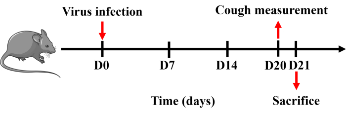
Figure 1: Protocol for establishing a mouse model of cough. Mice were anesthetized with pentobarbital sodium and intranasally instilled with 0.8 x LD50 of H1N1 virus dissolved in 50 µL of PBS once on day 0. Cough measurement was performed on day 20, and mice were sacrificed the following day. Please click here to view a larger version of this figure.
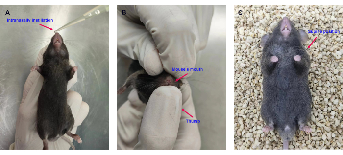
Figure 2: Intranasal instillation of the H1N1 virus. (A) Intranasal instillation of 0.8 x LD50 of H1N1 virus in 50 µL of PBS. (B) Holding the mouse's mouth closed with the thumb. (C) Placing the mice in the supine position. Please click here to view a larger version of this figure.
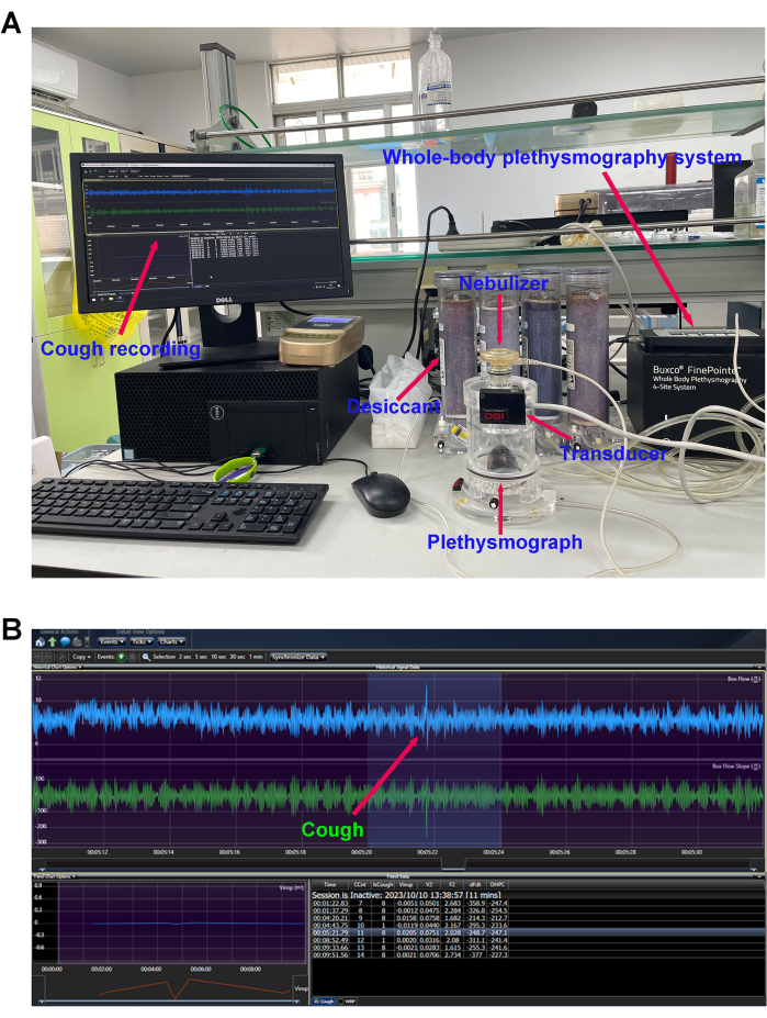
Figure 3: Cough sensitivity measurement. (A) Mouse cough detection equipment and (B) cough reflex curve. The number of cough events in response to the nebulized citric acid solution (0.4 M) was detected using the whole-body plethysmography (WBP) system after modeling. Please click here to view a larger version of this figure.
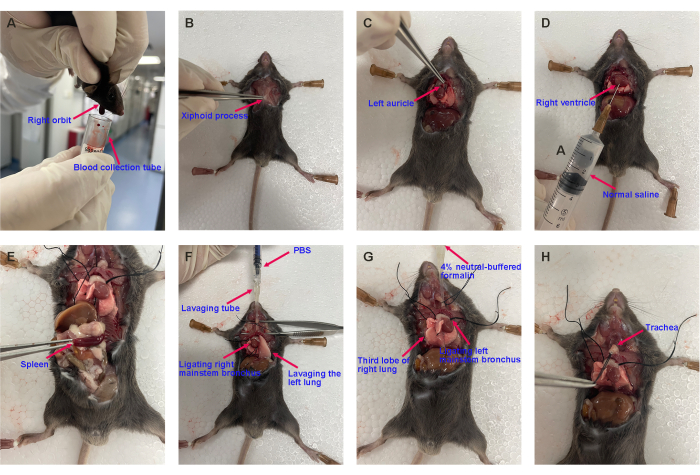
Figure 4: Representative images of harvesting the mouse's blood, spleen, BALF, lung, and trachea. (A) Blood collection, (B) opening the chest, (C) cutting off the left auricle, (D) pulmonary circulation perfusion, (E) harvesting the spleen, (F) bronchoalveolar lavage, and (G) harvesting the lung lobe and (H) the trachea for histopathological analysis. Please click here to view a larger version of this figure.
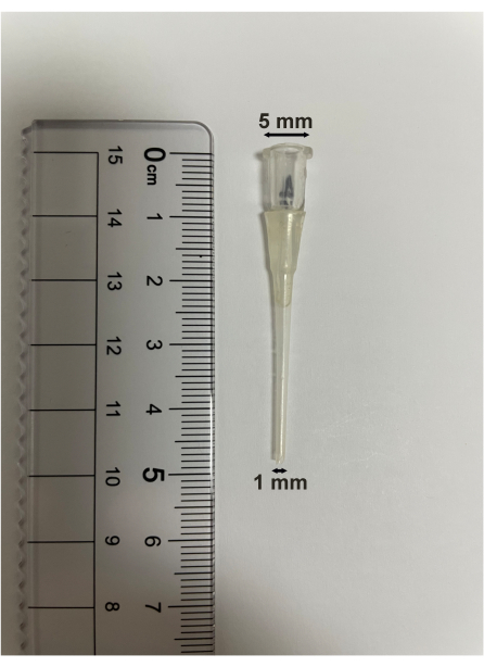
Figure 5: Bronchoalveolar lavage tube specification. The length of the bronchoalveolar lavage tube is 5 cm. The upper diameter is 5 mm, and the lower diameter is 1 mm. Please click here to view a larger version of this figure.
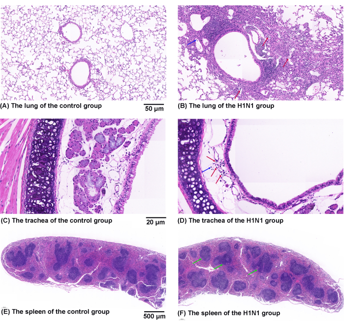
Figure 6: Effects of H1N1 virus on pathological changes in the mouse's lung, trachea, and spleen. (A,B) Representative figures of pathological changes in HE-stained lung sections from the (A) control and (B) H1N1 groups. The symbol of "↑" marks the infiltration of lymphocytes (red) and neutrophils (blue). Scale bars: 50 µm. (C,D) Representative figures of pathological changes in HE-stained trachea sections from the (C) control and (D) H1N1 groups. The symbol of "↑" marks the infiltration of lymphocytes (red) and neutrophils (blue). Scale bars: 20 µm. (E,F) Representative figures of pathological changes in HE-stained spleen sections from the (E) control and (F) H1N1 groups. The symbol of "↑" marks the white pulp (green). Scale bars: 500 µm. Please click here to view a larger version of this figure.
Some chronic refractory and postinfectious coughs are common conditions associated with respiratory virus infection27. In order to better evaluate cough sensitivity and airway inflammation, a mouse model of cough was established using the H1N1 virus in this study. Appropriate mouse cough models should be selected for other studies according to the purpose of the study. Most previous studies used the guinea pig as an animal model in mechanistic studies or new drug trials for cough28,29,30. Recent studies have suggested that mice might be used to assess cough pathophysiology owing to their shorter reproductive cycle, more reagents, and readiness for genetic manipulations despite their cough behavior still being debated16,31. The citric acid was used as a tussive agent to induce cough in this study. The mechanisms of cough reflex induced by citric acid may be related to the activation of jugular C-fibers and nodose Aδ-fibers32. Besides, the WBP system measures the changes in cough reflex in mice in a noninvasive manner and minimizes the effects of psychological stress. The effect of the external environment on mice should be minimized when cough sensitivity is detected. The mouse should be placed in the testing room and then covered with a plastic bag to reduce the irritation caused by the external environment.
This study details the normative procedures for harvesting blood, spleen, BALF, lung, and trachea tissues of mice and introduces some measurements to assess airway inflammation. To detect airway inflammation, the lung and trachea sections are stained with hematoxylin and eosin to assess general histopathology24. The increased concentration of total protein and uric acid in the supernatant of BALF are associated with inflammation and cell injury in the airway33. LDH activity in the supernatant of BALF reflects cell damage and necrosis34. The leukocytes in BALF and blood can reflect the degree of inflammation of the disease35. Differential cell counts of BALF are widely used in assessing airway inflammation in chronic airway diseases and provide important information in studying pathogenesis, making diagnoses, and management strategies for chronic respiratory diseases36.
The limitation of this experiment is that collecting a large number of tissue samples makes mice be operated for a long time, which may affect the activity of the tissue samples. Therefore, mouse tissue samples need to be placed on ice immediately after collection. In addition, the bronchoalveolar lavage tube used in this study is suitable for mice but not for bigger animals.
In summary, we provide a detailed description of the detection methods of cough and airway inflammation in mice. These methods provide researchers with tools to study the complex pathophysiology of cough.
The authors have nothing to disclose.
This work was supported by the Guangzhou Science and Technology Planning Project (202002030151), the Major Project of Guangzhou National Laboratory (GZNL2024A02001), and the grant of State Key Laboratory of Respiratory Disease (SKLRD-Z-202202).
4% paraformaldehyde | Biosharp | BL539A | |
Buxco Small Animal Whole Body Plethysmography System | DSI | — | |
Calcium-free and magnesium-free Hank’s Balanced Salt Solution | Beyotime | C0219 | |
Citric acid | Sigma-Aldrich | C2404 | |
Hematoxylin-Eosin | BASO Biotechnology | BA-4098 | |
Heparin sodium | Alfa Aesar | A16198 | |
Influenza A/California/7/2009 (H1N1) virus | ATCC | VR-1894 | |
Isoflurane | RWD | R510-22 | |
Lactate dehydrogenase assay kit | Nanjing Jiancheng Bioengineering Institute | A020-2-2 | |
Normal saline | Guangzhou Zhongbo Biotechnology | 1234-1 | |
Pasteur pipet | NEST | 318415 | |
Pentobarbital sodium | Merck | P3761 | |
Phosphate buffered saline | Meilunbio | MA0015 | |
Total protein assay kit | Nanjing Jiancheng Bioengineering Institute | A045-3 | |
Uric acid assay kit | Thermo Fisher Scientific | A22181 |
.