Intradermal microdialysis is a minimally invasive technique used to investigate microvascular function in health and disease. Both dose-response and local heating protocols can be utilized for this technique to explore mechanisms of vasodilation and vasoconstriction in the cutaneous circulation.
The cutaneous vasculature is an accessible tissue that can be used to assess microvascular function in humans. Intradermal microdialysis is a minimally invasive technique used to investigate mechanisms of vascular smooth muscle and endothelial function in the cutaneous circulation. This technique allows for the pharmacological dissection of the pathophysiology of microvascular endothelial dysfunction as indexed by decreased nitric oxide-mediated vasodilation, an indicator of cardiovascular disease development risk. In this technique, a microdialysis probe is placed in the dermal layer of the skin, and a local heating unit with a laser Doppler flowmetry probe is placed over the probe to measure the red blood cell flux. The local skin temperature is clamped or stimulated with direct heat application, and pharmacological agents are perfused through the probe to stimulate or inhibit intracellular signaling pathways in order to induce vasodilation or vasoconstriction or to interrogate mechanisms of interest (co-factors, antioxidants, etc.). The cutaneous vascular conductance is quantified, and mechanisms of endothelial dysfunction in disease states can be delineated.
Cardiovascular disease (CVD) is the leading cause of death in the United States1. Hypertension (HTN) is an independent risk factor for stroke, coronary heart disease, and heart failure and is estimated to affect upward of ~50% of the United States population2. HTN can develop as an independent CVD (primary HTN) or as a result of another condition, such as polycystic kidney disease and/or endocrine disorders (secondary HTN). The breadth of etiologies of HTN complicates investigations into the underlying mechanisms and end-organ damage observed with HTN. Diverse and novel research approaches into the pathophysiology of the end-organ damage associated with HTN are needed.
One of the earliest pathological signs of CVD is endothelial dysfunction, as characterized by impaired nitric oxide (NO)-mediated vasodilation3,4,5. Flow-mediated dilation is a common approach used to quantify the endothelial dysfunction associated with CVD, but endothelial dysfunction in microvascular beds can be both independent of and precursory to that of large conduit arteries6,7,8. Furthermore, resistance arterioles are more directly acted on by local tissue than conduit arteries and have more immediate control over the delivery of oxygen-rich blood. Microvascular function is predictive of adverse cardiovascular event-free survival9,10,11. The cutaneous microvasculature is an accessible vascular bed that can be used to examine responses to physiological and pharmacological vasoconstrictive or vasodilatory stimuli. Intradermal microdialysis is a minimally invasive technique, the goal of which is to investigate the mechanisms of both vascular smooth muscle and endothelial function in the cutaneous microvasculature with targeted pharmacological dissection. This method contrasts with other techniques, such as post-occlusive reactive hyperemia, which does not allow for pharmacological dissection, and iontophoresis, which allows for pharmacological delivery but is less precise in its mechanism of action (reviewed thoroughly elsewhere12).
The rationale behind the development and use of this technique is extensively reviewed elsewhere13. This approach was originally developed for use in neurological research in rodents and then was first applied to humans to investigate the mechanisms underlying active vasodilation from a thermoregulatory standpoint. In the late 1990s, this method was used to examine both neural and endothelial mechanisms with regard to local heating of the skin. Since that time, the technique has been utilized to investigate a number of neurovascular signaling mechanisms in the skin.
Using this technique, our group and others have interrogated the mechanisms of endothelial dysfunction in the microvasculature of several clinical populations, including, but not limited to, dyslipidemia, primary aging, diabetes, chronic kidney disease, polycystic ovary syndrome, preeclampsia, major depressive disorder14,15,16,17,18,19, and hypertension20,21,22,23,24. For example, a previous study found that normotensive women with a history of preeclampsia, who are at an increased risk for CVD, had reduced NO-mediated vasodilation in the cutaneous circulation compared with women with a history of normotensive pregnancy20. In another study, adults diagnosed with primary HTN demonstrated increased angiotensin II sensitivity in the microvasculature compared with healthy controls21, and chronic sulfhydryl-donating antihypertensive pharmacotherapy in primary HTN patients has been shown to decrease blood pressure and improve both hydrogen sulfide- and NO-mediated vasodilation22. Wong et al.23 found impaired sensory-mediated and NO-mediated vasodilation in prehypertensive adults, coinciding with our finding of a progression of endothelial dysfunction with increasing HTN stages, as categorized by the 2017 American Heart Association and American College of Cardiology guidelines24.
The intradermal microdialysis technique allows for tightly controlled mechanistic investigations into microvascular function in health and disease states. Therefore, this paper aims to describe the intradermal microdialysis technique as applied by our group and others. We detail the procedures for both pharmacological stimulation of the endothelium with acetylcholine (ACh) to examine the dose-response relationship and physiological stimulation of endogenous NO production with either a 39 °C or 42 °C local heating stimulus protocol. We present representative results for each approach and discuss the clinical implications of the findings that have arisen from this technique.
All procedures are approved by the Institutional Review Board of the Pennsylvania State University prior to participant recruitment.
1. Equipment setup
2. Microdialysis fiber placement
3. Hyperemia
4. Acetylcholine dose-response protocol
5. Local heating protocol
6. Removing the microdialysis probes
Acetylcholine dose-response protocol
Figure 1A depicts a schematic detailing the ACh dose-response protocol. Figure 1B illustrates representative tracings of the RBC flux values (perfusion units, PU; 30 s averages) from the standardized ACh dose-response protocol for one subject over time. Figure 1C illustrates a raw data file of an ACh dose-response protocol. Additional baseline measurements were maintained in the raw data file, but only ~10 min of baseline were utilized for the data analysis.
Following hyperemia resolution and a stable RBC flux for 5 min, the 10 min baseline data collection may begin. The baseline is depicted as a relatively stable horizontal RBC flux line, where any cause for deviations (e.g., movement artifacts, probe adjustments) should be logged as data acquisition software comments for analysis purposes. The dose-response protocol follows the baseline period, and the syringes must be changed with each dose, from 10−10 M to 10−1 M ACh. Before starting the 5-10 min perfusion of each dose, one must ensure that the pharmacological agent has fully perfused through the length of the fiber. In the data acquisition software, there will be an initial rise in the RBC flux due to the perfusion, but this is not included in the analyses, as the 5 min data collection for that concentration has not begun. Once the perfusion for each dose has begun, there will be a continual increase in the RBC flux to a peak, followed by a steady decline. This curvilinear response to pharmacological agents will be replicated throughout the protocol, but the RBC flux will be relatively greater with increasing concentrations of ACh. With lower concentrations of ACh, the curvilinear response may not be as prominent. Examples of non-optimal RBC flux include the following: 1) a non-curvilinear response, where the RBC flux does not increase and remains plateaued, or 2) increasing ACh concentrations having a minimal impact on the RBC flux, where the RBC flux does not relatively increase with each concentration of ACh. This is dependent on the research question and the clinical cohort being tested.
Following the final concentration of ACh, lactated Ringer's is perfused, and the local heaters are increased to 43 °C. During this phase, a plateau must be obtained before sodium nitroprusside is perfused. This may take up to ~45 min, depending on the previous agents perfused. This phase is not included in the analyses. Once the plateau has been obtained for 5 min, sodium nitroprusside is perfused to produce maximal local vasodilation. This maximal local vasodilation is depicted as a rise in the RBC flux, where a plateau is obtained after ~20 min of perfusion, or by the RBC flux reaching a peak and declining immediately after. Once a plateau or a decline in the RBC flux has been obtained for sodium nitroprusside, the protocol is complete. A common example of non-optimal RBC flux is the highest value of RBC flux being obtained at a different phase of the protocol (e.g., during the dose-response protocol) rather than during the maximal local vasodilation.
Acetylcholine dose-response protocol: Nitric Oxide synthase inhibition
To quantify the contribution of NO to cutaneous blood flow in response to ACh, NG-nitro-l-arginine methyl ester (L-NAME), a NO synthase inhibitor, is perfused in combination with ACh through an additional fiber. Figure 2A depicts a schematic detailing the ACh dose-response protocol with L-NAME. Figure 2B illustrates representative tracings of the RBC flux (30 s averages) from the standardized ACh dose-response protocol for one subject over time with L-NAME. Figure 2C illustrates a raw data file of an ACh dose-response protocol with L-NAME. Additional baseline measurements were maintained in the raw data file, but only ~10 min of baseline were utilized for the data analysis.
Following hyperemia resolution, a stable RBC flux for 5 min, and adequate time to fully block the enzymatic pathway of interest (e.g., NO synthase) and/or deliver adequate concentrations of co-factors, the 10 min of baseline data collection may begin (depicted as a relatively stable horizontal line). The dose-response protocol follows baseline, and the syringes must be changed with each dose starting from 1010 M to 101 M ACh with the NO synthase inhibitor (e.g., 15 mM L-NAME). In the presence of a NO synthase inhibitor, the curvilinear response is not well replicated until higher concentrations of ACh. A relatively lower RBC flux, in comparison to a site without NO synthase inhibition, will be observed. A common example of non-optimal RBC flux is the NO synthase inhibition, as compared to the conditions without NO synthase inhibition, resulting in a higher RBC flux. This indicates that the protocol has failed.
Following perfusion of the final concentration of ACh, lactated Ringer's is perfused, and the local heaters are increased to 43 °C. During this phase, a plateau should be obtained before sodium nitroprusside is perfused. This phase is not included in the analyses. Once the plateau has been obtained for 5 min, sodium nitroprusside is perfused, producing maximal local vasodilation. During maximal local vasodilation, there will be an exponential rise in the RBC flux due to the previous NO synthase inhibition. A plateau will be obtained after ~20 min of perfusion, or the RBC flux will reach its absolute peak and decline immediately after. Once a plateau or a decline in the RBC flux has been obtained for sodium nitroprusside, the protocol is complete. A common example of non-optimal RBC flux is obtaining the highest RBC flux value at a different phase of the protocol (e.g., during the dose-response protocol) rather than during the maximal local vasodilation.
Local heating protocol
Figure 3A depicts a schematic detailing the local heating protocol. Figure 3B illustrates representative tracings of the RBC flux (30 s averages) for the standardized local heating protocol for one subject over time. Figure 3C illustrates a raw data file of a local heating protocol. Following hyperemia resolution and a stable RBC flux for 5 min, the 10 min of baseline data collection may begin (depicted as a relatively stable horizontal line). The local heaters are set to either 39 °C or 42 °C, and an initial peak and nadir response will occur in the RBC flux. To quantify the contribution of NO to the cutaneous blood flow in response to a local heat stimulus, L-NAME is perfused after a stable plateau in the RBC flux has been achieved. There will be a rapid decline in the RBC flux until it reaches a new plateau in response to L-NAME. After 5 min of stable RBC flux values, lactated Ringer's is perfused, and the local heaters are increased to 43 °C. The heating will produce an additional peak and nadir response in the RBC flux. During this phase, one must ensure that a plateau has been obtained before sodium nitroprusside is perfused. This phase is not included in the analyses. To induce local maximal vasodilation, sodium nitroprusside is perfused, and a rapid increase in the RBC flux will occur. Once a plateau or a decline in the RBC flux has been observed in response to sodium nitroprusside, the protocol is complete.
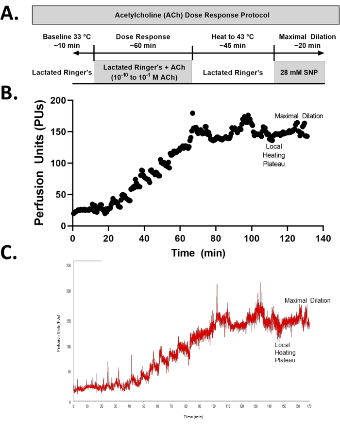
Figure 1: Acetylcholine (ACh) dose-response protocol. (A) Schematic of an ACh dose-response protocol. (B) Representative tracing (30 s averages) of an ACh dose-response protocol. (C) Raw data of an ACh dose-response protocol. Additional baseline measurements are maintained in the raw data file to demonstrate the fluctuations prior to stabilization, but only ~10 min of stable resting data were utilized for the data analysis. Please click here to view a larger version of this figure.
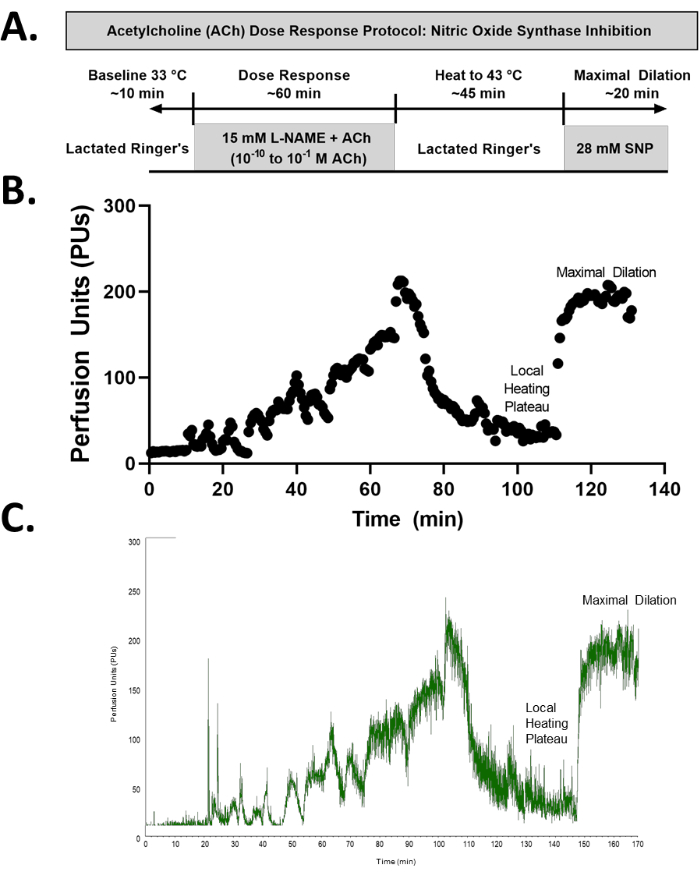
Figure 2: ACh dose-response protocol with Nitric Oxide (NO) synthase inhibition. (A) Schematic of an ACh dose-response protocol with NO synthase inhibition. (B) Representative tracing of an ACh dose-response protocol with NO synthase inhibition. (C) Raw data of an ACh dose-response protocol with NO synthase inhibition. Additional baseline measurements are maintained in the raw data file to demonstrate the fluctuations prior to stabilization, but only ~10 min of stable resting data were utilized for the data analysis. Please click here to view a larger version of this figure.
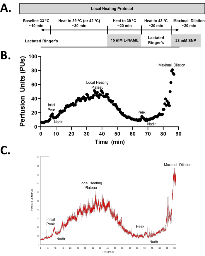
Figure 3: Local heating protocol. (A) Schematic of a local heating protocol. (B) Representative tracing of a local heating protocol. (C) Raw data of a local heating protocol. Please click here to view a larger version of this figure.
The intradermal microdialysis technique is a versatile tool in human vascular research. Investigators may alter the protocol to further diversify its applications. For example, we describe an ACh dose-response protocol, but other investigations into the mechanisms of vasoconstriction or vasomotor tone, rather than vasodilation alone, have utilized norepinephrine or sodium nitroprusside dose-response approaches26,27,28,29,30,31. Other mediators of vasodilation, such as menthol or metachlorine chloride, have also been employed in the dose-response protocol31,32. The dose-response protocol as a pharmacological assessment of vascular function is a more targeted, mechanistic technique to isolate specific signaling mechanisms compared to the local heating protocol, as it removes variations in the sympathetic response to thermal stimuli. However, local heating is a cost-effective approach that uses a physiological stimulus to induce vasodilation via both neurogenic and endothelium-dependent mechanisms. It is also important to consider the mechanism of interest when choosing between a 39 °C or 42 °C local heating protocol. The 39 °C protocol has been suggested to better isolate NO-mediated vasodilation, whereas the 42 °C protocol allows for the investigation of both NO-mediated vasodilation and endothelial-derived hyperpolarizing factor-mediated vasodilation33,34. Additionally, the CVC increase in response to 42 °C local heating tends to be more robust (i.e., reaching a higher %CVCmax34). However, when utilizing a new agent to interrogate a specific signaling pathway, rigorous methods must be employed to evaluate the efficacy (i.e., fully block) and/or saturate concentrations of the co-factors.
Endothelial function is often measured using the flow-mediated dilation technique, but endothelial dysfunction in microvascular beds can occur prior to or independent of endothelial dysfunction in large conduit arteries, especially in pathologies such as HTN6,7,8. Furthermore, the flow-mediated dilation technique does not allow for the pharmacological dissection of the pathophysiology of endothelial dysfunction in isolation from systemic effects. Other methods for investigating microvascular endothelial function, such as iontophoresis or post-occlusive reactive hyperemia, are unable to precisely target the mechanisms of endothelial function with pharmacological intervention12. Therefore, intradermal microdialysis uniquely allows for targeted investigations into the mechanisms of vascular function, and its use, together with flow-mediated dilation outcomes, may provide a more holistic picture of systemic vascular function.
Whichever intradermal microdialysis approach is utilized, certain precautions must be taken to ensure the validity and reproducibility of the responses. While the specifics of the experimental protocol can be adjusted to answer specific research questions, the precise placement of the microdialysis probe is absolutely critical. Care must be taken to insert the probe into the dermis and to avoid the larger visible or palpable blood vessels of the skin. Puncturing these vessels will result in abnormally low perfusion unit values; in this case, the laser Doppler flowmetry will measure the formation of a hematoma rather than the flow of red blood cells through an intact vessel. After this, the next most critical step of this protocol is the resolution of the hyperemic response to the needle puncture. If the hyperemic response is not allowed to fully subside, the recorded perfusion units throughout the baseline and early portions of the protocol will be greater than the true resting values. If sufficient recovery time has been allowed but the perfusion units remain abnormally high, a recalibration of the probes may be required prior to beginning the baseline data collection phase.
A limitation of the intradermal microdialysis technique is that it cannot specifically isolate a tissue type to evaluate vascular signaling pathways. As the vessels of the skin cannot be dissected and visualized in vivo, there is no way to ensure the semipermeable portion of a microdialysis probe is immediately adjacent to the tissue of interest (e.g., the vascular endothelium). Therefore, the results obtained from this technique are representative of the integrative nature of human physiology and provide insights into the collective function of the endothelial, vascular smooth muscle, and neural influences on local blood flow. However, if using a local heating protocol, allowing for the RBC flux to reach a plateau at the initial increase in heat to 39 °C or 42 °C allows for the resolution of the axonal reflex to the heat, which then allows for a primarily endothelium-mediated response, as discussed elsewhere35. An additional limitation of this technique is the use of laser Doppler flowmetry as an index of the skin blood flow. Laser Doppler flowmetry quantifies the red blood cell flux, which does not account for the changes in the vessel diameter (i.e., dilation of the microvasculature), as would be necessary to quantify the absolute flow. It may be sensitive to between-participant or between-condition differences36. Future applications of intradermal microdialysis may incorporate techniques to quantify the absolute microvascular blood flow. For example, the recent development of optical coherence tomography allows for the quantification of the vessel diameter using three-dimensional imaging of the skin microvasculature13. The intradermal microdialysis technique is contraindicated in very few but important instances, which include, but are not limited to, participants with skin conditions, participants with allergies related to the substances described here, participants with severe trypanophobia, and participants with tattoos covering the entirety of the ventral aspect of the forearm (but small tattoos in this area are not exclusionary).
The unique ability of the microdialysis approach to aid in isolating and delineating underlying pathophysiological mechanisms makes it advantageous for investigating the variable etiology of HTN, among other CVDs. Following protocol optimization, this technique allows for evaluating the efficacy of novel CVD treatments. Furthermore, intradermal microdialysis provides a method for assessing the off-target effects of hypothetically unrelated pharmacotherapies, making it a highly valuable tool for informing larger-scale clinical trials. Taken together, this technique is an invaluable asset in microvascular research.
The authors have nothing to disclose.
None.
1 mL syringes | BD Syringes | 302100 | |
Acetlycholine | United States Pharmacopeia | 1424511 | Pilot data collected in our lab indicate drying acetylcholine increases variability of CVC response; do not dry, store in desiccator |
Alcohol swabs | Mckesson | 191089 | |
Baby Bee Syringe Drive | Bioanalytical Systems, Incorporated | MD-1001 | In this study the optional 3-syringe bracket (catalg number MD-1002) was utilized |
CMA 30 Linear Microdialysis Probes | Harvard Apparatus | CMA8010460 | |
Connex Spot Monitor | WelchAllyn | 74CT-B | automated blood pressure monitor |
Hive Syringe Pump Controller | Bioanalytical Systems, Incorporated | MD-1020 | Controls up to 4 Baby Bee Syringe Drives |
LabChart 8 | AD Instruments | **PowerLab hardware and LabChart software must be compatible versions | |
Lactated Ringer's Solution | Avantor (VWR) | 76313-478 | |
Laser Doppler Blood FlowMeter | Moor Instruments | MoorVMS-LDF | |
Laser Doppler probe calibration kit | Moor Instruments | CAL | |
Laser Doppler VP12 probe | Moor Instruments | VP12 | |
Linear Microdialysis Probes | Bioanalytical Systems, Inc. | MD-2000 | |
NG-nitro-l-arginine methyl ester | Sigma Aldrich | 483125-M | L-NAME |
Povidone-iodine / betadine | Dynarex | 1202 | |
PowerLab C Data Acquisition Device | AD Instruments | PLC01 | ** |
PowerLab C Instrument Interface | AD Instruments | PLCI1 | ** |
Probe adhesive discs | Moor Instruments | attach local heating unit to skin | |
Skin Heater Controller | Moor Instruments | moorVMS-HEAT 1.3 | |
Small heating probe | Moor Instruments | VHP2 | |
Sterile drapes | Halyard | 89731 | |
Sterile gauze | Dukal Corporation | 2085 | |
Sterile surgical gloves | Esteem Cardinal Health | 8856N | catalogue number followed by the initials of the glove size, then the letter "B" (e.g., 8856NMB for medium) |
Surgical scissors | Cole-Parmer | UX-06287-26 |