A subscription to JoVE is required to view this content. Sign in or start your free trial.

Method Article

Bacterial Co-Incubation Assay: A Fluorescence Microscopy-Based Technique to Visualize Intraspecific Bacterial Competition at the Single-Cell Level

1.7K views

⸱

July 8th, 2025

In This Article

Abstract

Source: Smith, S., et al., Quantification of Interbacterial Competition using Single-Cell Fluorescence Imaging. J. Vis. Exp. (2021).

This video demonstrates a fluorescence microscopy-based method to study bacterial competition at the single-cell level. This method provides insights into the structure and function of bacterial communities.
Protocol

1. Coincubate bacterial strains

  1. Choose two bacterial strains for single-cell bacterial competition assays. Here, two strains of V. fischeri are used: a target strain (ES11424) and an inhibitor strain (MJ1125) that is known to kill the target strain using the type VI secretion system on chromosome II (T6SS2)1, which is a contact-dependent killing mechanism.
  2. Transform strains with stable plasmids encoding genes for different fluorescent proteins (e.g., GFP or RFP) to visually distinguish strain types on the microscope. Here, the inhibitor strain is tagged with a GFP-encoding plasmid (pVSV102), and the target strain is tagged with a dsRed-e....

Access restricted. Please log in or start a trial to view this content.

Results

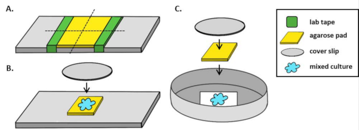
Agarose pad setup diagram for microbial culture under cover slip: lab tape, mixed culture.
Figure 1: Agarose pad preparation and slide setup for coincubation assays. (A) Setup for making 2% agarose pads. Five layers of lab tape (green) are wrapped around a cover slip at two points approximately 20 mm apart. Next, warm 2% agarose in mar.......

Access restricted. Please log in or start a trial to view this content.

Disclosures

No conflicts of interest declared.

Materials

List of materials used in this article
NameCompanyCatalog NumberComments
1.5 mL Microcentrifuge tubeFisher05-408-129
10 uL single channel pipette
1000 uL single channel pipette
20 uL single channel pipette
200 uL single channel pipette
AgaroseFisherBP165-25Low melting agarose
Cellvis 35 mm DishFisherNC0409658#1.5 cover glass bottom
ChloramphenicolSigmaC0378stock (20 mg/mL in Ethanol); final concentration in media (2 μg /mL LBS)
FIJI image analysis sofwareImageJhttps://imagej.net/Fiji/Downloadsopen-source software
Fisherbrand Cover Glasses: CirclesFisher12-545-81P#1.5 cover glass; 12 mm diameter
Kanamycin SulfateFisherBP906-5stock (100 mg/mL in water, filter sterilize); final concentration in media (1 μg/mL LBS)
Lens Cleaning Tissue PaperFisherS24530
Petri PlatesFisherFB0875713sterile with lid
Razor BladesFisherS65921
Semi-micro CuvettesVWR97000-586
Spectrophotometer
Thermo Scientific Gold Seal Plain Microscope SlidesFisher12-518-100B
Thermo Scientific Richard-Allan Scientific Cover GlassFisher22-050-235#1.5 cover glass, 25 mm2
Type F Immersion OilFisherNC0297589
Upright or inverted fluorescence microscope with camera and imaging softwareImages in this article were acquired on a Nikon TI-2 inverted fluorescent microscope outfitted with an ORCA-Fusion Digital CMOS camera using NIS-Elements software.
Vortex
Water bathUsed to keep agarose warm prior to pipetting
<strong>LBS media</strong>
1M Tris Buffer (pH ~7.5)50 mL 1 M stock buffer (62 mL HCl, 938 mL DI water, 121 g Trizma Base)
Agar TechnicalFisherDF0812-17-915 g (Add only for plates)
DI water950 mL
Sodium ChlorideFisherS640-320 g
TryptoneFisherBP9726510 g
Yeast ExtractFisherBP9727-25 g
<strong>mPBS (marine PBS)</strong>Phosphate buffered saline with marine salts added; used for making agarose pad
10X PBSFisherICN1960454
Instant Ocean Sea SaltInstant OceanSS1-160PAdjust concentration to appropriate salinity; 20 psu used here
Sterile Vacuum Filter UnitsFisherSCGVU01REUsed to filter-sterilize mPBS
Vacuum pumpUsed to filter-sterilize mPBS

Explore More Articles

Type Six Secretion SystemSingle-Cell ImagingFluorescent ProteinsOptical DensityAgarose PadCoverslip PreparationDIC Imaging