All steps should be performed under BSL-2 conditions in a Laminar flow hood.
1. Preparing the STLV Bioreactor
- Assemble the STLV bioreactor according to manufacturer’s protocol and perform detoxification protocol to ensure sterility of the bioreactor. Cover open ports with luer caps and fill the STLV with 95% ethanol for 24 h.
- Remove ethanol and fill the STLV with sterilized distilled water for 24 h.
- Repeat step 1.2 with sterilized distilled water only.
- With the tool supplied by vendor, loosen all screws, caps and center plug from the STLV, and autoclave at 110 °C for 20 min in a sterilization pouch.
- Once the STLV is cooled, tighten screws and repeat steps 1.1-1.4 to ensure sterility and complete detoxification.
- Tighten screws of cooled the STLV and screw on pre-sterilized one-way stopcocks to each port.
- Open stopcock by positioning the knobs vertically.
- Remove the plungers from a 10 mL and 5 mL luer-lock syringe and re-sleeve plungers back in wrappers to maintain sterility. Screw syringes onto stopcocks (luer-lock tip syringes are required for this step).
- Add Dulbecco’s Phosphate-Buffered Saline (DPBS) to the 10 mL syringe until the STLV is full and begins to fill the 5 mL syringe.
- Replace the sterile plungers into each syringe and remove residual bubbles occurring in bioreactor by alternating between driving plungers of the syringes.
- Leave the syringes attached to bioreactor after removing all bubbles. Close stopcocks and attach the STLV to the rotating vertical platform and rotate for a minimum of 24 h to monitor for leaks.
2. Preparing Microcarrier Beads
- Add 15 mL DPBS to ~250 mg cytodex microcarrier beads in a 50 mL conical tube and autoclave under same conditions as the STLV (1.4).
- After cooling, remove DPBS, add media used to grow epithelial cell of interest, and swirl tube to resuspend beads.
- Repeat wash steps with media two more times. After final wash add 15 mL growth media to beads.
3. Seeding Epithelial Cells and Beads into the STLV Bioreactor
- Grow epithelial cells of interest as monolayers in tissue culture flasks until you obtain ≥1×10⁷ cells. Remove cells from the flask (i.e. trypinsization, EDTA), count cells to establish cellular concentration, and determine cellular viability by trypan blue exclusion staining1.
- To the conical tube containing prepared beads (see 2.3), add desired concentration of cells (2×10⁵-1×10⁷ epithelial cells)2, 8, depending on your epithelial cell type of intent (Figure 1A). Ensure all cells are transferred by rinsing the conical tube.
- Remove DPBS and syringes from the STLV.
- Remove plug from center port and add the epithelial cell/bead suspension into the STLV using 10 mL serological pipette. Rinse conical tube to ensure all cells/beads are transferred to the STLV and replace center port plug.
- Remove plungers from a 5 and 10 mL syringes and re-sleeve plungers back in wrappers to maintain sterility. Screw syringes into ports with open stopcocks. Fill the STLV with media, replace plungers, and close stopcocks.
- Place the newly seeded STLV in a 37 °C incubator for 30 min with no rotation.
- Open stopcocks, remove bubbles using plungers, and then close stopcocks.
- Place the STLV in a 37 °C, 5% CO2 humidified incubator rotating at 20 rpm (Figure 1B). Cultures must be rotated continuously 24 h a day except for when sampling, changing media, or harvesting aggregates (see next section).
4. Culturing, Sampling, and Photo Documentation of Cultures
- After an initial 96-120 h of culturing cells in the bioreactor, change media by tilting the STLV and allowing cells/beads to settle. Remove syringes, open both stopcocks, and pour off ~75% of the media from side port (Figure 1C). Based on cellular metabolism of media, change media every day or every other day after initial seeding and 96-120 h culture period.
- Screw on 5 and 10 mL syringes void of plungers and follow refeeding protocol as described above (1.7-1.11), then screw closed the STLV back in place on rotating platform.
- Monitor growth and viability of aggregates every 5-7 days after seeding into the STLV by carefully removing a small amount of aggregates (~200 μL) from the center port into two 1.5 mL tubes. To avoid aggregate shearing, for all aggregate transfers use a 1000 μL pipette tip that has been cut ~2 cm from the point and sterilized.
- Replace center plug, exchange with new syringes, add fresh media to the STLV as described above (1.7-1.11), and place the STLV back in the incubator with rotation (see 3.8).
- For monitoring cellular viability, use one of the two 1.5 mL tube aggregates aliquoted in step 4.3, and remove cells from beads in the same manner the epithelial cells are removed from tissue culture flasks (i.e. trypsinization). Monitor viability by trypan blue exclusion staining1.
- For imaging cellular aggregates, add 0.5-1 mL of media to the second 1.5 mL aggregate aliquot and transfer to a small Petri dish using a cut-off 1000 μL pipette tip. Image aggregates using an inverted light microscope (Figure 1D).
5. Transferring and Harvesting Aggregates
- For some epithelial cell models it is necessary to transfer the aggregates to a disposable HARV to increase culture aeration2. Approximately one week prior to harvesting aggregates for analysis, remove syringes and stopcocks from the STLV and pour off ~50% of the media from side port.
- Remove the STLV’s center plug and carefully pour all contents into a 50 mL conical tube from central port (Figure 1E). Rinse the STLV two times with 5 mL media and combine rinse with rest of aggregates.
- Continue with transferring the aggregates as described in 5.2, but transfer aggregates from the 50 mL conical tube to the HARV.
- Carefully harvest aggregates from the HARV after ~1 week or from the STLV (based on cell type) as was performed above (5.2). See below for potential assay formats, assays and analysis following aggregate harvest.
6. Potential Analysis, Applications and Assays Conducted with Aggregates
- Seeding aggregates for various experimental formats. Transfer desired amount of aggregates from the 50 mL conical tube to a 1.5 mL tube, 24 well plate, or 96 well plate using a sterilized cut-off 1000 μL pipette tip (Figure 1F).
- Washing and preparing aggregates for analysis. Remove media after aggregates have settled. Dispense DPBS directly to center of tube or well so all aggregates are stirred up. Once aggregates have settled to bottom, remove DPBS and repeat as many times as needed.
- Shipping aggregates for off-site experimentation and analysis. Add desired amount of aggregates to a 50 mL tube and wash aggregates as in 6.2. Completely fill 50 mL tube with media, cap, and parafilm around cap to avoid leakage. Securely ship aggregates to desired location overnight at ambient temperature.
- Fixing aggregates for electron microscopy (Figure 2). Scanning, transmission or immuno-electron microscopy can be conducted by transferring 150-300 μL aggregates to a 1.5 mL tube and washing aggregates 3 times with DPBS (6.2). Add 200 μL of application-specific (SEM, TEM, immuno-EM, etc.) electron microscopy fixative for the desired incubation period. Remove fix, wash aggregates 2 times with DPBS, and proceed as outlined in Hjelm et al. 2010 for scanning and transmission electron microscopy2.
- Immunofluorescence microscopy imaging (Figure 3). Transfer ~100 μL aggregates to a 1.5 mL tube and wash aggregates (6.2). Fix and label aggregates with antibody under similar conditions as with monolayers, except in a 1.5 mL tube. To mount, place 1 drop of mounting media on microscope slide, transfer labeled aggregates on top of mounting media with a cut-off 1000 μL pipette tip, place coverslip over aggregates, seal coverslip with nail polish, and dry slide overnight2.
- Measuring cellular viability/proliferation using MTT assay (Figure 4). Transfer aggregates to a 24 well plate and replace media with 625 μL phenol red free media. Add 62.5 μL of 5 mg/mL Thiazolyl Blue Tetrazolium Bromide (MTT) to each well and incubate for 4 h at 37 °C. Add 625 μL of 100 mg/mL SDS-0.01 M HCl to each well and incubate overnight. Read absorbance at 570 nm and obtain % cell viability using equation:

- Toxicology studies. Transfer aggregates to 24 well plate and perform trypan blue exclusion on ≥2 wells for initial cell viability/concentration. Add test compound at ranging concentrations to duplicate wells. For cell viability (Figure 5A), wash aggregates and perform trypan blue exclusion after treatment. For TC50 (Figure 5B), take cellular viability of duplicate wells for each concentration over time and use Reed Muench method to determine toxic concentration 50%2, 15.
- Cytometric bead array (CBA) or ELISA. Cellular supernatants can be taken after seeding cells in any experimental format diagrammed. Stimulate seeded aggregates as with cells grown as monolayers, collect supernatants (≥120 μL), and store at -80 °C for analysis by ELISA or CBA (Figure 6).
- RNA analysis. Transfer ≥500 μL of aggregates to 1.5 mL tube and wash aggregates with DPBS. Continue extraction using Qiagen RNAeasy kit and protocol for animal cells with homogenization of lysate using 20-gauge needle. Make sure to let beads settle at the bottom of the tube, prior to transferring supernatant, and DO NOT transfer empty beads to the spin column (beads will clog column membrane resulting in low RNA yield) (Figure 7).
- Protein analysis. Harvest aggregates under the same methods as with monolayers using a 1.5 mL tube or larger experimental format. However, after lysis of the aggregates, allow the beads to settle and transfer lysate to a separate tube before running a protein gel or other form of protein analysis (Figure 8).
- Infection studies. Seed aggregates into desired experimental format. Use at least two wells/samples for trypsinizing cells from beads to enumerate cell number and quantify cell viability. Infect aggregates with pathogen of interest at selected multiplicity of infection as performed with cells grown as monolayers (Figure 9). Wash steps must be performed as in 6.2.
- Flow cytometry: Seed aggregates in 50 mL tube, wash aggregates (6.2), and add 2mM EDTA for 5-10 min at 37 °C. Add 5-10 mL media to cells and pass cells through a cell strainer. Aliquot 1×10⁶ cells into a polypropylene tube and wash cells in cold blocking solution (1%FBS in PBS). Resuspend cells with antibody and incubate according to manufacturer. Wash cells by centrifugation, resuspend in PBS, and transfer cells to filter tube for analysis (Figure 10).
7. Representative Results
An example of a differentiated human epithelial aggregate grown in the STLV bioreactor system can be seen in Figure 2. The SEM and TEM images are collected from human vaginal cells grown in the STLV for 39 days, where microridges, invaginations, intracellular secretory vesicles, and microvilli on the apical surface can be observed. Further demonstration of in vivo-like characteristics of epithelial cells grown in the bioreactor can be observed by confocal immunofluorescence microscopy images (Figure 3) of 3-D vaginal cells expressing mucin (MUC1) and markers specific for epithelial cells (ESA) and terminal differentiation (Involucrin; INV).
As shown, 3-D aggregates display ultrastructural features similar to tissues, however their more physiologically relevant phenotypes also serve as an excellent platform for evaluating toxicity of compounds. The MTT assay, a commonly used assay to measure cell viability, metabolism or proliferation, can be used to measure toxicity to various chemicals and compounds (Figure 4). Triton X-100, a detergent that destroys cellular membranes, was used as a positive control for validating the MTT assay. An additional method to measure toxicity following treatment with test compounds is trypan blue exclusion (Figure 5). Following treatment with the nonoxynol-9 (N-9), the vaginal aggregates demonstrated a toxicity response similar to that of cervical explant models, but a different profile compared to monolayers 2, 16.
Additional studies that demonstrate the ability of epithelial cells, grown in the bioreactor, to function and signal similarly to tissues in vivo, can be observed in Figure 6. Upon stimulation with molecules derived from microbes that are recognized by the Toll-like Receptors (TLR), the aggregates respond and secrete pro-inflammatory cytokines, including IL-61. Expression of the progesterone receptor (PR), a receptor that responds to hormone stimulation, can also be observed in the vaginal cells both at the RNA and protein levels (Figure 7 and Figure 8, respectively). The 3-D aggregates not only have the capability to respond to pathogen and hormone molecules, but are also able to functionally support a herpes simplex virus type 2 (HSV-2) infection. A confocal microscopy image of HSV-2 infected 3-D vaginal aggregates (Figure 9) demonstrates infection. Parallel 2-D vaginal epithelial cell cultures are not shown as a result of the severe cellular destruction caused by HSV-2 infection. RWV-derived 3-D aggregates have also been used to quantify bacterial and viral replication through intracellular growth curves and plaque assays, respectively8, 9. Lastly, the physical and functional properties of individual cells within the aggregates can also be quantified by flow cytometry. For example, we employed a flow cytometry assay to measure MUC1 surface expression on individual vaginal cells (Figure 10). Vaginal 3-D aggregates expressed a lower percentage of MUC1 (27.7%) compared to monolayers (67.2%). The lower percentage of MUC1 on the 3-D aggregates surface compared to monolayers may be a result of the majority of MUC1 being secreted from the cells as observed in the vaginal tract 17, 18.
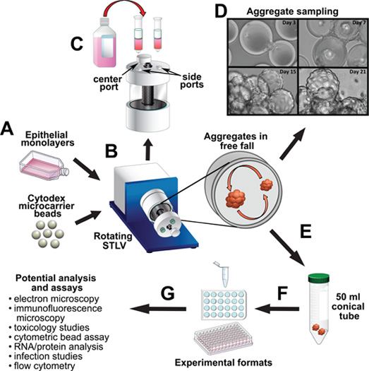
Figure 1. Schematic for culturing human epithelial cells in RWV and potential applications using RWV-derived aggregates. A) Epithelial cells are initially grown as monolayers in a tissue culture flask. Once confluent, cells are removed from flask and combined with microcarrier beads in the STLV bioreactor. Scale bar: 80 μm. B) The STLV rotates on a platform to create a constant free fall environment of low fluid shear, allowing the cells to attach and grow on beads thereby forming visible cellular aggregates. C) After 96 h, the media in the STLV must be changed to accommodate for cellular metabolism. The media is poured out of a side port, fresh media is replaced through an attached 10 mL syringe (plunger removed), and syringe plungers are replaced and used to extrude bubbles. D) After ~5 days (d) the aggregates begin to form. The aggregates are sampled every 7 days and imaged by light microscopy to monitor their developmental progression (20x magnification of 3-D human epithelial vaginal cells). E) Once complete aggregate formation and cellular differentiation has occurred, the aggregates are harvested from the bioreactor and transferred into a 50 mL conical tube. F) Aggregates can be seeded into a 1.5 mL tube or a multi-well plate format to carry out experimental analysis and assays. G) Outline of potential assays and analyses that can be conducted on the aggregates. This representative list of analyses is not meant to be an exhaustive catalog of all potential downstream applications.
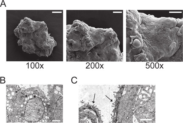
Figure 2. Examining morphological and structural characteristics of 3-D epithelial cell aggregates using electron microscopy. (A) Scanning electron microscopy (SEM) image of a 3-D human epithelial vaginal cell aggregate. Scale bars: 200 μm (100x), 100 μm (200x), 50 μm (500x). Transmission electron microscopy (TEM) image of a 3-D human epithelial vaginal cell aggregate with arrows pointing to (B) intracellular secretory vesicles and microvilli or (C) “cytoplasmic processes”. Scale bars: 2 μm (modified from Hjelm et al. 2010)2.
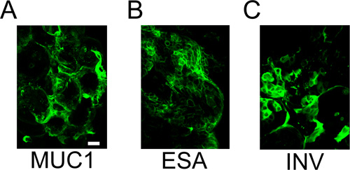
Figure 3. Confocal immunofluorescence microscopy used to identify junctional differentiation and protein markers in 3-D epithelial cell aggregates. Human 3-D vaginal cells harvested from bioreactor after 32 days, fixed, and stained with (A) mucin antibody MUC1, (B) antibody specific for epithelial cells, ESA, or (C) anti-Involucrin (INV) antibody that recognizes terminally differentiated epithelial cells. Aggregates were indirectly labeled with Alexa 488 secondary antibody (green). Scale bar: 60 μm (modified from Hjelm et al. 2010)2.
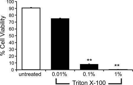
Figure 4. MTT assay is used to measure cellular viability. Human 3-D vaginal aggregates were seeded in a 24 well plate and treated with media alone (untreated) or 0.01%, 0.1% or 1.0% Triton X-100 detergent for 1 h at 37 °C. Following treatment, media was removed and the MTT assay was performed to measure cellular viability. ** represents p<0.001; one-tailed Student’s t-test comparing Triton X-100 treated cells to untreated control.
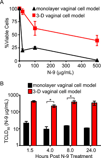
Figure 5. Utilizing 3-D epithelial cell aggregates to screen compound toxicity. (A) Nonoxynol-9 (N-9) dose-dependent viability curve 24 h post-treatment of 3-D human vaginal epithelial cell model (red line/bars) compared to same cell type grown as confluent monolayers (black line/bars). (B) N-9 TC50 levels of vaginal epithelial cell cultures in (A) at 1.5 h, 4 h, 8 h, and 24 h post treatment. * represents p<0.05; one-tailed Student’s t-test comparing monolayers to 3-D cells at each exposure time. (Modified from Hjelm et al. 2010)2.
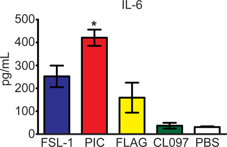
Figure 6. Analysis of cytokine production in 3-D epithelial cells in response to toll-like receptor (TLR) agonist stimulation. Three-D human vaginal cells were stimulated with FSL-1 (TLR2/6), PIC (TLR3), FLAG (TLR5), and CL097 (TLR7/8) for 24 h and secreted cytokines were measured by cytometric bead array. IL-6 is shown as a representative proinflammatory cytokine produced and measured. * represents p<0.05; one-tailed Student’s t-test comparing stimulated samples to PBS group. (Modified from Hjelm et al. 2010)2.
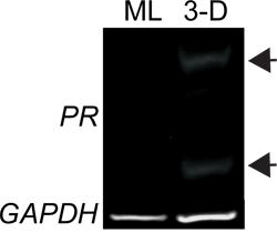
Figure 7. Monitoring gene expression in 3-D epithelial cells. Semi-quantitative RT-PCR analysis of progesterone receptor (PR) expression in monolayer (ML) or 3-D human vaginal epithelial cells. GAPDH is shown as a loading control. Amplification products shown are a result of transcript expression occurring after 30 PCR cycles. Arrow heads point to PR bands only occurring in the 3-D sample.
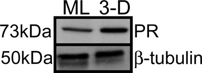
Figure 8. Protein expression analysis of 3-D epithelial cells. Western blot analysis of monolayer and 3-D human vaginal epithelial cell whole cell lysates (30μg) probed with an anti-progesterone receptor (PR) antibody. β-tubulin was used as a probe for loading control.
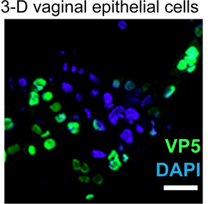
Figure 9. Three-D human epithelial cell model supports a productive viral infection. Confocal immunofluorescence microscopy image of 3-D human vaginal epithelial cell aggregate infected with HSV-2. Cells were infected at an MOI of 1.0 for two hours, washed, fixed 24 h post infection (4% PFA), and stained with a HSV-2 specific VP5 capsid antibody (green) and the nuclear stain DAPI (blue). Scale bar: 50 μm.
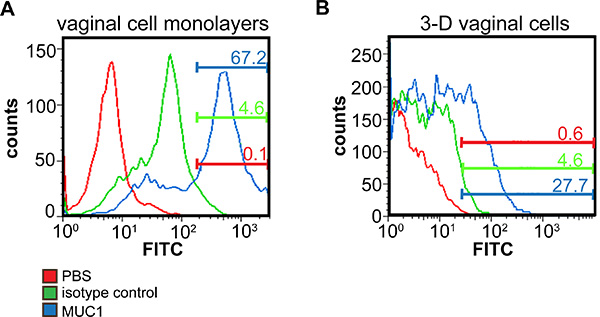
Figure 10. Individual cell analysis in 3-D human epithelial cell model by flow cytometry. (A) Monolayer or (B) 3-D human vaginal epithelial aggregates were dissociated with EDTA, labeled with a FITC conjugated MUC1 antibody (blue), an isotype control (green), or PBS (red), and FITC expression was quantified by flow cytometry. Numbers represent the percentage of cells that stained positive for mucin antibody that remained after gating out background fluorescence from the isotype control stained cells. Non-uniform peaks are a result of the multitude of glycosolation patterns and varying levels of MUC1 on the surface of these cells.
