Because many zebrafish models of neurological and non-neurological diseases are studied in the adult fish rather than the embryo/larvae, we developed a quantitative lateral line regenerative assay that can be applied to adult zebrafish disease models. The assay involved resolution at the 1) neuromast and 2) individual hair cell levels.
Due to the clinical importance of hearing and balance disorders in man, model organisms such as the zebrafish have been used to study lateral line development and regeneration. The zebrafish is particularly attractive for such studies because of its rapid development time and its high regenerative capacity. To date, zebrafish studies of lateral line regeneration have mainly utilized fish of the embryonic and larval stages because of the lower number of neuromasts at these stages. This has made quantitative analysis of lateral line regeneration/and or development easier in the earlier developmental stages. Because many zebrafish models of neurological and non-neurological diseases are studied in the adult fish and not in the embryo/larvae, we focused on developing a quantitative lateral line regenerative assay in adult zebrafish so that an assay was available that could be applied to current adult zebrafish disease models. Building on previous studies by Van Trump et al.17 that described procedures for ablation of hair cells in adult Mexican blind cave fish and zebrafish (Danio rerio), our assay was designed to allow quantitative comparison between control and experimental groups. This was accomplished by developing a regenerative neuromast standard curve based on the percent of neuromast reappearance over a 24 hr time period following gentamicin-induced necrosis of hair cells in a defined region of the lateral line. The assay was also designed to allow extension of the analysis to the individual hair cell level when a higher level of resolution is required.
The lateral line (LL) system is a mechanosensory organ found in both fish and amphibians that is responsible for hearing, balance, rheotaxis and mediating behaviors such as schooling and predator avoidance1-5. It is composed of clusters of hair cells surrounded by supporting cells, both of which are positioned in structures called neuromasts6. These neuromasts are typically organized into vertical lines (called stitches) along the longitudinal axis of the body and tail with some horizontal stitches observed in the head of the fish. In the adult, neuromasts are significantly greater in number within the stitches as compared to embryonic or larval fish6. Biomedical studies in zebrafish have focused on the effect of antibiotic treatment, noise-induced trauma, chronic infection, etc. on hair cells7,8 in an attempt to better understand their effects in humans.
Unlike most vertebrates, teleosts, such as the zebrafish (Danio rerio), have the ability to regenerate lost hair cells. Zebrafish are particularly useful because of their rapid development time and high regenerative capacity. To date, however; zebrafish studies on lateral line development and/or regeneration have mainly utilized the embryonic and larval stage fish due to the reduced number of lateral line neuromasts which allows for easier counting and analysis6,9,10.
However, as many zebrafish models of neurological and non-neurological diseases11-16 are studied in the adult fish and not the larvae, we focused on developing a lateral line regenerative assay in adult zebrafish using gentamicin (an aminoglycoside previously used in zebrafish larvae and more recently used with adult fish17) so that an assay was available that could be applied to current adult zebrafish disease models. While previously published procedures by Van Trump et al.17 established the conditions for hair cell ablation in the adult fish, they did not establish a standard curve for neuromast regeneration which is required for quantitative comparison between control and experimental groups such as when using transgenic zebrafish lines or pharmacologically-induced disease states in zebrafish18. We therefore followed the procedures of Van Trump et al.17 for hair cell ablation, but built on their work to establish a standard curve of neuromast regeneration to enable investigators to use our data when comparing control and experimental groups such as with adult zebrafish disease models. The assay was also designed to allow extension of the analysis to the individual hair cell when a higher level of resolution is required.
All procedures are performed following the guidelines described in “Principles of Laboratory Animal Care” (National Institutes of Health publication no. 85-23, revised 1985) and the approved Rosalind Franklin University Institutional Animal Care and Use Committee animal protocol 08-19.
1. Gentamicin-induction of Hair Cell Necrosis
2. Vital Staining of Hair Cells
3. Anesthetizing Fish and Fluorescent Counting of Neuromasts
4. Fluorescent Counting of Individual Hair Cells for Obtaining Higher Resolution of the Quantitative Analysis if the Neuromast Analysis is Not Statistically Significant
Optimization of the procedures for quantifying neuromast regeneration of the lateral line in adult zebrafish.
The neuromasts of larval zebrafish are readily quantifiable; however, the lateral line of the adult zebrafish has a much greater number of neuromasts per stitch making quantitative analyses more difficult6,17,19,20. As seen in Figure 1A, the head has a significantly higher number of neuromasts compared to either the mid-section or tail; with the tail region having the least number of neuromasts as shown in Figure 1D. Because the pattern of stitches in the head is complicated and significantly greater in the number of neuromasts, it did not lend itself as a region for quantitative analysis. In addition, regardless of the gentamicin concentration we tested, complete ablation of neuromasts throughout the head was rarely attainable; leaving spots of neuromasts observed after gentamicin treatment as previously reported by Van Trump et al.17 In contrast, the tail has too few neuromasts, and as such, we selected the mid-body region (Figure 1B) to quantitatively analyze neuromast regeneration in the adult. In this region, we identified four stitches just posterior to the lateral pectoral fin that were consistent in neuromast number among all adults [61.45 (n = 95)] (Figures 1B and 1E). Importantly, we were able to consistently and completely ablate the neuromasts of this region by a 24 hr 0.004% gentamicin treatment (as previously reported in Van Trump et al.17) allowing for a subsequent accurate determination of neuromast regeneration (compare Figures 1B and 1E and inset with Figure 2, 0 hr).
Figure 1. The fluorescent pattern of neuromast within stitches of the adult zebrafish is shown along the longitudinal axis. Panel A is the head region, Panel B is the Mid-body region with the four stitches used for quantitative analysis outlined with a box. Panel C is the Posterior-body region. Panel D is the caudal fin region. A higher magnification of the 4 stitches of the Mid-body region used for quantitative analysis is shown in Panel E. Magnification Power of 1X and 2X.
It has been reported that copper sulfate treatment is an effective chemical method to induce rapid necrosis of hair cells in embryos and larvae21. Here we tested copper sulfate treatment with the hope that it might shorten the time to induce neuromast ablation. Copper sulfate concentrations ranging from 5-50 mM for various exposure times up to 48 hr were utilized as was previously reported by Liang et al.21 It was found that copper sulfate was lethal at the higher concentrations and not effective at lower concentrations in adult fish (data not shown). Please click here to view a larger version of this figure.
Parameters used for fluorescent analysis of lateral line regeneration in adult zebrafish.
Regeneration was monitored after all neuromasts within the four mid-body stitches were ablated following 24 hr gentamicin-treatment (Figure 2, compare 0 hr with control) and positive regeneration was determined by the appearance of a minimum of three neuromasts within a stitch. (Figure 2, 0 hr). By 8 hpg, approximately one third of the fish had some sign of recovery (n = 34); although the intensity of neuromasts was faint in the regenerating stitches (Figure 2, 8 hr, faint stitches outlined by boxes). The number of neuromasts and their intensity continued to increase in a linear fashion until regeneration reached a plateau at 16 hpg (compare Figure 2, 16 hr with Figure 2, 8 hr). It was not until at least 24 hpg that all fish treated with gentamicin had fully recovered with both equal numbers and intensities of the neuromasts within the lateral line stitches as compared to controls (Figure 2, 24 hr). A time-line for neuromast regeneration following gentamicin withdrawal is shown in Figure 3 which shows the linear and plateau phases of the recovery curve. We note that in less than 5% of cases, the regenerating stitches did not appear as individual entities but instead appeared as a smear of fluorescence.
Figure 2. Fluorescent images of neuromasts. Control fish, 0 Hr fish (immediately following 24 hr of 0.004% gentamicin treatment), 8 Hr fish (8 hpg) with some faint staining of neuromasts within the 4 stitches used for quantitative analysis (only 30% of all fish showed this pattern of staining at 8 hpg; 70% showed no neuromast staining at this time point; the white boxes outline the faintly stained neuromasts that were seen in the 30% of fish that showed some degree of regeneration at 8 hpg), 16 Hr fish, and 24 Hr fish. Complete regeneration of neuromasts within the 4 stitches in regard to 1) the number of neuromasts and 2) intensity of staining of neuromasts was observed by 24 hpg. Please click here to view a larger version of this figure.
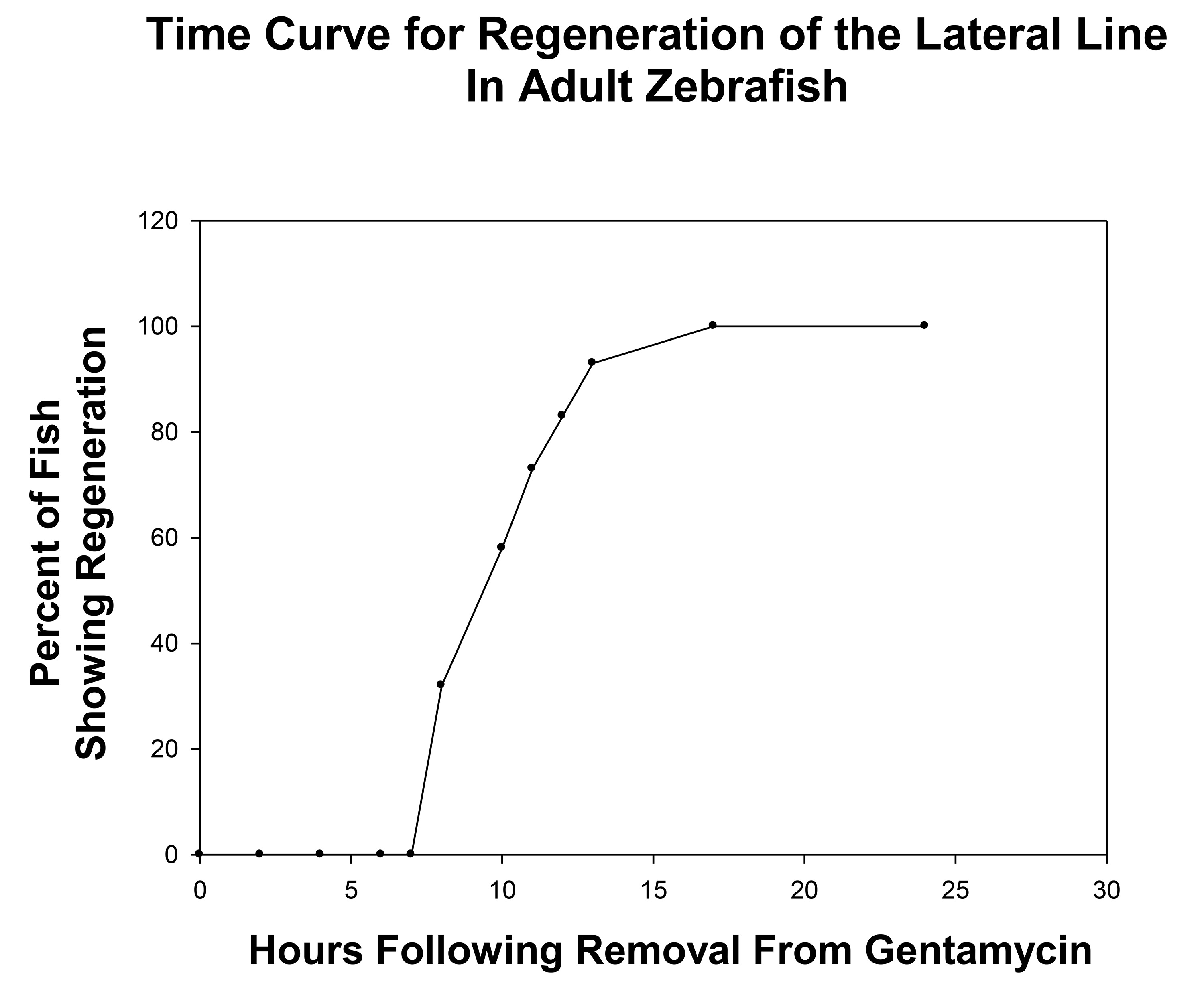
Figure 3. Graph showing the time course of neuromasts regeneration following withdrawal from 24 hr treatment of adult zebrafish with 0.004% gentamicin. As shown, recovery begins at the 8 hpg time point and reached a plateau at the 16 hpg time point. This defined the liner phase of neuromast regeneration between 8-16 hr and time points within this linear phase should be used to quantitatively compare control and experiment groups.
Neuromast toxicity is induced by prolonged exposure to fluorescent staining dyes.
In our estimation an ideal way to perform these experiments would be to stain the fish with the fluorescent dye prior to treatment, stain again at 0 hr then stain again at regeneration time points. However, we encountered a complication to these studies which is the fact that hair cell fluorescent staining dyes such as 4-Di-2-Asp, as can also be the case with other mitochondrial stains22 can have a toxic affect on hair cells23. This fact required us to use separate groups of fish since repeated staining of the same fish could not be employed. In all cases the experimental fish including controls were treated in parallel to eliminate experimental variability.
Confocal analysis at the level of individual hair cells.
If the results obtained from the neuromast analysis are not statistically significant between the control and experimental groups, one may extend these studies to the level of the individual hail cells to obtain a higher degree of resolution for quantitative comparisons. As indicated in Figure 4, neuromasts from a control group (a neuromast at 12 hr of regeneration is shown in this figure) can be viewed by confocal microscopy of skin preparations from the mid body region. At 8 hr, 10 hr, and 12 hr of regeneration time, we found that control groups (7 animals/group) had a range of 0-4 hair cells/neuromast. As expected for control groups, when quantitatively analyzed, no statistical difference between neuromasts was detected in terms of the number of hair cells per neuromast at the time points indicated above (P values ranged from 0.230-0.472). Such an approach may be taken between any control and experiment group when needed to extend or confirm the data obtained from the first phase of neuromast studies.
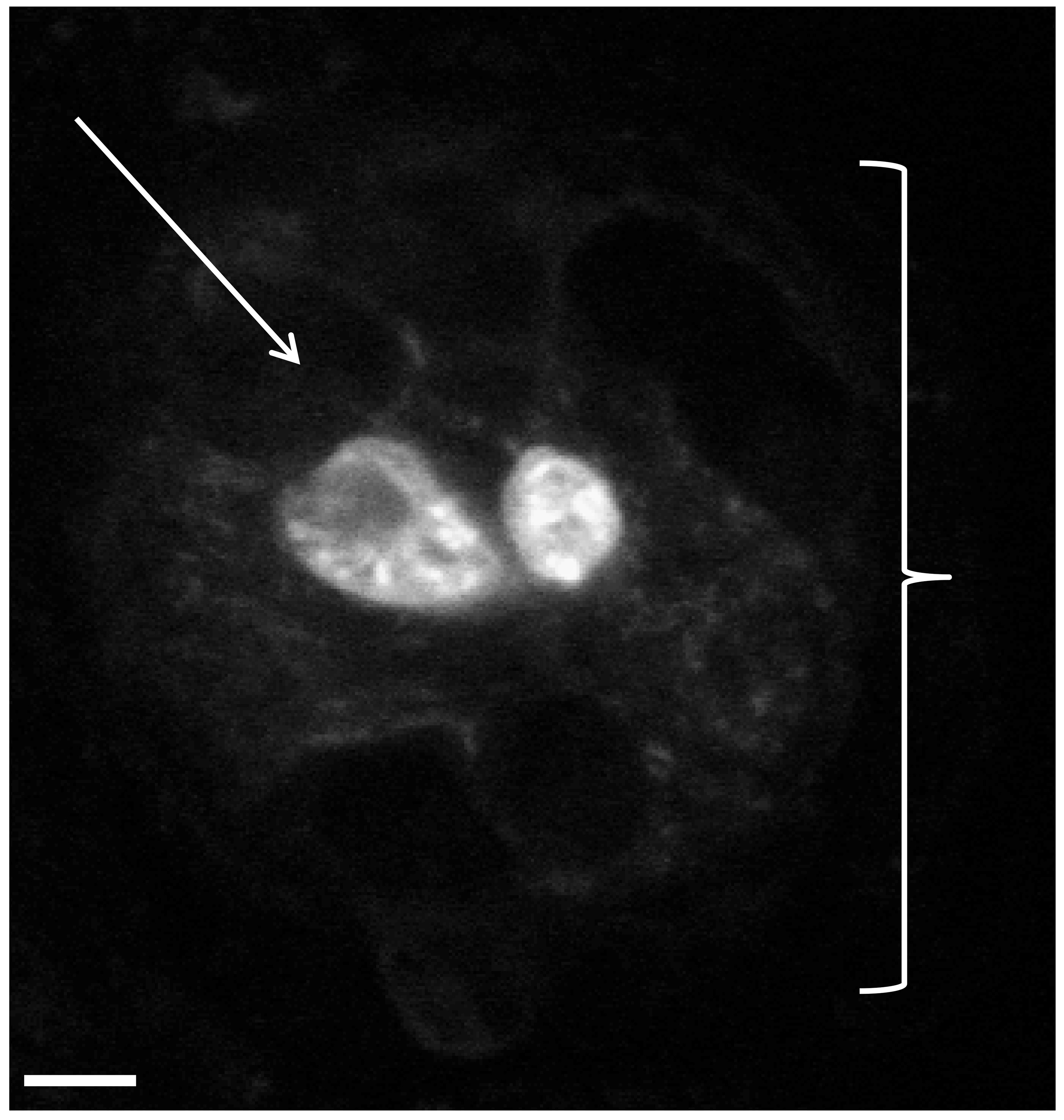
Figure 4. Analysis of hair cell/neuromast regeneration using skin preparations of the defined lateral line region described in protocol step 3.3. Fluorescent confocal image of hair cells within a neuromast obtained from a zebrafish skin preparation. Two vital dye stained hair cells are shown within a neuromast of a control fish (Figure 4). This image was obtained 12 hr post-removal of gentamicin (linear regeneration phase). The white bracket symbol ( ) indicates an individual neuromast while the white arrow indicates a supporting cell surrounding the hair cells. Supporting cells do not stain under these conditions and appear as black spaces. Magnification, 60X.
1 | 1. Gentamicin treatment [0.004% (4.32 mM)] of control and experimental fish for 24 hr at 28 °C using an incubator. |
2 | 2. Wash out of gentamicin to initiate regeneration of hair cells. Return fish to the 28 °C incubator for investigator selected time periods between 8-16 hr. |
2 | 3. Vital dye stain [0.08% 4-4-diethylaminostyryl-N-methylpyridinium iodide (4-Di-2-Asp)] control and experimental fish for 1 hr at room temperature and then wash out stain with fish water for fluorescent imaging. |
1 or 2 | 4. If necessary due to nonsignificant results from analysis of neuromasts, repeat Protocols 1-3 with a separate group of control and experimental fish, but then obtain a skin preparation for confocal analysis of individual hair cells. |
Table 1. A summary of the protocol outlined above.
Based on the extensive body of literature that has been established for analysis of lateral line (LL) regeneration in embryonic and larval zebrafish8,24,25, the goal of our study was to develop a quantitative assay for lateral line regeneration in zebrafish that could be applied to disease models that are best studied in the adult fish. We found that certain critical points are important when applying procedures developed for embryonic/larval fish to the adult fish. The most important of these points regarded: 1) the number of lateral line neuromasts along the longitudinal axis of the fish, 2) the duration of staining of neuromast hair cells, 3) the concentration and duration of treatment of the aminoglycoside, and 4) the timing of lateral line regeneration following aminoglycoside treatment. These points will be addressed in the following discussion.
In regard to the number of neuromasts along the lateral line of zebrafish, embryonic and larval zebrafish have the distinct advantage that the neuromasts have a simple pattern because of their lower number within a given stitch as compared to adult fish. This low number has allowed investigators to clearly identify each neuromast and assign a name to it6. In this way, regeneration studies can quantify the reappearance of a particular neuromast by name at any time following withdrawal from an aminoglycoside agent. The greater number of neuromasts per stitch in the adult introduces significant difficulty in accurate counting and quantification during the reappearance of regenerating neuromasts when compared to that of embryos or larvae. As shown, we assessed the pattern of lateral line neuromasts within stitches of the adult and determined that the mid-body (Figures. 1 and 2) provided the optimal region of stitches for analysis.
Staining of neuromasts with hair cells dyes such as 2-[4-(dimethylamino)styryl]-N-ethylpyridinium iodide (DASPEI) or 4-Di-2-Asp allows one to visualize neuromasts of the lateral line using fluorescent stereomicroscopy26,27 in larval and juvenile fish. These same stains are also effective in the adult fish and our quantitative analysis indicated that no significant difference in the number of neuromasts within the mid-body region was observed among normal control zebrafish (with an average of 61.45 neuromasts in normal control adult fish).
It has been reported that hair cell dyes such as 2-[4-(dimethylamino) styryl]-N-ethylpyridinium iodide (DASPEI) or 4-Di-2-Asp can themselves be toxic to neuromast cells24, and fish cannot be repeatedly stained with these fluorescent agents if one is to monitor aminoglycoside-induced regeneration. Repeated staining of the same fish introduces multiple toxicity events (by both the stain and the aminoglycoside) that makes the experiment un-interpretable23. Accordingly, all experiments in our study required parallel 4-Di-2-Asp staining of multiple sets of fish in order to show that neuromasts were 1) present in the nontreated gentamicin control condition, 2) fully ablated immediately following gentamicin exposure, and 3) regenerating at some hour post gentamicin-treatment following withdrawal and washing out of this aminoglycoside. In this way, all fish was stained only once with the neuromast dye.
It should be pointed out that while the procedures of Van Trump et al.17 establish the conditions for hair cell ablation in the adult fish, they do not establish a standard curve for neuromast regeneration which is required for quantitative comparison between control and experimental groups. We therefore followed the procedures of Van Trump et al.17 for hair cell ablation (gentamicin concentration of 0.004% using a 24 hr exposure time, see Figure 2 for hair cell ablation results at 24 hr) but extended their work to establish a standard curve of neuromast regeneration. This allows for comparative analysis of LL regeneration in the adult zebrafish using the four stitches of the mid body region that we established for our assay conditions (see Figures 1 and 2). In order to determine if a shorter period for hair cell ablation could be obtained, we also tested the effect of copper sulfate which has been effectively used in larval fish for periods as short as 2 hr. Our studies indicated that copper sulfate (5-50 mM for various exposure times up to 48 hr as was previously reported by Liang et al.21 for larvae) was not found to be effective in adult fish as an agent for hair cell ablation. This highlights the fact that conditions used for ablation of hair cells in embryos and larvae cannot always be directly transferred for use with the adult fish.
As pertaining to the lateral line regeneration in the adult, we found similar time frames for regeneration of neuromasts between that of embryo/larval and adult fish. As reported previously by others, zebrafish embryos and larvae show neuromast regeneration at 12-24 hr post aminoglycoside wash-out8. We observed the linear phase of neuromast regeneration appearing in the 8-12 time-frame (not seen as full stitches for the 8 hr time point as shown in Figure 2) with a plateau reached at 16 hpg. Complete control-like appearance of neuromasts within stitches was not observed until 24 hpg as reported for embryos and larvae. Complete control-like appearance denotes both the number and intensity of neuromasts within all four stitches of the mid-body region in adult zebrafish. Additionally, if the quantitative results obtained at the level of the neuromasts are not statistically significant, the investigator can extend their studies to the level of the individual hair cells within the neuromasts using confocal microscopy as described by our procedures.
The neuromasts/hair cell regeneration assay described in this article can be applied to disease states that are best manifested in the adult zebrafish rather than in the early larval/embryonic stages. A limitation of the assay concerns the affect of the experimental condition whether it be 1) the transgenic strain that mimics a particular disease state or 2) a pharmacologically-induced disease state] on stem cells within the lateral line system of adult zebrafish. In this regard, a particular disease state of the adult zebrafish may or may not affect stem cells of the hair cell lineage, and it is important to note that neuromast regeneration is completely dependent on these stem cell proliferation/differentiation processes8,24,25.
As an example of this limitation, we will describe experiments performed on an adult zebrafish model of Type I diabetes. This particular disease model was developed in adult zebrafish in order to study the long term secondary complication induced by hyperglycemia28. For a number of reasons described previously28,29, these studies can only be performed using adult zebrafish. Because peripheral nerves along with the specialized cellular structures they innervate are adversely affected in patients with diabetes, we wanted to determine if lateral line regeneration of neuromasts/hair cells was also impaired in diabetic zebrafish. Using the lateral line regeneration assay no statistically significant delay was detected in neuromast regeneration. To confirm this negative result, the experiments were repeated at the more refined level of the individual hair cell. Again, no statistically significant difference was observed in hair cell regeneration between control and diabetic groups. Therefore, the data were inconsistent with the supposition that hyperglycemia impedes neuromast/hair cell regeneration; possibly due to the resistance of stem cells of the hair cell lineage to hyperglycemic conditions. With this limitation in mind, the neuromasts/hair cell regeneration assay described in this article does provide a means to test whether any particular adult zebrafish disease model involves dysfunction in the hair cell regenerative process as monitored using the lateral line system. Positive results would imply stem cell involvement and further studies would therefore be warranted.
The authors have nothing to disclose.
Gentamicin sulfate solution (50mg/ml) | Sigma Aldrich | G1397 | |
2 Phenoxyethanol | Sigma Aldrich | P1126 | |
4-4-diethylaminostryryl-N-methylpyridinium iodide (4-Di-2-Asp) | Aldrich | D-3418 | 485 nm excitation λ and 603 nm emission λ |
in methanol | |||
6 well plates | Mid Sci | TP92006 | |
Petri Dishes | Fisher Scientific | 08-757-13 | |
Glass Bottom Microwell Dishes | Matek Corporation | P35G-1.5-14-C | |
Sodium Chloride | Sigma Aldrich | S3014 | |
Dissecting Microscope | Nikon | TMZ-1500 | Any dissecting microscope is fine. |
Camera for Imaging | Nikon | Q imaging | Any camera is suitable. |
Image J software | National Institutes of Health | NIH Image | |
NIS Elements | Nikon | Any imaging software is suitable. | |
Confocal microscope | Olympus | FV10i | Any high resolution fluorescent microscope is suitable |
Aquatic System | KG Aquatics ZFS Rack System. | Any aquatic system can be used |