We present a protocol for engineering the corona phase of near infrared fluorescent single walled carbon nanotubes (SWNTs) using amphiphilic polymers and DNA to develop sensors for molecular targets without known recognition elements.
Semiconducting single-wall carbon nanotubes (SWNTs) are a class of optically active nanomaterial that fluoresce in the near infrared, coinciding with the optical window where biological samples are most transparent. Here, we outline techniques to adsorb amphiphilic polymers and polynucleic acids onto the surface of SWNTs to engineer their corona phases and create novel molecular sensors for small molecules and proteins. These functionalized SWNT sensors are both biocompatible and stable. Polymers are adsorbed onto the nanotube surface either by direct sonication of SWNTs and polymer or by suspending SWNTs using a surfactant followed by dialysis with polymer. The fluorescence emission, stability, and response of these sensors to target analytes are confirmed using absorbance and near-infrared fluorescence spectroscopy. Furthermore, we demonstrate surface immobilization of the sensors onto glass slides to enable single-molecule fluorescence microscopy to characterize polymer adsorption and analyte binding kinetics.
Single-walled carbon nanotubes (SWNTs) are atomically thin layers of carbon atoms rolled into long, thin cylinders that exhibit unique electronic and optical properties.1 Such properties include a band-gap producing near infrared (nIR) fluorescence emission via exciton recombination that is highly sensitive to its local environment. The nIR emission of SWNTs falls within the near infrared window in which the penetration depth of light is maximal for biological tissue.2,3 Additionally, SWNTs exhibit several unique features atypical in contrast to organic fluorophores: SWNT exhibit a large Stokes shift, do not photobleach, and do not blink.4 Recently, exploiting these characteristics has led to the development of an assortment of novel molecular sensors with applications to biology.5,6 Unmodified, however, SWNTs are insoluble in water, and obtaining suspensions of individual SWNTs can be a challenge.7,8 Bundling and aggregation of SWNTs in solution can obfuscate their band-gap fluorescence,2 rendering them unsuitable for sensing applications.
Dispersing individual carbon nanotubes in aqueous solution requires modifying their surface to prevent hydrophobicity-driven aggregation.9 While covalent modification can render SWNTs water-soluble,10 as well as impart specific binding chemistry, defect sites in the SWNT lattice reduce or abate their fluorescence emission. Instead, SWNT functionalization can be accomplished by using surfactants, lipids, polymers and DNA9,11–13 that adsorb to the nanotube surface through hydrophobic and pi-pi stacking interactions. The resulting chemical environment surrounding surface-functionalized SWNTs is referred to as its corona phase. Perturbations to the corona phase can have a large impact on excitons traveling on the nanotube surface, causing modulations to SWNT fluorescence emission. It is this sensitive relationship between the corona phase and SWNT fluorescence that can be exploited to develop new molecular sensors by incorporating specific binding modalities onto the large surface area of SWNT. Perturbations to the SWNT corona phase upon binding analyte can lead to changes in the local dielectric environment, charge transfer, or introduce lattice defects, all of which can modulate the fluorescence emission of the SWNTs to serve as a signal transduction mechanism.14 This approach is used in the development of novel fluorescent sensors for the detection of many different classes of molecules including DNA,15,16 glucose17 and small molecules such as ATP,18 reactive oxygen species19 and nitric oxide.20,21 However, these approaches are limited in that they rely on the existence of a known binding modality for the target analyte.
Recently, a more generic approach to designing fluorescent sensors was developed using SWNTs non-covalently functionalized with amphiphilic heteropolymers, phospholipids, and polynucleic acids. These molecules adsorb to carbon nanotube surfaces to produce highly stable suspensions of individual SWNTs22–25 with unique corona phases that can specifically bind proteins26,27 or small molecules including the neurotransmitter dopamine.28–30 Engineering the corona phase to disperse SWNTs and specifically bind target analytes is referred to as corona phase molecular recognition (CoPhMoRe).28 The small size, low toxicity, high stability and unbleaching nIR fluorescence of CoPhMoRe SWNT sensors make them excellent candidates for in vivo sensing for extended time-resolved measurements.6 Recent work has shown their applications in plant tissues for optical detection of reactive nitrogen and oxygen species.31 A particularly exciting application for CoPhMoRe SWNT sensors is the potential for label free detection of neurotransmitters such as dopamine in vivo, where other techniques, such as electrochemical sensing or immunohistochemistry, suffer from a lack of spatial resolution, temporal resolution, and specificity.
Designing and discovering CoPhMoRe SWNT sensors has so far been restrained by the size and chemical diversity of the dispersant library, limiting the likelihood of finding a sensor for a particular target. To date, researchers have only scratched the surface of available conjugated, co-block, biological and biomimetic polymers that could serve as functionally active dispersants for SWNT sensors. Here, we present different methods for both dispersing SWNTs and characterizing their fluorescence for high throughput screening and for single SWNT sensor analysis. Specifically, we outline the procedure for coating SWNTs with polynucleic acid oligomers using direct sonication as well as how to functionalize SWNT with amphiphilic polymers through surfactant exchange by dialysis. We use (GT)15-DNA and polyethylene glycol functionalized with rhodamine isothiocyanate (RITC-PEG-RITC) as examples. We demonstrate the use of (GT)15-DNA SWNTs as a CoPhMoRe sensor for the detection of dopamine. Lastly, we outline procedures for performing single molecule sensor measurements, which can be used for characterization or single molecule sensing.
Caution: Please consult all relevant material safety data sheets (SDS) before use. Nanomaterials may have additional hazards compared to their bulk material counterpart. Use all appropriate safety practices including engineering controls (fume hood, noise enclosure) and personal protective equipment (safety glasses, goggles, lab coat, full length pants, closed-toe shoes).
1. Preparation of Buffer, Surfactant, and Polymer Solutions
2. Preparation of Single Walled Carbon Nanotube (SWNTs) Suspensions
3. Preparation of Surface Immobilized SWNT Sensors
4. Fluorescence Spectroscopy and Microscopy of SWNT Sensors
SWNTs were suspended in aqueous solution using both surfactants and amphiphilic polymers by direct sonication and by dialysis exchange. Figure 1 shows SWNTs, grown using the iron carbonyl catalyzed method (HiPCO), suspended using SC, RITC-PEF20-RITC, and (GT)15-DNA. The optical density of a SWNTs with SDS (or polymer) increases dramatically after sonication and decreases upon removal of aggregates and contaminants through purification by centrifugation (Figure 1). Measurements of absorbance at 632 nm quantified the concentration of suspended SWNT.28
The photophysical properties of the SWNT suspensions are characterized using absorbance and fluorescence spectroscopy. Figure 2 shows the absorbance and fluorescence emission spectra of SWNTs suspended using (GT)15-DNA and RITC-PEG20-RITC. The absorbance spectra are a superposition of the individual absorbance peaks for each distinct chirality of nanotube present in the sample. Similarly, each chirality exhibits its unique fluorescence emission peak. Differences in relative emission peak intensity are a result of differences in the population distribution of chiralities as well as differences in excitation efficiency using the 721 nm laser.
The fluorescence response of (GT)15DNA-SWNTs (where I0 is the initial SWNT fluorescence intensity and I is the SWNT intensity after dopamine addition) in the presence of different concentrations of dopamine is measured by monitoring the fluorescence of a sample using a spectrophotometer and InGaAs linear array (Figure 3). The total fluorescence of (GT)15-DNA-SWNTs increased with increasing dopamine concentration (Figure 3a). The fluorescence response is a function of the emission peak (Figure 3b), indicating that the response may be chirality specific. The fluorescence of the 1,044 nm and 1,078 nm peaks increase in intensity 2-fold as dopamine concentration approaches 2 µM. Figure 3e shows the intensity of the entire emission spectra of PEG-(GT)15 DNA-SWNT increase in response to the addition of dopamine.
Individual SWNTs coated using an alternative DNA sequence, C26-DNA (prepared using the same methods at (GT)15), tethered to the surface of a microscope slide are measured under laser illumination using an InGaAs camera and 100X oil immersion objective (Figure 4). Monitoring single emitters tethered to a surface can be used to verify the reversibility of sensor response by washing target molecules away using buffer solution. Total internal reflection fluorescence (TIRF) microscopy can also be used to image dye-conjugated DNA adsorbed on the SWNTs to quantify the number of DNA molecules attached to each tube through photobleaching experiments. Figure 4 shows 3 distinct bleaching events of Cy3-labeled DNA inferred from the quantized steps of the fluorescence intensity trace of a single emitter. These results indicate that three DNA molecules are attached to the SWNT.
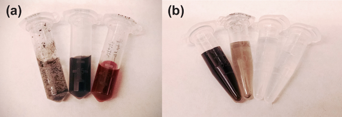
Figure 1: Polymer and Surfactant Suspended SWNTs. (a) Photograph of RITC-PEG20-RITC SWNTs suspended using 2% SC at various points of preparation. Left: SWNTs added directly to SC solution prior to sonication. Center: After 10 min of bath sonication followed by 10 min probe tip sonication at 90% amplitude followed by centrifugation. The optical density of the solution increases as SWNTs bundles are dispersed (~100 mg/L SWNT concentration). Right: After dialysis with RITC-PEG20-RITC polymer, the final concentration of RITC-PEG-RITC suspended SWNTs is ~20 mg/L. (b) SWNT suspension yield can vary depending on the polymer used to suspend the SWNT. The optical density of the solution provides a good estimate of solution-phase SWNT concentration. Pictured are different concentrations of (GT)15-DNA suspended tubes at different concentrations. From left to right: 100 mg/L, 10 mg/L, 1 mg/L, 0 mg/L. Please click here to view a larger version of this figure.
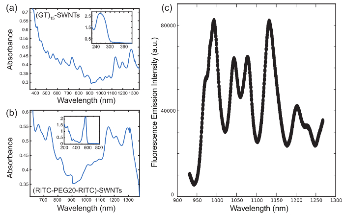
Figure 2: Absorption and fluorescence emission spectra of surfactant and polymer suspended SWNTs. (a) Representative absorbance spectra of SWNTs suspended using (GT)15-DNA by direct sonication. The concentration of SWNT is 10 mg/L. Inset: The UV region of the absorbance spectra shows the characteristic DNA absorbance peak at 260 nm. (b) Absorbance spectra of RITC-PEG20-RITC SWNTs after exchange of SC by dialysis. Inset: Absorbance of a 10x diluted sample of RITC-PEG-RITC SWNTs shows the characteristic absorbance of rhodamine. (c) Representative nIR emission spectra of SWNTs suspended using (GT)15-DNA by direct sonication (785 nm excitation). Please click here to view a larger version of this figure.
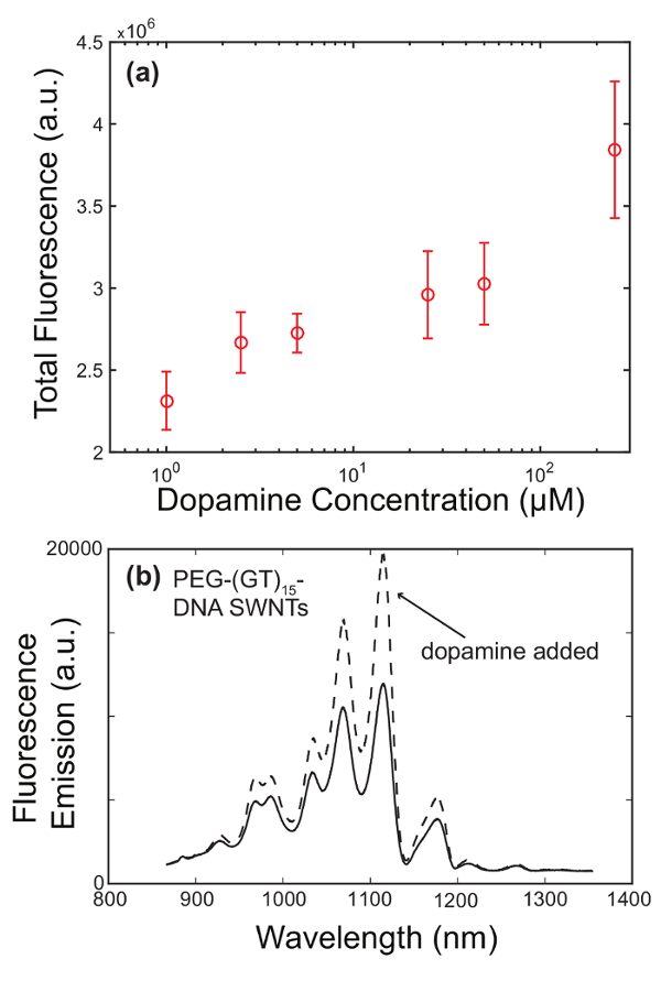
Figure 3: Fluorescence detection of dopamine using (GT)15-DNA wrapped SWNTs. (a) Fluorescence response of (GT)15-DNA wrapped SWNTs to the addition of dopamine. Samples of sensors at a concentration of 5 mg/L were excited using a 500 mW, 721 nm CW laser. The integrated fluorescence of sensor emission from 900-1,350 nm increases with added dopamine concentration 1 µM to 250 µM. (b) Fluorescence emission spectra of PEG-(GT)15-DNA wrapped SWNTs before and after addition of dopamine. The concentration of sensor is 10 mg/mL to which dopamine was added to a final concentration of 100 µM. The samples were excited using a 500 mW, 721 nm CW laser. The two highest intensity peaks approximately double in intensity after addition of dopamine. Please click here to view a larger version of this figure.
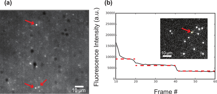
Figure 4: Fluorescence imaging of single surface-immobilized SWNTs. (a) Fluorescence emission of individual C26-DNA-SWNT (red arrows) immobilized onto a silica cover slip (#1.5) using an APTES silanization procedure and imaged using a 2D InGaAs sensor array, inverted microscope with a 100X oil immersion objective (plan apochromat, 1.4 NA), and a 500 mW, 721 nm CW laser. (b) Fluorescence bleaching experiment of C26-Cy3 DNA-SWNTs tethered to a surface using APTES. The DNA strands are 3' terminally labeled with Cy3 prior to tube suspension. Tracking the incremental step-wise photobleaching (red fitted trace) of individual sensors is used to determine the number of DNA molecules adsorbed onto the surface of the SWNTs. Images were acquired using an inverted microscope in TIRF mode with a 100X oil immersion objective (plan apochromat, 1.4 NA), and 561 nm laser excitation. Scale bar: 10 µm. Please click here to view a larger version of this figure.
SWNTs are readily suspended in aqueous solution via direct sonication with SDS or ssDNA, as indicated by an increase in optical density provided by the colloidal dispersion of the resulting SWNT-polymer hybrid. SDS and ssDNA disperses and solubilizes bundles of SWNTs by adsorbing onto the SWNT surface through hydrophobic or pi-pi interactions. Additionally, other polymers, such as genomic DNA, amphiphilic polymers, conjugated polymers and lipids, can be adsorbed onto the surface of SWNTs by dialysis of samples suspended using SC or SDS. Hydrophilic polymers, such as PEG, can be end-modified with hydrophobic "anchors" such as RITC or FITC to enable surface adsorption of the block copolymer. For polymers that are susceptible to shearing or degradation upon exposure to the high powers of probe-tip sonication, or for polymers with low binding affinities to SWNT, dialysis is the best method to produce a stable SWNT-polymer suspension. Following polymer encapsulation, centrifugation removes large SWNT bundles, amorphous carbon, residual metal catalyst, and other insoluble contaminants to leave a uniformly dispersed sample. Typical final concentrations of dispersed SWNTs after centrifugation range between 10-100 mg/L.
Sonication power and duration can be adjusted and optimized for the particular choice of dispersant. This is a critical step in the procedure for coating SWNTs because too little or weak sonication can result in poor dispersal, while too much or too powerful sonication can lead to poor fluorescence. Typically, lower powers are necessary to minimize damage when using polymers susceptible to shearing such as DNA. Longer sonication durations or higher intensities can lead to reduction in the length of SWNTs, where SWNTs below the ~100 nm exciton recombination length become non-fluorescent. The size of the sonicator probe tip should also match the sample volume to avoid splashing and foaming for optimal results (typically provided by manufacturer). Avoid touching the probe tip to the sides of the container and place the solution on an ice block to minimize heating of the solution. Once dispersed, solutions of SWNTs are stable at room temperature indefinitely.
Absorbance and emission spectra of solutions of dispersed generated SWNTs contain multiple peaks, indicating the presence of a mixture of dispersed SWNTs of different chiralities. Alternative methods of SWNT generation or purification methods can change the chirality distribution, leading to different peak excitation and emission fluorescence spectra. Additionally, different synthesis methods can yield samples of SWNTs with different chirality population distributions. For example: CoMoCAT (cobalt-molybdenum catalyst) grown SWNTs are rich in (6,5) chirality, while HiPCO (high pressure with iron carbonyl catalyst) grown SWNTs are rich in (7,6) chirality, leading to differences in the absorption and photoluminescence spectra.
Certain adsorbed polymers enable the specific detection of analytes by modulating the fluorescence emission of the SWNT by changing the local environment at the tube surface. This approach offers the distinct advantage over covalent attachment of binding moieties by not permanently disrupting the SWNT lattice, which can reduce fluorescence emission intensity.6,32–34 Additionally, the CoPhMoRe approach has the potential for developing antibody-free sensors for targets where there may not already exist a known binding moiety.28 Specifically, (GT)15-DNA has been shown to selectively enhance the fluorescence emission of SWNTs in the presence of dopamine, enabling its use as a dopamine sensor. Bulk fluorescence measurements show an increase of as much as 80% in peak emission for certain SWNT chiralities. Immobilizing (GT)15-DNA SWNTs on a glass slide enables fluorescence response measurements of individual (GT)15-DNA functionalized SWNT, showing that fluorescence can increase by over 3-fold for single SWNT sensors in the presence of dopamine, without any appreciable photobleaching, under continuous laser illumination. The interaction between dopamine and the SWNT sensor is reversible, as evident by the recovery of the original fluorescent signal after washing dopamine out of the microfluidic chamber with buffer. Additionally, single-molecule measurements can be a powerful characterization technique for quantifying polymer adsorption (Figure 4) or the kinetics of binding events. Also, ratiometric sensing can be achieved by chemically isolating a singular SWNT chirality with a unique excitation-emission peak, functionalizing it with (GT)15-DNA, and isolating a second SWNT chirality to be insensitive to dopamine. Monitoring both SWNT chiralities provides a steady control fluorescence channel that can be compared to the modulating fluorescence of the (GT)15-DNA SWNTs. Combining different polymers (e.g., polyethylene glycol and (GT)15-DNA) can add additional functionality such as modifying diffusivity or cellular uptake characteristics, properties that are critical when performing in vivo experiments.
Currently, a limitation of the CoPhMoRe approach to sensor development include polymer library development. Because binding moieties are not known a priori, developing a sensor for a particular target can be time intensive and require a large number of chemically varied polymers for constructing the sensor library for screening. Additionally, stability and compatibility of sensors in in vivo environments can vary from sensor to sensor. However, once a candidate sensor has been identified, further modification strategies can be employed to optimize properties as necessary for in vivo applications.
Herein, we have demonstrated a methodology for dispersing SWNTs in aqueous solutions applicable to a wide variety of dispersing agents. This approach can be used to create libraries of dispersed SWNTs for the discovery of novel new nIR sensors for small molecules and biological markers. Of particular interest are sensors for the detection of neurotransmitters, which could enable real time, spatially precise detection of these molecules in complex biological environments.
The authors have nothing to disclose.
This work was supported by Burroughs Wellcome Fund Career Award at the Scientific Interface (CASI), a Simons Foundation grant, and a Brain and Behavior Research foundation young investigator grant.
sodium chloride | Fisher Scientific | S271-1 | |
sodium dodecyl sulfate | Sigma Aldrich | L6026 | |
sodium cholate hydrate | Sigma Aldrich | C6445 | |
tris base (Trizma base) | Sigma Aldrich | 93362 | |
hydrochloric acid | Fisher Scientific | A144-212 | |
Amine-PEG-amine,NH2-PEG-NH2 | Nanocs Inc | PG2-AM-5k | |
rhodamine B isothiocyanate | Sigma Aldrich | 283924 | |
fluorescein isothiocyanate | Sigma Aldrich | F7250 | |
dichloromethane | Sigma Aldrich | 676853 | |
dimethylformamide | Sigma Aldrich | D4551 | |
N,N-diisopropylethylamine | Sigma Aldrich | D125806 | |
diethyl ether | Sigma Aldrich | 673811 | |
Tris(2-carboxyethyl)phosphine hydrochloride | Sigma Aldrich | C4706 | |
5’-thiol-modified DNA | Integrated DNA Technologies | ||
methoxypolyethylene glycol maleimide | Sigma Aldrich | 63187 | |
100k Da spin filters | Millipore | ||
HiPCO Super purified single walled carbon nanotubes | Integris | HiPco SuperPurified | |
phosphate buffered saline | Sigma Aldrich | P5493 | |
anti static gun | Milty | Milty Zerostat 3 | |
centrifuge | Eppendorf | 5415 D | |
ultra sonicator | Cole Parmer | CV18 | |
dialysis cassettes | Thermo scientific | Slide-A-Lyzer G2 87722 | |
BSA-biotin | Thermo scientific | 29130 | |
Neutravidin protein | Thermo scientific | 31000 | |
(3-Aminopropyl)triethoxysilane (APTES) | Sigma Aldrich | 440140 | |
inverted microscope | Zeiss | Axio Observer.Z1 | |
kinematic mirrors | ThorLabs | KM200-E03 | |
periscope | ThorLabs | RS99 | |
immersion oil | Zeiss | Immersol 518f | |
100X objective | Zeiss | Plan-apochromat 100X oil, 1.4NA, PH3, 420791-9911-000 | |
20X objective | Zeiss | N-Achroplan 0.45 NA, 420953-9901-000 | |
cover glass | Healthrow Scientific | HS159879H | |
dopamine hydrochloride | Sigma Aldrich | H8502 | |
infrared 2d array camera | Princeton Instruments | NIRvana | |
infrared 1d sensor array | Princeton Instruments | PyLoN IR | |
nIR spectrograph | Princeton Instruments | SCT-320 | |
planoconvex lens | ThorLabs | LA1384 | |
wellplates (glass bottom) | Corning | 4580 |