This protocol describes confocal microscopy detection of G protein-coupled receptor (GPCR) internalization in mammalian cells. It includes the basic cell culture, transfection, and confocal microscopy procedure and provides an efficient and easily interpretable method to detect the subcellular localization and internalization of fusion-expressed GPCR.
Confocal laser scanning microscopy (CLSM) is an optical imaging technique for high-contrast imaging. It is a powerful approach to visualize fluorescent fusion proteins, such as green fluorescent protein (GFP), to determine their expression, localization, and function. The subcellular localization of target proteins is important for identification, characterization, and functional analyses. Internalization is one of the predominant mechanisms controlling G protein-coupled receptor (GPCR) signaling to ensure the appropriate cellular responses to stimuli. Here, we describe an experimental method to detect the subcellular localization and internalization of GPCR in HEK293 cells with confocal microscopy. In addition, this experiment provides some details about cell culture and transfection. This protocol is compatible with a variety of widely available fluorescent markers and is applicable to the visualization of the subcellular localization of a majority of proteins, as well as of the internalization of GPCR. This technique should enable researchers to efficiently manipulate GPCR gene expression in mammalian cell lines and should facilitate studies on GPCR subcellular localization and internalization.
Cells possess cargo trafficking machinery to transport extracellular materials-such as ligands, microorganisms, nutrients, and transmembrane proteins-into the cell for information, energy, and other purposes1,2. It is critical for cellular homeostasis, tissue function, and overall cell survival. The cell-based expression of fluorescent fusion proteins is a powerful way to study the localization and internalization of transmembrane receptors, such as G protein-coupled receptors (GPCR), in signaling pathways3.
The expression of fusion proteins tagged with green fluorescent protein (GFP) from jellyfish Aequorea victoria, combined with direct fluorescence detection techniques, has gained wide acceptance in protein-targeting research4,5,6. GFP-based detection has the advantage of allowing the real-time imaging of living cells while avoiding fixation artifacts7. A series of versatile cloning vectors has been constructed to facilitate the expression of protein fusions to GFP in various cells8,9. The pEGFP-N1 vector is a widely used and commercialized plasmid vector encoding an enhanced green fluorescent protein (EGFP), which has been optimized from wild-type GFP for brighter fluorescence and higher expression in mammalian cells. To observe the fluorescent signal of EGFP expressed in mammalian cells, the fluorescence microscopy should be conducted with the excitation at 488 nm and the emission at 507 nm, preferably with confocal microscopy.
Monitoring the subcellular localization and ligand-mediated internalization of receptors is a common technique to investigate the signal transduction and function of GPCRs10,11. In most cases, internalization leads to desensitization of the GPCRs (the attenuation of the stimulus response in spite of the presence of agonists)12,13. The internalized receptors can be incorporated into the lysosome and degraded, or they can be recycled back to the cell surface, depending on the nature of the receptors and the cell lines used in the experiments.
The gonadotropin-releasing hormone receptors (GnRHRs) belong to the GPCR family and have a typical GPCR protein structure, with an extracellular amino terminal, an intracellular -COOH terminal, and seven transmembrane (TM) domains14. The transfection of recombinant plasmid SjGnRHR-EGFP (with the GnRHR gene from Sepiella japonica) and the confocal microscopy detection of EGFP-tagged SjGnRHR (expressed in HEK293 cells) was reported previously, and GFP fluorescence was established as a reporter for transmembrane protein localization assays15. Now, the internalization of SjGnRHR, activated by S. japonica gonadotropin-releasing hormone (SjGnRH), was demonstrated in time- and dose-dependent manners by confocal microscopy.
1. Cell Preparation
2. Cell Seeding
3. Cell Transfection
4. Cell Culture on Cover Slips
5. Confocal Microscopy Detection
Figure 1 shows an example of a confocal microscope system. Figure 2 presents the expression of pEGFP-N1 in HEK293 cells. The GFP signal was detected in the cytoplasm and nucleus. Figure 3 shows the subcellular localization of GnRHR from S. japonica in HEK293 cells, consistent with our previously published results15. The fusion protein with the EGFP tag at the C-terminus of SjGnRHR (SjGnRHR-EGFP) appeared green in this experiment when under CLSM. The nucleus stained with DAPI was revealed in blue. DiI staining highlighted the cell membrane in red. The yellow signal on the cell surface in the merged image indicated the coincidence of red and green, demonstrating the plasma membrane localization of SjGnRHR.
Figures 4 and 5 display the results of the SjGnRHR internalization assays in HEK293 cells. To visualize the internalization of SjGnRHR, cells were treated with gonadotropin-releasing hormone (SjGnRH)15 at different concentrations for 30 min (Figure 4), or they were treated with a single concentration (10−6 M) for different durations(Figure 5). In the absence of SjGnRH, the control (CTL) receptor fusion had plasma membrane localization. As shown in Figure 4, at different ligand concentrations, the internalization of the SjGnRHR proceeded in a dose-dependent manner, and the receptor was completely internalized from the cell surface with the 10−6 M ligand. As shown in Figure 5, the results of the time course analysis of receptor internalization with 10−6 M ligand demonstrated that internalized SjGnRHR was detectable 10 min after agonist stimulation, and extreme internalization occurred by 60 min. Observation with confocal microscopy from these two figures revealed that the fluorescent SjGnRHR-EGFP fusion protein was primarily localized in the plasma membrane and was dramatically and rapidly internalized into the cell in response to the SjGnRH peptide in HEK293 cells.
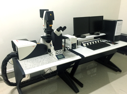
Figure 1. An Example of a Confocal Microscope System. Please click here to view a larger version of this figure.
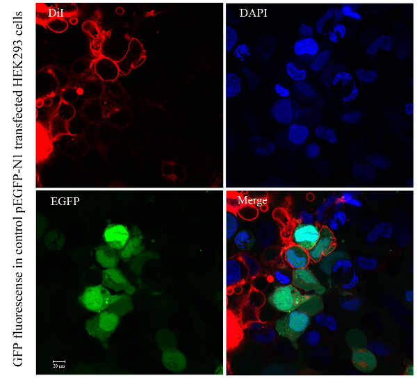
Figure 2. Confocal Microscopy of GFP Protein in Control pEGFP-N1-transfected HEK293 Cells. Please click here to view a larger version of this figure.
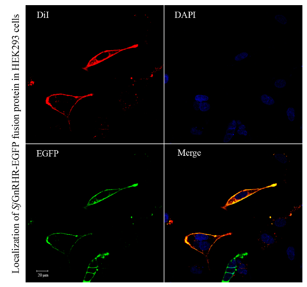
Figure 3. Localization of SjGnRHR-EGFP Fusion Protein in HEK293 Cells by Confocal Microscopy. The nucleus stained with DAPI is shown in blue. DiI staining, shown in red, highlights the cell membrane. SjGnRHR-EGFP fusion protein is shown in green. In the merged image, yellow indicates the coincidence of the red and green signals. All images represent at least three independent experiments. Please click here to view a larger version of this figure.
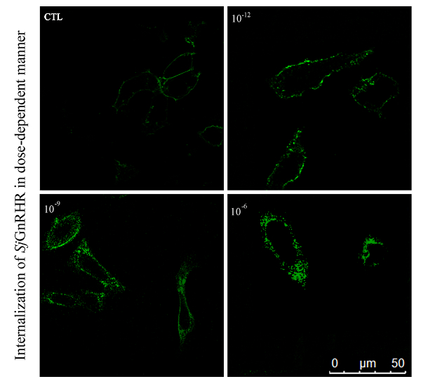
Figure 4. Internalization of SjGnRHR-EGFP Fusion Protein in HEK293 Cells in a Time Course by Confocal Microscopy. HEK293 cells transfected with SjGnRHR-EGFP plasmid were activated by treatment with different concentrations of GnRH for 30 min. The control case was without GnRH stimulation (CTL). All images represent at least three independent experiments. Please click here to view a larger version of this figure.
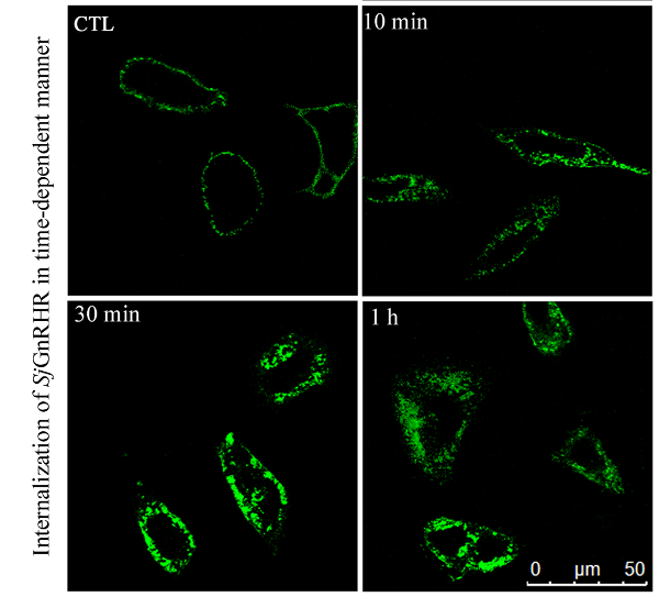
Figure 5. Internalization of SjGnRHR-EGFP Fusion Protein in HEK293 Cells in a Dose Dependent Manner by Confocal Microscopy. HEK293 cells transfected with SjGnRHR-EGFP plasmid were activated by treatment with 10−6 M ligand for different durations. The control case was without GnRH stimulation (CTL). All images represent at least three independent experiments. Please click here to view a larger version of this figure.
The protocol presented here provides an efficient and easily interpretable method to detect the subcellular localization and internalization of expressed GPCR fusion protein in HEK293 cells. The technique can be easily adapted for many different genes and cell types, such as for the cellular localization and internalization of the corazonin receptor from Bombyx mori in HEK293 and BmN cells16.
The successful application of this powerful assay relies on a few key factors. Foremost is the good health of the cells, which is essential for efficient transfection and data quality. The transfection incubation time, the interval between transfection and other experiments, and the passage of transfected cells to cover slips before imaging, are all steps where damage to the cultured cells is possible. Therefore, maintaining the good health of the cells is an important prerequisite for all steps in this experiment. Moreover, avoiding tightly compressed cells in dense cultures is vital in order to acquire high-quality images with confocal microscopy. To minimize the influence of the fluorescent tag on the localization of the GPCRs, we generated receptors with EGFP fused to the intracellular C-terminus. The successful and efficient transfection of the gene into the cells is also important. Transfection efficiency may vary greatly in different cell lines. Therefore, the optimization of certain steps in the protocol may be necessary, depending on the particular cell type used. For example, in transfection method 1, the incubation duration of transfected cells in DMEM without FBS could be optimized according to the cell condition. In addition to the dosage of the DNA plasmid, the dosage of the liposome reagent and reduced serum media should be adjusted according to specific cell lines. Otherwise, the electrotransfection technique could be conducted instead of liposome reagent mediated transfection for acceptable transfection efficiency in a specific cell line. However, additional hardware is required, and cells in suspension and in a small volume are difficult to transfect by electrotransfection. The true strength of the current assay, however, lies in the comparison between changes in the same cells under identical culture conditions after acute manipulations, such as the internalization of SjGnRHR in response to the SjGnRH ligand.
Internalization is one of the predominant mechanisms controlling GPCR signaling to ensure the appropriate cellular responses to stimuli11. We can easily observe changes in internalization through imaging with confocal microscopy. The internalization of SjGnRHR in a dose- and time-dependent manner is clearly revealed in Figure 4.
The protocol we have described can be easily adapted to the subcellular tracing of other eukaryotic proteins, such as the nuclear estrogen receptor17. However, the optimization of certain steps in the protocol will be necessary, depending on the particular cell types used, which will require further exploration and practice. Nevertheless, considering that the subcellular location of proteins has a great influence on the identification of genes, protein modifications, and signaling pathways, this protocol, based on the published research, can provide reliable data and information.
The authors have nothing to disclose.
The authors of this paper would like to thank Prof. Jiayan Xie for technical assistance and equipment usage. This work was financially supported by the National Natural Science Foundation of China (41406137).
HEK293 cell line | |||
DMEM/High glucose | Hyclone | SH30022.01 | High glucose 4.0 mM L-glutamine |
Foetal Bovine Serum (FBS) | PuFei | 1101-100 | |
Phosphate Buffered Saline (PBS) | Genom | GNM10010 | |
Penicillin-Streptomycin | Genom | GNM15140 | |
0.25% Trypsin & 0.02% EDTA | Genom | GNM25200 | |
Opti-MEM (1X) | GIBCO, by Life Technologies | 31985-062 | reduced serum media |
Lipofectamine 2000 Transfection Reagent | Invitrogen | 11668027 | Reagent 1 |
X-tremeGENE HP DNA Transfection Reagent | Roche | 6366236001 | Reagent 2 |
DAPI Staining Solution | Beyotime | C1006 | |
DiI | Beyotime | C1036 | |
Antifade Mounting Medium | Beyotime | P0126 | |
Paraformaldehyde | Sinopharm Chemical Reagent Co.(SCRC) | 80096618 | |
100 mm cell culture | CORNING | 430167 | |
6-well plate | ExCell Bio | CS016-0092 | |
12-well plate | ExCell Bio | CS016-0093 | |
Cover Glass | NEST | 801007 | |
Microscope Slides | Citoglas | 10127105P-G | |
Thermo Scientific Forma CO2 Incubator | Thermo | 3111 | |
TCS SP5II laser scanning confocal microscope | Leica | 5100001311 |