Summary

Schnelle und spezifische Immunomagnetic Isolierung der Maus primären Oligodendrozyten

Published: May 21, 2018
doi:

Summary

Wir beschreiben die Immunomagnetic Isolierung der primäre Maustaste Oligodendrozyten, ermöglicht die schnelle und spezielle Isolierung der Zellen für in-vitro- Kultur.

Abstract

Die effiziente und robuste Isolierung und Kultur der primären Oligodendrozyten (OLs) ist ein wertvolles Werkzeug für die in-vitro- Studie über die Entwicklung der oligodendrogliazellen sowie die Biologie der demyelinisierenden Erkrankungen wie der multiplen Sklerose und Pelizaeus-Merzbacher-ähnliche Erkrankung (PMLD). Hier präsentieren wir Ihnen eine einfache und effiziente Auswahlmethode für die Immunomagnetic Isolierung der Stufe drei O4+ Preoligodendrocytes Zellen von Neugeborenen Mäusen Welpen. Da unreif OL bilden mehr als 80 % der Nagetier-Gehirn weißen Substanz bei postnatalen 7.Tag (P7) dieser Isolationsmethode sorgt nicht nur für zelluläre Hochzinsanleihen, sondern auch die spezielle Isolierung des OLs bereits verpflichtet, die oligodendroglial Linie, Verringerung der Möglichkeit der Isolierung von kontaminierenden wie Astrozyten und andere Zellen aus dem Gehirn der Maus. Diese Methode ist eine Modifikation der zuvor berichteten Techniken und bietet Oligodendrozyt Vorbereitung Reinheit über 80 % in etwa 4 h.

Introduction

Oligodendrozyten (OLs) sind die Myelinating Zellen des zentralen Nervensystems (ZNS)1. Die Isolation und die Kultur der primären Oligodendrozyten in einem streng regulierten Umfeld ist ein wertvolles Werkzeug für die in-vitro- Studie über die Entwicklung der oligodendrogliazellen sowie die Biologie der demyelinisierenden Erkrankungen wie der multiplen Sklerose2 . Dies erfordert eine effiziente und robuste Oligodendrozyt Isolation und Kultur Methode3. In dieser Studie nutzten wir den Ausdruck einer unverwechselbaren Oligodendrozyt Zelle Oberfläche Markierung eine modifizierte Isolierung Technik zu implementieren, die schnellen und spezifischen ist.

Vier verschiedene Stadien der Reifung Oligodendrozyt identifiziert worden, jeweils geprägt durch den Ausdruck des unverwechselbaren Zelle oberflächenmarker für jede Entwicklungsstufe (Abbildung 1). Diese Zelle oberflächenmarker durch spezifische Antikörper4,5erkannt werden und können verwendet werden, um die OLs in bestimmten Phasen zu isolieren. In der ersten Stufe haben Oligodendrozyt Vorläuferzellen (OPCs) die Fähigkeit, sich vermehren, migrieren und speziell express Platelet-derived Wachstumsfaktor-Rezeptor (PDGF-Rα)6, Gangliosid A2B5, Proteoglykan NG27,8 , Polysialic Säure-neuronale Zelle Adhäsion Molekül9 und 7 (FABP7)-Fett-Säure-bindendes Protein10. OPCs haben bipolare Morphologie mit wenigen kurzen Prozesse ausgehend von den entgegengesetzten Polen der Zellkörper charakteristisch von neuronalen Vorläufer Zellen11ist.

Figure 1
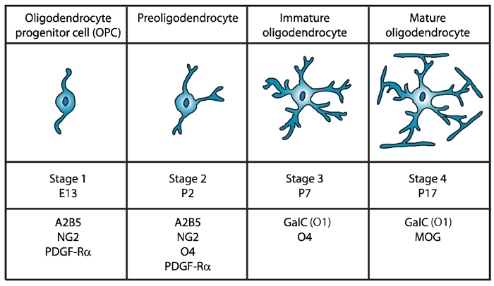
Abbildung 1: Ausdruck der Zelle oberflächenmarker während der Maus Oligodendrozyt Entwicklung. OLs Zelle oberflächenmarker wie z. B. A2B5, GalC (O1) NG2, O4 und PDGF-Rα können verwendet werden, um speziell Oligodendrozyten in bestimmten Entwicklungsstadium isolieren mit spezifischen Antikörpern.   Klicken Sie bitte hier, um eine größere Version dieser Figur.

In der zweiten Stufe OPCs geben Anlass zu Preoligodendrocytes und an der Zellmembran express nicht nur OPC Marker, sondern auch die Sulfatide (sulfatierten Galactolipid) von O4 Antikörper12,13, und das GPR17 Protein14anerkannt die weiterhin erst im Stadium der Unreife Oligodendrozyt (OL). Zu diesem Zeitpunkt verlängern Preoligodendrocytes multipolaren kurze Prozesse. Preoligodendrocytes sind die Hauptphase OL postnatale Tag 2 (P2) in der zerebralen weißen Substanz von Ratte und Maus mit einer kleineren Bevölkerung von unreifen OLs15.

Während der dritten Phase weiterhin die unreifen OLs express O4, Ausdruck von A2B5 und NG2 Markern zu verlieren und beginnen Galactocerebroside C16zum Ausdruck bringen. In diesem Stadium OLs oligodendroglial Abstammung verpflichtet und werden nach dem mitotischen Zellen mit langen verzweigten Ästen17,18. Unreife OL bilden mehr als 80 % der Nager weißen Substanz auf P7 und zu diesem Zeitpunkt die ersten Zellen der MBP+ 15,19,20,21eingehalten werden. Daher könnte Isolierung des OLs auf P7 zellulären Hochzinsanleihen sicherstellen.

In der vierten und letzten Ausbaustufe OL express Reife OLs Myelinating Proteine (Myelin basic Protein (MBP), Proteolipid-Protein (PLP), Myelin verbundenen Glykoprotein (MAG) und Myelin Oligodendrozyt Glykoprotein (MOG)22,23 ,24,25,26. In diesem Stadium Reifen OLs Membranen dieser Form kompakt Messgutes Hüllen um die Axone zu erweitern und sind in der Lage zu myelinisieren. Dies deckt sich mit der Beobachtung, dass im Gehirn der Ratte und Maus MBP+ Zellen immer reichlich bei P1419,20,21geworden.

Seit der ersten Isolierung von Oligodendrozyt von Fewster und Kollegen in 196727wurden mehrere Methoden zur Isolierung des OLs von Nagetier CNS einschließlich Immunopanning28,29,30umgesetzt, Fluoreszenz-aktivierte Zellsortierung (FACS) Nutzung Zelle Oberfläche-spezifische Antigene28,31, Differentielle Zentrifugation gradient32,33,34,35 und eine schütteln Methode basiert auf differenzielle Einhaltung von verschiedenen CNS Glia36,37. Bestehende Kultur Verfahren haben jedoch Einschränkungen, insbesondere in Bezug auf Reinheit, Ertrag und Zeitaufwand Verfahren38. Daher sind effizientere Isolationsmethoden für Oligodendrozyten erforderlich.

In diesem Papier stellen wir eine einfache und effiziente Auswahlmethode für die Immunomagnetic Isolierung der Stufe drei O4+ Preoligodendrocytes Zellen von Neugeborenen Mäusen Welpen. Diese Methode ist eine Abwandlung der Techniken von Emery Et Al. berichtet 39 und Dincman Et al. 40 und bietet eine Oligodendrozyt Vorbereitung Reinheit über 80 % in etwa 4 h.

Protocol

Die Mäuse, die in dieser Studie verwendet wurden nach den Richtlinien der SUNY Downstate Medical Center Division von Labor Tier Ressourcen (DLAR)-Protokoll-Nummer 15-10492 betreut. Hinweis: Primären Oligodendrozyten wurden isoliert von Neugeborenen (P5-P7 Wildtyp C57Bl/6N) Mäuse. In diesem Stadium bilden unreifen OLs mehr als 80 % der Nager weißen Substanz zellulären ertragreich zu gewährleisten. Alle Puffer und Reagenz Kompositionen stehen am Ende der Tabelle der Materialien</st…

Representative Results

Der Zweck dieser Studie war es, eine verbesserte Isolationsmethode für O4 etablieren+ primäre Maustaste Oligodendrozyten erfordern die geringst mögliche Manipulation von den Zielzellen. Der gesamte Vorgang von Euthanasie der Welpen auf Beschichtung der Zellen im Deckgläsern dauert ca. 4 Stunden und Darstellung von Daten, die hier gezeigten drei unabhängige Experimenten. Nach Gewebe Dissoziation wurden durchschnittlich 4,3 ± 0,46 x 10⁷ Zellen isoliert für jedes…

Discussion

In dieser Mitteilung präsentieren wir eine Methode für die effiziente Isolierung hochgereinigte unreifen Maus Oligodendrozyt Kulturen. Im Vergleich zu früher veröffentlichten Protokolle39,40, ergab diese Methode eine höhere Reinheit mit einem viel niedrigeren Niveau der Astroglia-positiven Astrozyten und einen sehr geringen Prozentsatz von anderen Zellen nicht gekennzeichnet. Es ist wichtig, darauf hinzuweisen, dass diese unreifen OLs bereits verpflichtet, d…

Disclosures

The authors have nothing to disclose.

Acknowledgements

Diese Studie wurde unterstützt durch Zuschüsse aus dem National Multiple Sclerosis Society (RG4591A1/2) und die National Institutes of Health (R03NS06740402). Die Autoren danken Dr. Ivan Hernandez und seinem Labor-Mitglieder für die Laborfläche, Ausrüstung und Beratung.

Materials

10ml serological pipets Fisher Scientific 13-676-10J
10ml syringe Luer-Loc tip BD, Becton Dickinson 309604
15ml conical tubes Falcon 352097
24-well tissue culture plates Falcon 353935
40µm cell strainer Fisher Scientific 22368547
50ml conical tubes Falcon 352098
5ml serological pipets Fisher Scientific 13-676-10H
60mm tissue culture plates Falcon 353002
70µm cell strainer Fisher Scientific 22363548
Alexa Fluor 488 goat anti-mouse IgG (H+L) secondary antibody Invitrogen A11001
Alexa Fluor 488 goat anti-rabbit IgM (H+L) secondary antibody Invitrogen A21042
Alexa Fluor 488 goat anti-rabbit IgM (H+L) secondary antibody Invitrogen A11008
Alexa Fluor 594 goat anti-chicken IgG (H+L) secondary antibody Invitrogen A11042
Anti-O4 beads- Anti-O4MicroBeads Miltenyi Biotec 130-094-543
Apo-Transferrin human Sigma T1147
Autofil complete bottle top filter assembly, 0.22um filter, 250ml USA Scientific 6032-1101
Autofil complete bottle top filter assembly, 0.22um filter, 250ml USA Scientific 6032-1102
B27 Supplement Invitrogen 17504-044
Boric acid Sigma B7660
Bovine Growth Serum (BGS) GE Healthcare Life Sciences SH30541.03
BSA Fisher Scientific BP-1600-100
CNTF Peprotech 450-50
d-Biotin Sigma B4639
Desoxyribonuclease I (DNAse I) Worthington LS002007
EDTA Fisher Scientific S311
Epifluorescence microscope with an Olympus DP70 camera Olympus Bx51
Feather disposable scalpels Andwin Scientific EF7281C
Forskolin Sigma F6886
German glass coverslips, #1 thickness, 12mm diameter round NeuVitro GG-12-oz
GFAP antibody Aves GFAP
Glucose Fisher Scientific D16-1
GlutaMAX Invitrogen 35050-61
Insulin Invitrogen 12585-014
Magnetic separator stand – MACS multistand Miltenyi Biotec 130-042-303
Magnetic separator-MiniMACS separator Miltenyi Biotec 130-042-302
Millex PES 0.22µm filter unit Millipore SLG033RS
Mounting media- Prolong Gold with DAPI Thermo Fisher P36930
N-acetyl-cysteine (NAC) Sigma A8199
Natural mouse laminin Invitrogen 23017-015
Neurobasal Medium A Invitrogen 10888-022
Neurotrophin-3 (NT-3) Peprotech 450-03
NG2 antibody Millipore AB5320
Papain Worthington LS003126
PBS without Ca2+ and Mg2+ Sigma D5652
PDGF Peprotech 100-13A
Petri dishes Falcon 351029
Poly-D-Lysine Sigma P6407
Primocin Invivogen ant-pm-2
Progesterone Sigma P8783
Putrescine Sigma P5780
Selection column-LS columns Miltenyi Biotec 130-042-401
Sodium Selenite Sigma S5261
Trace elements B Corning 25-000-CI
Triiodothyronine (T3) Sigma T6397
Triton-X Sigma T8787
Trypan Blue Solution Corning 25-900-CI
Tween 20 Sigma P1379
B27NBMA 487.75 mL Neurobasal Medium A; 10 mL B27 Supplement; 1 mL Primocin; 1.25 mL Glutamax; Filter sterilize and store at 4 °C until use.
B27NBMA + 10% BGS 27 mL B27NBMA; 3 mL Bovine growth serum
CNTF solution stock (10 µg/ml; 1000X) Order from Peprotech (450-50). Make up at 0.1 to 1 mg/ml according to Manufacturer’s instruction (may vary from lot to lot) in buffer (e.g. DPBS + 0.2% BSA). Store at -80 °C.
Working solution (10 µg/ml, 1000X)
1. Make on 0.2% BSA (Fisher scientific BP-1600-100) in DPBS solution and filter sterilize.
2. Dilute master stock aliquot to 10µg/ml in sterile, chilled 0.2% BSA/DPBS.
3. Aliquot (20µl/tube) and snap freeze in liquid nitrogen.
4. Store aliquots at -80 °C.
d-Biotin stock solution (50 µg/ml; 5000X) Resuspend d-Biotin (Sigma-B4639) in double-distilled H2O at 50 µg/ml (e.g. 2.5 mg in 50 ml of ddH2O). Resuspension might take fair amount of agitation/vortexing, or mild warming briefly at 37°C. If the d-Biotin still will not solubilize, it is fine to make up a less concentrated (e.g. 10µg/ml), and to add a higher volume to the B27NBMA (1/1000), instead of 1/5000). Store at 4°C.
DNase I stock solution 1. Dissolve at 12,500 U Deoxyribonuclease I / ml in HBSS chilled on ice.
2. Filter sterilize on ice
3. Aliquot at 200 µl and freeze overnight at -20°C.
4. Store aliquots at -20 to -30°C.
Dulbecco’s Phosphate Buffered Saline (w/o Ca2+ and Mg2+) Dissolve pouch in 1 Liter of water to yield 1 liter of medium at 9.6 grams of powder per liter of medium. Store at 2-8 °C.
Forskolin stock solution (4.2 mg/ml; 1000X) Add 1 ml of sterile DMSO to 50 mg Forskolin in bottle (Sigma-F6886) and pipette until resuspended. Transfer to a 15 ml centrifuge tube and add 11 ml of sterile DMSO to bring to 4.2 mg/ml. Aliquot (e.g. 20 µl) and store at -20°C.
Hank’s balanced salts (HBSS) (Sigma 1. Measure 900 ml of water (temperature 15-20 °C) in a cylinder and stir gently.
2. Add the power and stir until dissolved.
3. Rinse original package with a small amount of water to remove all traces of the powder.
4. Add to the solution in step 2.
5. Add 0.35 gr of sodium bicarbonate (7.5% w/v) for each liter of final volume.
6. Keep stirring until dissolved.
7. Adjust the pH of the buffer while stirring to 0.1-0.3 units below pH= 7.4 since it may rise during filtration. The use of 1N HCl or 1N NaOH is recommended to adjust the pH.
8. Add additional water to bring the final volume to 1L.
9. Sterilize by filtration using a membrane with a porosity of 0.22 microns.
10. Store at 2-8 °C.
Insulin stock solution (4000 µg/ml) Thaw the bottle and aliquot 25 µl per microcentrifuge tube and store at -20°C.
Laminin solution Slowly thaw laminin in the cold (2°C to 8°C) to avoid gel formation. Then, aliquot into polypropylene tubes. Store at 5° C to -20° C in aliquots (e.g. 20 µl) and do not freeze/thaw repeatedly. Laminin may be stored at these temperatures for up to six months.
Magnetic Cell Sorting (MCS) Buffer Prepare the solution containing phosphate-buffered saline (PBS), pH 7.2, and 0.5% bovine serum albumin (BSA), 0.5 mM EDTA, 5µg/ml Insulin, 1 g/L Glucose. Sterilize and degas by filtration the buffer by passing it through a 0.22 µm Millex filter. Store the buffer at 4°C until use
N-Acetyl-L-cysteine (NAC) stock solution (5mg/ml; 1000X) Dissolve N-Acetyl-L-cysteine (Sigma-A8199) at 5 mg/ml in DMEM (e.g. 50 mg NAC in 10 ml B27NBMA). Filter sterilize and aliquot (e.g. 20 µl). Store at -20°C.
NT3 stock solution (1 µg/ml; 1000X) Master stock:
Order from Peprotech (450-03). Make up at 0.1 to 1 mg/ml according to manufacturer’s instructions (may vary from lot to lot), in buffer (e.g. DPBS + 0.2% BSA). Store at -80°C.

Working stock (1µg/ml; 1000X):
1. Make on 0.2% BSA in DPBS solution and filter sterilize.
2. Dilute master stock aliquot to 1 µg/ml in sterile, chilled 0.2% BSA/DPBS.
3. Aliquot (e.g. 20µl/tube) and snap freeze in liquid nitrogen.
4. Store aliquots at -80°C.
PDGF stock solution (10 µg/ml; 1000X) Master stock:
Order from Peprotech (100-13A). Make up at 0.1 to 1 mg/ml according to manufacturer’s instructions (may vary from lot to lot) in buffer (e.g. DPBS) + 0.2% BSA). Store at -80°C.

Working stock (1µg/ml; 1000X):
1. Make on 0.2% BSA in DPBS solution and filter sterilize.
2. Dilute master stock aliquot to 1µg/ml in sterile, chilled 0.2% BSA/DPBS.
3. Aliquot (e.g. 20µl/tube) and snap freeze in liquid nitrogen.
4. Store aliquots at -80°C.
Poly-D-lysine (1mg/ml; 100X) Resuspend poly-D-lysine, molecular weight 70-150 kD (Sigma P6407) at 0.5mg/ml in 0.15M boric acid pH 8.4 (e.g. 50mg in 50ml borate buffer). Filter sterilize and aliquot (e.g. 100µl/tube). Store at -20°C. Prior to use, dilute the 100X stock (1mg/ml) to 50 µg/ml in sterile water.
Oligodendrocyte proliferation media see Supplementary Table 1
Oligodendrocyte differentiation media see Supplementary Table 1
Sato supplement (100X) see Supplementary Table 1
References: the list of reagents and recipes were adopted from the protocols previously described by Emery et. al. 2013 (Emery, B. & Dugas, J. C. Purification of oligodendrocyte lineage cells from mouse cortices by immunopanning. Cold Spring Harb Protoc. 2013 (9), 854-868, doi:10.1101/pdb.prot073973, (2013)) and Dincman et. al. (Dincman, T. A., Beare, J. E., Ohri, S. S. & Whittemore, S. R. Isolation of cortical mouse oligodendrocyte precursor cells. J Neurosci Methods. 209 (1), 219-226, doi:10.1016/j.jneumeth.2012.06.017, (2012))

References

  1. Emery, B. Regulation of oligodendrocyte differentiation and myelination. Science. 330 (6005), 779-782 (2010).
  2. Yang, Z., Watanabe, M., Nishiyama, A. Optimization of oligodendrocyte progenitor cell culture method for enhanced survival. J Neurosci Methods. 149 (1), 50-56 (2005).
  3. Niu, J., et al. An efficient and economical culture approach for the enrichment of purified oligodendrocyte progenitor cells. J Neurosci Methods. 209 (1), 241-249 (2012).
  4. Zhang, S. C. Defining glial cells during CNS development. Nat Rev Neurosci. 2 (11), 840-843 (2001).
  5. Pfeiffer, S. E., Warrington, A. E., Bansal, R. The oligodendrocyte and its many cellular processes. Trends Cell Biol. 3 (6), 191-197 (1993).
  6. Hart, I. K., Richardson, W. D., Heldin, C. H., Westermark, B., Raff, M. C. PDGF receptors on cells of the oligodendrocyte-type-2 astrocyte (O-2A) cell lineage. Development. 105 (3), 595-603 (1989).
  7. Nishiyama, A., Lin, X. H., Giese, N., Heldin, C. H., Stallcup, W. B. Interaction between NG2 proteoglycan and PDGF alpha-receptor on O2A progenitor cells is required for optimal response to PDGF. J Neurosci Res. 43 (3), 315-330 (1996).
  8. Pringle, N. P., Mudhar, H. S., Collarini, E. J., Richardson, W. D. PDGF receptors in the rat CNS: during late neurogenesis, PDGF alpha-receptor expression appears to be restricted to glial cells of the oligodendrocyte lineage. Development. 115 (2), 535-551 (1992).
  9. Grinspan, J. B., Franceschini, B. Platelet-derived growth factor is a survival factor for PSA-NCAM+ oligodendrocyte pre-progenitor cells. J Neurosci Res. 41 (4), 540-551 (1995).
  10. Sharifi, K., et al. Differential expression and regulatory roles of FABP5 and FABP7 in oligodendrocyte lineage cells. Cell Tissue Res. 354 (3), 683-695 (2013).
  11. Chittajallu, R., Aguirre, A., Gallo, V. NG2-positive cells in the mouse white and grey matter display distinct physiological properties. J Physiol. 561 (Pt 1), 109-122 (2004).
  12. Bansal, R., Warrington, A. E., Gard, A. L., Ranscht, B., Pfeiffer, S. E. Multiple and novel specificities of monoclonal antibodies O1, O4, and R-mAb used in the analysis of oligodendrocyte development. J Neurosci Res. 24 (4), 548-557 (1989).
  13. Sommer, I., Schachner, M. Monoclonal antibodies (O1 to O4) to oligodendrocyte cell surfaces: an immunocytological study in the central nervous system. Dev Biol. 83 (2), 311-327 (1981).
  14. Boda, E., et al. The GPR17 receptor in NG2 expressing cells: focus on in vivo cell maturation and participation in acute trauma and chronic damage. Glia. 59 (12), 1958-1973 (2011).
  15. Dean, J. M., et al. Strain-specific differences in perinatal rodent oligodendrocyte lineage progression and its correlation with human. Dev Neurosci. 33 (3-4), 251-260 (2011).
  16. Yu, W. P., Collarini, E. J., Pringle, N. P., Richardson, W. D. Embryonic expression of myelin genes: evidence for a focal source of oligodendrocyte precursors in the ventricular zone of the neural tube. Neuron. 12 (6), 1353-1362 (1994).
  17. Armstrong, R. C., Dorn, H. H., Kufta, C. V., Friedman, E., Dubois-Dalcq, M. E. Pre-oligodendrocytes from adult human CNS. J Neurosci. 12 (4), 1538-1547 (1992).
  18. Gard, A. L., Pfeiffer, S. E. Oligodendrocyte progenitors isolated directly from developing telencephalon at a specific phenotypic stage: myelinogenic potential in a defined environment. Development. 106 (1), 119-132 (1989).
  19. Bjelke, B., Seiger, A. Morphological distribution of MBP-like immunoreactivity in the brain during development. Int J Dev Neurosci. 7 (2), 145-164 (1989).
  20. Hardy, R. J., Friedrich, V. L. Progressive remodeling of the oligodendrocyte process arbor during myelinogenesis. Dev Neurosci. 18 (4), 243-254 (1996).
  21. Hartman, B. K., Agrawal, H. C., Kalmbach, S., Shearer, W. T. A comparative study of the immunohistochemical localization of basic protein to myelin and oligodendrocytes in rat and chicken brain. J Comp Neurol. 188 (2), 273-290 (1979).
  22. Wei, Q., Miskimins, W. K., Miskimins, R. Stage-specific expression of myelin basic protein in oligodendrocytes involves Nkx2.2-mediated repression that is relieved by the Sp1 transcription factor. J Biol Chem. 280 (16), 16284-16294 (2005).
  23. Stolt, C. C., et al. Terminal differentiation of myelin-forming oligodendrocytes depends on the transcription factor Sox10. Genes Dev. 16 (2), 165-170 (2002).
  24. Emery, B., et al. Myelin gene regulatory factor is a critical transcriptional regulator required for CNS myelination. Cell. 138 (1), 172-185 (2009).
  25. Reynolds, R., Wilkin, G. P. Development of macroglial cells in rat cerebellum. II. An in situ immunohistochemical study of oligodendroglial lineage from precursor to mature myelinating cell. Development. 102 (2), 409-425 (1988).
  26. Scolding, N. J., et al. Myelin-oligodendrocyte glycoprotein (MOG) is a surface marker of oligodendrocyte maturation. J Neuroimmunol. 22 (3), 169-176 (1989).
  27. Fewster, M. E., Scheibel, A. B., Mead, J. F. The preparation of isolated glial cells from rat and bovine white matter. Brain Res. 6 (3), 401-408 (1967).
  28. Gard, A. L., Williams, W. C., Burrell, M. R. Oligodendroblasts distinguished from O-2A glial progenitors by surface phenotype (O4+GalC-) and response to cytokines using signal transducer LIFR beta. Dev Biol. 167 (2), 596-608 (1995).
  29. Gard, A. L., Pfeiffer, S. E. Glial cell mitogens bFGF and PDGF differentially regulate development of O4+GalC- oligodendrocyte progenitors. Dev Biol. 159 (2), 618-630 (1993).
  30. Barres, B. A., Raff, M. C. Proliferation of oligodendrocyte precursor cells depends on electrical activity in axons. Nature. 361 (6409), 258-260 (1993).
  31. Behar, T., McMorris, F. A., Novotny, E. A., Barker, J. L., Dubois-Dalcq, M. Growth and differentiation properties of O-2A progenitors purified from rat cerebral hemispheres. J Neurosci Res. 21 (2-4), 168-180 (1988).
  32. Vitry, S., Avellana-Adalid, V., Lachapelle, F., Baron-Van Evercooren, A. Migration and multipotentiality of PSA-NCAM+ neural precursors transplanted in the developing brain. Mol Cell Neurosci. 17 (6), 983-1000 (2001).
  33. Duncan, I. D., Paino, C., Archer, D. R., Wood, P. M. Functional capacities of transplanted cell-sorted adult oligodendrocytes. Dev Neurosci. 14 (2), 114-122 (1992).
  34. Goldman, J. E., Geier, S. S., Hirano, M. Differentiation of astrocytes and oligodendrocytes from germinal matrix cells in primary culture. J Neurosci. 6 (1), 52-60 (1986).
  35. Althaus, H. H., Montz, H., Neuhoff, V., Schwartz, P. Isolation and cultivation of mature oligodendroglial cells. Naturwissenschaften. 71 (6), 309-315 (1984).
  36. McCarthy, K. D., de Vellis, J. Preparation of separate astroglial and oligodendroglial cell cultures from rat cerebral tissue. J Cell Biol. 85 (3), 890-902 (1980).
  37. Szuchet, S., Yim, S. H. Characterization of a subset of oligodendrocytes separated on the basis of selective adherence properties. J Neurosci Res. 11 (2), 131-144 (1984).
  38. Chew, L. J., DeBoy, C. A., Senatorov, V. V. Finding degrees of separation: experimental approaches for astroglial and oligodendroglial cell isolation and genetic targeting. J Neurosci Methods. 236, 125-147 (2014).
  39. Emery, B., Dugas, J. C. Purification of oligodendrocyte lineage cells from mouse cortices by immunopanning. Cold Spring Harb Protoc. 2013 (9), 854-868 (2013).
  40. Dincman, T. A., Beare, J. E., Ohri, S. S., Whittemore, S. R. Isolation of cortical mouse oligodendrocyte precursor cells. J Neurosci Methods. 209 (1), 219-226 (2012).
  41. Buttery, P. C., ffrench-Constant, C. Laminin-2/integrin interactions enhance myelin membrane formation by oligodendrocytes. Mol Cell Neurosci. 14 (3), 199-212 (1999).
  42. Chun, S. J., Rasband, M. N., Sidman, R. L., Habib, A. A., Vartanian, T. Integrin-linked kinase is required for laminin-2-induced oligodendrocyte cell spreading and CNS myelination. J Cell Biol. 163 (2), 397-408 (2003).
  43. Colognato, H., Ramachandrappa, S., Olsen, I. M., ffrench-Constant, C. Integrins direct Src family kinases to regulate distinct phases of oligodendrocyte development. J Cell Biol. 167 (2), 365-375 (2004).
  44. ffrench-Constant, C., Colognato, H. Integrins: versatile integrators of extracellular signals. Trends Cell Biol. 14 (12), 678-686 (2004).
  45. Oh, L. Y., Yong, V. W. Astrocytes promote process outgrowth by adult human oligodendrocytes in vitro through interaction between bFGF and astrocyte extracellular matrix. Glia. 17 (3), 237-253 (1996).
  46. Besnard, F., Perraud, F., Sensenbrenner, M., Labourdette, G. Effects of acidic and basic fibroblast growth factors on proliferation and maturation of cultured rat oligodendrocytes. Int J Dev Neurosci. 7 (4), 401-409 (1989).
  47. Armstrong, R., Friedrich, V. L., Holmes, K. V., Dubois-Dalcq, M. In vitro analysis of the oligodendrocyte lineage in mice during demyelination and remyelination. J Cell Biol. 111 (3), 1183-1195 (1990).
  48. Grinspan, J. B., Stern, J. L., Franceschini, B., Pleasure, D. Trophic effects of basic fibroblast growth factor (bFGF) on differentiated oligodendroglia: a mechanism for regeneration of the oligodendroglial lineage. J Neurosci Res. 36 (6), 672-680 (1993).
  49. Mason, J. L., Goldman, J. E. A2B5+ and O4+ Cycling progenitors in the adult forebrain white matter respond differentially to PDGF-AA, FGF-2, and IGF-1. Mol Cell Neurosci. 20 (1), 30-42 (2002).
  50. Schildge, S., Bohrer, C., Beck, K., Schachtrup, C. Isolation and culture of mouse cortical astrocytes. J Vis Exp. (71), (2013).
check_url/kr/57543?article_type=t

Play Video

Cite This Article
Flores-Obando, R. E., Freidin, M. M., Abrams, C. K. Rapid and Specific Immunomagnetic Isolation of Mouse Primary Oligodendrocytes. J. Vis. Exp. (135), e57543, doi:10.3791/57543 (2018).

View Video