在糖尿病小鼠中创建慢性伤口的协议
Summary
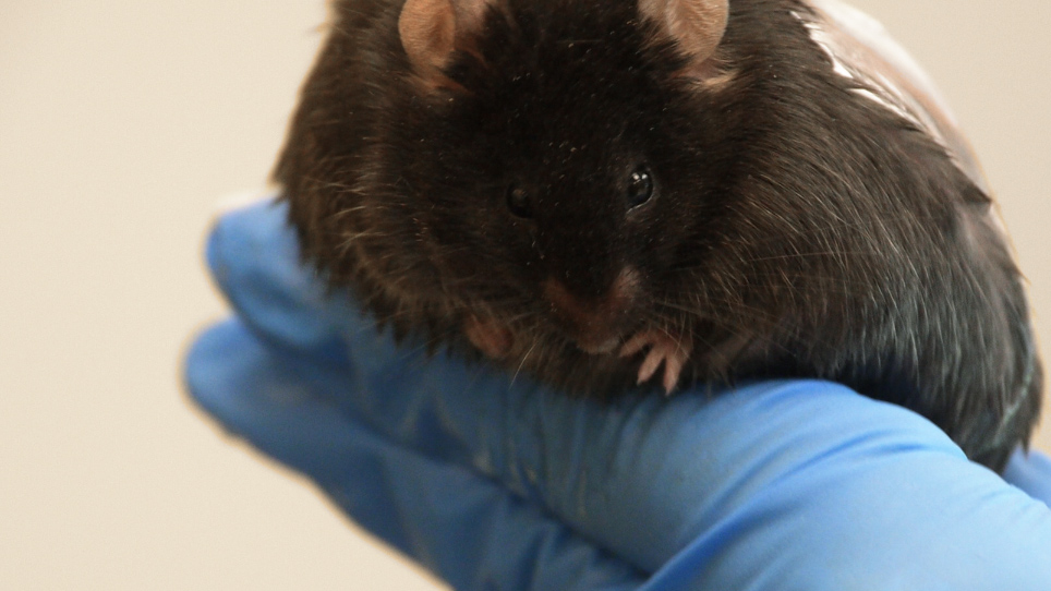
慢性伤口是从糖尿病小鼠模型上的急性伤口发展,通过在全厚皮伤后诱导高水平的氧化应激。伤口用催化酶和谷胱甘肽过氧化物酶抑制剂进行治疗,导致皮肤微生物群中细菌的愈合和生物膜发育受损。
Abstract
慢性伤口的发展,由于在一个或多个复杂的细胞和分子过程中涉及适当的愈合有缺陷的调节。仅在美国,它们就影响 650 万人,每年成本为 400 亿英镑。尽管在了解慢性伤口如何发展人类方面已经投入了大量精力,但基本问题仍未得到解答。最近,我们开发了一种新的小鼠模型,用于具有许多人类慢性伤口特征的糖尿病慢性伤口。使用db/db-/-小鼠,我们可以在伤人后立即在伤口组织中诱导高水平的氧化应激(OS),使用与抗氧化酶催化酶特有的抑制剂的一次性治疗,从而产生慢性伤口。谷胱甘肽过氧化物酶。这些伤口具有高水平的操作系统,自然发展生物膜,在治疗后20天内完全慢性,可以保持开放超过60天。这种新颖的模型具有人类糖尿病慢性伤口的许多特征,因此可以大大有助于促进对伤口如何成为慢性的基本理解。这是一个重大突破,因为人类慢性伤口给患者带来严重的疼痛和痛苦,如果得不到解决,则导致截肢。此外,这些伤口非常昂贵和费时治疗,并导致患者个人收入重大损失。通过使用我们的慢性伤口模型,这一研究领域的进步可以显著改善数百万在这种衰弱条件下受苦的人的医疗保健。在这个协议中,我们非常详细地描述了导致急性伤口成为慢性伤口的程序,这是以前没有做过的。
Introduction
伤口愈合涉及复杂的细胞和分子过程,这些过程在时间和空间上受到调节,在顺序和重叠阶段组织,涉及许多不同的细胞类型,包括但不限于免疫反应和血管系统1.皮肤受伤后,因子和血细胞立即聚集到伤口部位,并启动凝血级联形成血块。平衡后,血管分裂,让进入伤口部位的氧气、营养物质、酶、抗体和化学因子,使多态细胞清除伤口床的异物碎片,并分泌蛋白解酶2.激活的血小板分泌各种生长因子,以刺激伤口边缘的角蛋白细胞,使受伤区域重新上皮。被招募到伤口部位的单核细胞分化成巨噬细胞,其中噬菌体和死中性粒细胞和分泌其他因素,以保持角蛋白细胞增殖和亲迁移信号。在增殖阶段,在继续再上皮化的同时,由成纤维细胞、单核细胞/巨噬细胞、淋巴细胞和内皮细胞组成的新的造粒组织继续重建过程2。血管生成通过促进内皮细胞增殖和迁移来刺激,导致新的血管发育。细胞外基质的分体化和改造为环境设置了屏障。当伤口愈合和造粒组织演变为疤痕时,凋亡可消除炎症细胞、成纤维细胞和内皮细胞,而不会造成额外的组织损伤。成纤维细胞重塑细胞外基质的各种成分(如胶原蛋白)增强了组织的拉伸强度,使新形成的组织几乎与未受伤的皮肤2一样强壮和灵活。
任何偏离这种高度协调的进展到伤口关闭导致受损和/或慢性伤口3。慢性伤口的特点是氧化应激增加,慢性炎症,微血管受损,伤口中胶原蛋白基质异常。氧化应激,特别是在伤口,可以延迟伤口封闭2,5。当伤口愈合的第一阶段,炎症阶段变得不受管制时,宿主组织由于炎症细胞5的不断涌入而承担广泛的损害,释放细胞毒性酶,自由氧基增加,以及不受监管的炎症介质,导致细胞死亡6,7。
在这个破坏性的微环境中,生物膜形成细菌利用宿主营养物质,导致宿主组织2的损害。这些生物膜难以控制和去除,因为由蛋白质、DNA、RNA 和多糖组成的水合物细胞外聚合物物质使体内的细菌能够对传统抗生素疗法具有耐受性,并避开宿主的先天和适应性免疫反应2,8,9。
研究慢性伤口至关重要,因为它们影响650万人,仅在美国每年就花费400亿美元。糖尿病患者增加了患慢性伤口的风险,需要截肢以遏制感染的传播。这些患者在截肢后5年内有50%的死亡风险,这归因于糖尿病11的病理生理学机制。宿主的免疫系统和伤口愈合中的微生物群之间的关系是正在进行的研究的一个重要课题,因为慢性伤口的后果,如果得不到解决,包括截肢和死亡12。
尽管在了解慢性伤口如何发展人类方面已经投入了大量精力,但慢性伤口是如何形成的,为什么形成,目前还不清楚。研究受损愈合机制的实验很难在人类身上进行,伤口愈合专家只能看到慢性伤口患者已经达到慢性数周到数月。因此,专家无法研究哪些过程出错,导致伤口发展成慢性2。缺乏能够概括人类慢性创伤复杂性的动物模型。直到我们的模型被开发,没有慢性伤口研究模型存在。
慢性伤口模型是在瘦素受体(db/db-/–)13中发生突变的小鼠身上开发的。这些小鼠肥胖,糖尿病,有损伤愈合,但不开发慢性伤口14。血糖水平平均在200毫克/分升左右,但可以高达400毫克/分升15。当伤口组织中高水平的氧化应激(OS)在伤伤后立即诱发时,伤口就变成慢性16。db/db-/-伤口在20天内被认为是慢性的,并且保持开放60天或更长时间。细菌产生的生物膜在伤人后三天开始发育;成熟的生物膜在伤后20天可见,并持续到伤口关闭。我们在这些小鼠中发现的生物膜形成细菌也存在于人类糖尿病慢性伤口中。
氧化应激是通过两种抗氧化酶抑制剂,即催化酶和谷胱甘肽过氧化物酶来治疗伤口,这两种酶具有分解过氧化氢的能力。过氧化氢是一种活性氧,通过蛋白质、脂质和DNA的氧化会导致细胞损伤。催化过氧化氢分解成危害较少的化学物质氧和水。3-Amino-1,2,4-triazole (ATZ) 通过专门和共价地结合酶的活性中心来抑制催化酶,使其17、18、19起灭活。ATZ一直被用来研究氧化应激在体外和体内通过抑制催化酶20,21,22,23,24的影响。谷胱甘肽过氧化物酶通过抗氧化剂谷胱甘肽催化过氧化氢的减少,是保护细胞免受氧化应激的重要酶25。Mercaptosuccinic酸(MSA)通过与酸醇酶的selenocystein活性位点结合来抑制谷胱甘肽过氧化物酶,使其26日失活。MSA已用于研究氧化应激在体外和体内以及20,27,28的影响。
这种新型慢性伤口模型是一个强大的研究模型,因为它与人类糖尿病慢性伤口观察到的许多相同特征相同,包括由于OS增加引起的长期炎症和皮肤微生物群形成的天然生物膜。伤口有损皮肤-表皮相互作用,异常基质沉积,血管生成不良和血管受损。雄性小鼠和雌性小鼠都会出现慢性伤口,因此两性都可用于研究慢性伤口。因此,慢性伤口模型可以大大有助于促进对此类伤口如何开始的基本理解。使用这种慢性伤口模型可以回答有关如何通过受损伤口愈合的生理学和宿主的微生物群的贡献来启动/实现慢性的基本问题。
Protocol
Representative Results
Discussion
一旦在小鼠身上形成慢性伤口,该模型可用于研究慢性开始过程中受损的伤口愈合过程。该模型还可用于测试各种化学品和药物的疗效,这些化学物质和药物可以逆转慢性伤口发育和受损愈合,并导致伤口闭合和愈合。可以研究慢性发病后的不同时间点:例如,在慢性早期发病后受伤的第1-5天,以及20天及以后的全身性慢性伤口。
慢性伤口模型也是研究伤口愈合和并发…
Disclosures
The authors have nothing to disclose.
Acknowledgements
作者没有承认。
Materials
| B6.BKS(D)-Leprdb/J | The Jackson Laboratory | 00697 | Homozygotes and heterozygotes available |
| Nair Hair Remover Lotion with Soothing Aloe and Lanolin | Nair | a chemical depilatory | |
| Buprenex (buprenorphine HCl) | Henry Stein Animal Health | 059122 | 0.3 mg/ml, Class 3 |
| 3-Amino-1,2,4-triazole (ATZ) | TCI | A0432 | |
| Mercaptosuccinic acid (MSA) | Aldrich | 88460 | |
| Phosphate buffer solution (PBS) | autoclave steriled | ||
| Isoflurane | Henry Schein Animal Health | 029405 | NDC 11695-6776-2 |
| Oxygen | Tank must be compatible with vaporizing system | ||
| Isoflurane vaporizer | JA Baulch & Associates | ||
| Wahl hair clipper | Wahl | Lithium Ion Pro | |
| Acu Punch 7mm skin biopsy punches | Acuderm Inc. | P750 | |
| Tegaderm | 3M | Ref: 1624W | Transparent film dressing (6 cm x 7 cm) |
| Heating pad | Conair | Moist Dry Heating Pad | |
| Insulin syringes | BD | 329461 | 0.35 mm (28G) x 12.7 mm (1/2") |
| 70% ethanol | |||
| Kimwipes | |||
| Tweezers | |||
| Sharp surgical scissors | |||
| Thin metal spatula | |||
| Tubing | |||
| Mouse nose cone | |||
| Gloves | |||
| small plastic containers |
References
- Singer, A. J., Clark, R. A. F. Cutaneous wound healing. New England Journal of Medicine. 341 (10), 738-746 (1999).
- Nouvong, A., Ambrus, A. M., Zhang, E. R., Hultman, L., Coller, H. A. Reactive oxygen species and bacterial biofilms in diabetic wound healing. Physiological Genomics. 48 (12), 889-896 (2016).
- MacLeod, A. S., Mansbridge, J. N. The innate immune system in acute and chronic wounds. Advanced Wound Care. 5 (2), 65-78 (2016).
- Zhao, G., et al. Biofilms and Inflammation in Chronic Wounds. Advanced Wound Care. 2 (7), 389-399 (2013).
- Wlaschek, M., Scharffetter-Kochanek, K. Oxidative stress in chronic venous leg ulcers. Wound Repair and Regeneration. 13 (5), 452-461 (2005).
- Stadelmann, W. K., Digenis, A. G., Tobin, G. R. Physiology and healing dynamics of chronic cutaneous wounds. American Journal of Surgery. 176 (2), 26-38 (1998).
- Loots, M. A., Lamme, E. N., Zeegelaar, J., Mekkes, J. R., Bos, J. D., Middelkoop, E. Differences in cellular infiltrate and extracellular matrix of chronic diabetic and venous ulcers versus acute wounds. Journal of Investigative Dermatology. 111 (5), 850-857 (1998).
- Costerton, W., Veeh, R., Shirtliff, M., Pasmore, M., Post, C., Ehrlich, G. The application of biofilm science to the study and control of chronic bacterial infections. Journal of Clinical Investigation. 112 (10), 1466-1477 (2003).
- Fux, C. A., Costerton, J. W., Stewart, P. S., Stoodley, P. Survival strategies of infectious biofilms. Trends in Microbiology. 13 (1), 34-40 (2005).
- Sen, C. K., et al. Human skin wounds: A major and snowballing threat to public health and the economy. Wound Repair and Regeneration. 17 (6), 763-771 (2009).
- Armstrong, D. G., Wrobel, J., Robbins, J. M. Are diabetes-related wounds and amputations worse than cancer. International Wound Journal. 4 (4), 286-287 (2007).
- James, G. A., et al. Biofilms in chronic wounds. Wound Repair and Regeneration. 16 (1), 37-44 (2008).
- Chen, H., et al. Evidence that the diabetes gene encodes the leptin receptor: Identification of a mutation in the leptin receptor gene in db/db mice. Cell. 84 (3), 491-495 (1996).
- Coleman, D. L. Obese and diabetes: Two mutant genes causing diabetes-obesity syndromes in mice. Diabetologia. 14 (3), 141-148 (1978).
- Garris, D. R., Garris, B. L. Genomic modulation of diabetes (db/db) and obese (ob/ob) mutation-induced hypercytolipidemia: cytochemical basis of female reproductive tract involution. Cell Tissue Research. 316 (2), 233-241 (2014).
- Dhall, S., et al. Generating and reversing chronic wounds in diabetic mice by manipulating wound redox parameters. Journal of Diabetes Research. , (2014).
- Feinstein, R. N., Berliner, S., Green, F. O. Mechanism of inhibition of catalase by 3-amino-1,2,4-triazole. Archives of Biochemistry and Biophysics. 76 (1), 32-44 (1958).
- Margoliash, E., Novogrodsky, A. A study of the inhibition of catalase by 3-amino-1:2:4:-triazole. Biochemical Journal. 68 (3), 468-475 (1958).
- Margoliash, E., Novogrodsky, A., Schejter, A. Irreversible reaction of 3-amino-1:2:4-triazole and related inhibitors with the protein of catalase. Biochemical Journal. 74 (2), 339-348 (1960).
- Shiba, D., Shimamoto, N. Attenuation of endogenous oxidative stress-induced cell death by cytochrome P450 inhibitors in primary cultures of rat hepatocytes. Free Radical Biology and Medicine. 27 (9-10), 1019-1026 (1999).
- Ishihara, Y., Shimamoto, N. Critical role of exposure time to endogenous oxidative stress in hepatocyte apoptosis. Redox Report. 12 (6), 275-281 (2007).
- Valenti, V. E., de Abreu, L. C., Sato, M. A., Ferreira, C. ATZ (3-amino-1,2,4-triazole) injected into the fourth cerebral ventricle influences the Bezold-Jarisch reflex in conscious rats. Clinics. 65 (12), 1339-1343 (2010).
- Welker, A. F., Campos, E. G., Cardoso, L. A., Hermes-Lima, M. Role of catalase on the hypoxia/reoxygenation stress in the hypoxia-tolerant Nile tilapia. American Journal of Physiology. Regulatory, Integrative and Comparative Physiology. 302 (9), 1111-1118 (2012).
- Bagnyukova, T. V., Vasylkiv, O. Y., Storey, K. B., Lushchak, V. I. Catalase inhibition by amino triazole induces oxidative stress in goldfish brain. Brain Research. 1052 (2), 180-186 (2005).
- Falck, E., Karlsson, S., Carlsson, J., Helenius, G., Karlsson, M., Klinga-Levan, K. Loss of glutathione peroxidase 3 expression is correlated with epigenetic mechanisms in endometrial adenocarcinoma. Cancer Cell International. 10 (46), (2010).
- Chaudiere, J., Wilhelmsen, E. C., Tappel, A. L. Mechanism of selenium-glutathione peroxidase and its inhibition by mercaptocarboxylic acids and other mercaptans. Journal of Biological Chemistry. 259 (2), 1043-1050 (1984).
- Dunning, S., et al. Glutathione and antioxidant enzymes serve complementary roles in protecting activated hepatic stellate cells against hydrogen peroxide-induced cell death. Biochimica et Biophysica Acta. 1832 (12), 2027-2034 (2013).
- Franco, J. L., et al. Methylmercury neurotoxicity is associated with inhibition of the antioxidant enzyme glutathione peroxidase. Free Radical Biology and Medicine. 47 (4), 449-457 (2009).
- Sundberg, J. P., Silva, K. A. What color is the skin of a mouse. Veterinary Pathology. 49 (1), 142-145 (2012).
- Curtis, A., Calabro, K., Galarneau, J. R., Bigio, I. J., Krucker, T. Temporal variations of skin pigmentation in C57BL/6 mice affect optical bioluminescence quantitation. Molecular Imaging & Biology. 13 (6), 1114-1123 (2011).
- Kim, J. H., Martins-Green, M. Protocol to create chronic wounds in diabetic mice. Nature Protocols Exchange. , (2016).
- Aasum, E., Hafstad, A. D., Severson, D. L., Larsen, T. S. Age-dependent changes in metabolism, contractile function, and ischemic sensitivity in hearts from db/db mice. Diabetes. 52 (2), 434-441 (2003).
- Vannucci, S. J., et al. Experimental stroke in the female diabetic, db/db, mouse. Journal of Cerebral Blood Flow & Metabolism. 21 (1), 52-60 (2001).
- Janssen, B. J., et al. Effects of anesthetics on systemic hemodynamics in mice. American Journal of Physiology-Heart and Circulatory Physiology. 287 (4), 1618-1624 (2004).
- Osborn, O., et al. Metabolic characterization of a mouse deficient in all known leptin receptor isoforms. Cellular and Molecular Neurobiology. 30 (1), 23 (2010).
- Scales, B. S., Huffnagle, G. B. The microbiome in wound repair and tissue fibrosis. Journal of Pathology. 229 (2), 323-331 (2013).
- Gjødsbøl, K., et al. No need for biopsies: Comparison of three sample techniques for wound microbiota determination. International Wound Journal. 9 (3), 295-302 (2012).
- Wolcott, R. D., et al. Analysis of the chronic wound microbiota of 2,963 patients by 16S rDNA pyrosequencing. Wound Repair Regeneration. 24 (1), 163-174 (2016).
- Gjødsbøl, K., Christensen, J. J., Karlsmark, T., Jørgensen, B., Klein, B. M., Krogfelt, K. A. Multiple bacterial species reside in chronic wounds: a longitudinal study. International Wound Journal. 3 (3), 225-231 (2006).
- Dowd, S. E., et al. Survey of bacterial diversity in chronic wounds using Pyrosequencing, DGGE, and full ribosome shotgun sequencing. BMC Microbiology. 8 (43), (2008).
- Price, L. B., et al. Community analysis of chronic wound bacteria using 16S rrna gene-based pyrosequencing: Impact of diabetes and antibiotics on chronic wound microbiota. PLoS One. 4 (7), 6462 (2009).
- Scales, B. S., Huffnagle, G. B. The microbiome in wound repair and tissue fibrosis. Journal of Pathology. 229 (2), 323-331 (2013).
- Dowd, S. E., et al. Polymicrobial nature of chronic diabetic foot ulcer biofilm infections determined using bacterial tag encoded FLX amplicon pyrosequencing (bTEFAP). PLoS One. 3 (10), 3326 (2008).
- Price, L. B., et al. Macroscale spatial variation in chronic wound microbiota: A cross-sectional study. Wound Repair and Regeneration. 19 (1), 80-88 (2011).
- Gontcharova, V., Youn, E., Sun, Y., Wolcott, R. D., Dowd, S. E. Comparison of bacterial composition in diabetic ulcers and contralateral intact skin. Open Microbiology Journal. 4, 8-19 (2010).
- Smith, K., et al. One step closer to understanding the role of bacteria in diabetic foot ulcers: characterising the microbiome of ulcers. BMC Microbiologyogy. 16 (54), (2016).
- Gardner, S. E., Hillis, S. L., Heilmann, K., Segre, J. A., Grice, E. A. The Neuropathic diabetic foot ulcer microbiome is associated with clinical factors. Diabetes. 62 (3), 923-930 (2013).
- Loesche, M., et al. Temporal stability in chronic wound microbiota is associated with poor healing. Journal of Investigative Dermatology. 137 (1), 237-244 (2017).
- Kalan, L., et al. Redefining the chronic-wound microbiome: Fungal communities are prevalent, dynamic, and associated with delayed healing. MBio. 7 (5), 01058-01116 (2016).
- Blakytny, R., Jude, E. The molecular biology of chronic wounds and delayed healing in diabetes. Diabetic Medicine. 23 (6), 594-608 (2006).

