糖尿病マウスの慢性創傷を作成するためのプロトコル
Summary
慢性創傷は、全厚の皮下創傷の後に高レベルの酸化ストレスを誘発することによって、糖尿病マウスモデルの急性創傷から発症する。創傷は、カタラゼおよびグルタチオンペルオキシダーゼの阻害剤で治療され、皮膚微生物叢に存在する細菌による治癒およびバイオフィルムの発達を損なう結果生じる。
Abstract
慢性創傷は、適切な治癒に関与する1つ以上の複雑な細胞および分子プロセスにおける不完全な調節の結果として発症する。彼らは約6.5Mの人々に影響を与え、米国だけで〜$40B/年の費用がかかります。ヒトにおける慢性創傷の発症方法を理解するために多大な努力がなされているが、根本的な疑問は未解決のままである。最近では、ヒト慢性創傷の多くの特徴を持つ糖尿病性慢性創傷の新しいマウスモデルを開発した。db/–マウスを用いて、創傷直後に創傷組織に高レベルの酸化ストレス(OS)を誘導し、抗酸化酵素に特異的な阻害剤を用いた1回限りの治療を用いて、慢性創傷を生成することができる。グルタチオンペルオキシダーゼ。これらの創傷は、高レベルのOSを持ち、バイオフィルムを自然に開発し、治療後20日以内に完全に慢性になり、60日以上開いたままにすることができます。この新しいモデルは、ヒトにおける糖尿病性慢性創傷の多くの特徴を有し、したがって、創傷が慢性になる方法の基本的な理解を進めることに大きく貢献できる。これは、ヒトの慢性創傷が患者に重大な痛みや苦痛を引き起こし、未解決の場合は切断をもたらすため、大きなブレークスルーです。さらに、これらの創傷は治療に非常に高価で時間がかかり、患者の個人所得の大幅な損失につながります。私たちの慢性創傷モデルの使用を通じて研究のこの分野の進歩は、この衰弱状態の下で苦しむ何百万人もの医療を大幅に改善することができます。このプロトコルでは、急性創傷を慢性化させる手順を詳細に説明しますが、これはこれまで行われていません。
Introduction
創傷治癒は、時間的および空間的に調節される複雑な細胞および分子プロセスを含み、免疫応答および血管に限定されない多くの異なる細胞タイプを含む多くの異なる細胞タイプを含む連続的かつ重なり合う段階で組織されるシステム1.皮膚が傷害を持続した直後に、因子および血液細胞は創傷部位に凝集し、凝固カスケードを開始して血栓を形成する。恒常性が達成された後、血管は、異物の創傷床をクリアし、プロテオ分解酵素を分泌するために多形態性細胞を化学的に引き付ける創傷部位酸素、栄養素、酵素、抗体および化学的要因に入るように拡張する。2.活性化血小板は、傷部のケラチノサイトを刺激するために様々な成長因子を分泌し、傷部を再上皮化させる。創傷部位に募集された単球は、食細胞細胞細菌と死んだ好中球を循環させるマクロファージに分化し、ケラチノサイト増殖およびプロ渡虫シグナルを維持するための追加因子を分泌する。増殖段階では、再上皮化が続く間、線維芽細胞、単球/マクロファージ、リンパ球、および内皮細胞からなる新しい造粒組織が再建プロセス2を継続する。血管新生は、内皮細胞の増殖および移動を促進することによって刺激され、新しい血管発達をもたらす。細胞外マトリックスの上皮化および改造は、環境に対する障壁を構築する。創傷が治癒し、造粒組織が瘢痕に進化するにつれて、アポトーシスは、追加の組織損傷を引き起こすことなく、炎症細胞、線維芽細胞、および内皮細胞を排除する。組織の引張強度は、コラーゲンのような細胞外マトリックスの様々な成分を改造する線維芽細胞によって増強され、新たに形成された組織が傷ついていない皮膚2とほぼ同じくらい強く、柔軟であるようにする。
創傷閉鎖に向かうこの高度に協調した進行からの逸脱は、障害および/または慢性創傷3につながる。慢性創傷は、酸化ストレスの増加、慢性炎症、損傷した微小血管系、および創傷4における異常なコラーゲンマトリックスによって特徴付けられる。酸化ストレスは、特に創傷において、創傷閉鎖2、5を遅らせることができる。創傷治癒の第1段階において、炎症期が調節されなくなると、宿主組織は細胞傷害性酵素を放出する炎症細胞5の連続的な流入による広範な損傷を想定し、フリー酸素ラジカルの増加、無秩序な炎症メディエーターは、細胞死6、7をもたらす。
この破壊的な微小環境では、バイオフィルム形成細菌は宿主栄養素を利用し、宿主組織2の損傷に寄与する。これらのバイオフィルムは、タンパク質、DNA、RNA、および多糖類からなる水和された細胞外高分子物質が、従来の抗生物質療法に耐性を有し、回避できるため、制御および除去が困難である。ホストの生来性および適応免疫応答2、8、9。
慢性創傷の研究は、米国だけで年間650万人に影響を与え、年間400億ドルの費用がかかるため、非常に重要です。糖尿病患者は、感染の広がりを抑えるために切断を必要とする慢性創傷を発症するリスクが高まっている。これらの患者は、糖尿病11の病態生理学メカニズムに起因する切断の5年以内に50%の死亡リスクを有する。創傷治癒における宿主の免疫系とマイクロバイオームとの関係は、慢性創傷の結果が未解決の場合、切断および死亡12を含むため、進行中の研究の重要なトピックである。
ヒトにおける慢性創傷がどのように発症するかを理解するために多大な努力がなされているが、慢性創傷がどのように形成されるのか、またその理由は依然として不明である。障害のある治癒のメカニズムを研究する実験はヒトでは行うことが困難であり、創傷治癒の専門家は、すでに数週間から数ヶ月間慢性に達している慢性創傷を有する患者だけを見る。したがって、専門家は、慢性2になるために創傷を開発するために導くプロセスが間違っていたかを研究することができません。ヒトの慢性創傷の複雑さを要約する動物モデルの欠如がある。我々のモデルが開発されるまで、慢性創傷研究のためのモデルは存在しなかった。
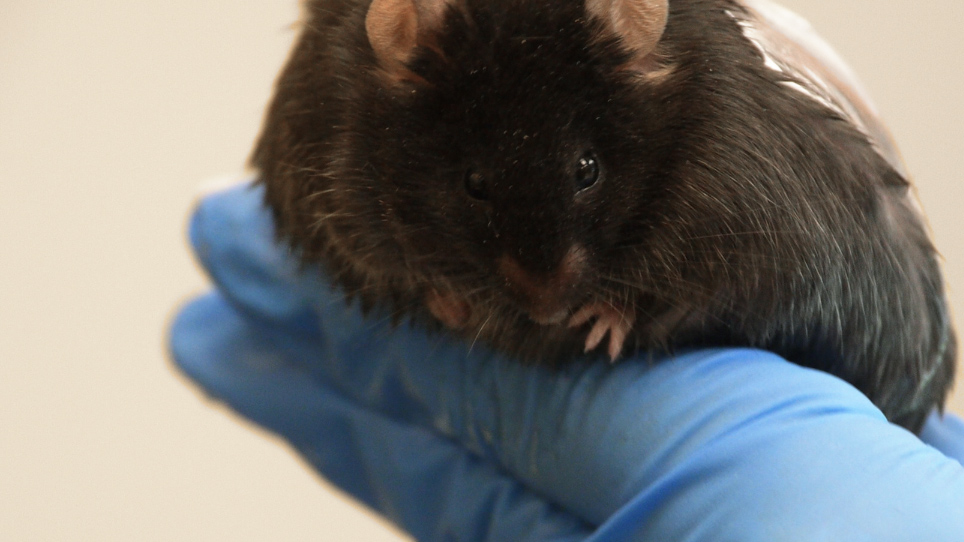
慢性創傷モデルは、レプチン受容体(db/db-/–)13に変異を有するマウスで開発された。これらのマウスは肥満、糖尿病、治癒を損なっているが、慢性創傷14を発症しない。血糖値は平均約200 mg/dL, しかし、400 mg/dL15と高くすることができます.創傷組織中の高レベルの酸化ストレス(OS)が創傷直後に誘発されると、創傷は慢性16になる。db//-創傷は20日までに慢性と見なされ、60日以上開いたままである。細菌によって産生されるバイオフィルムは、創傷の3日後に発症するのを見ることができる。成熟したバイオフィルムは、創傷後20日後に見ることができ、どちらかの創傷閉鎖まで持続する。これらのマウスで見つかったバイオフィルム形成細菌は、ヒト糖尿病性慢性創傷にも見られる。
酸化ストレスは、抗酸化酵素、カタラゼおよびグルタチオンペルオキシダーゼの2つの阻害剤、過酸化水素を分解する能力を有する2つの酵素で創傷を治療することによって誘導される。過酸化水素は活性酸素種であり、タンパク質、脂質、DNAの酸化を通じて細胞損傷を引き起こす可能性があります。カタラゼは、過酸化水素の分解を有害性の低い化学物質に触媒酸素と水にします。3-アミノ-1,2,4-トリアゾール(ATZ)は、酵素の活性中心に特異的かつ共生的に結合することによってカタラーゼを阻害し、それを不活性化する17、18、19。ATZは、カタラゼ20、21、22、23、24の阻害を通じてインビトロとインビボの両方の酸化ストレスの効果を研究するために使用されています。グルタチオンペルオキシダーゼは、抗酸化剤、グルタチオンを介した過酸化水素の還元を触媒し、酸化ストレスから細胞を保護する重要な酵素である25。メルカプトスチシン酸(MSA)は、チオールと酵素のセレノシステイン活性部位に結合することによりグルタチオンペルオキシダーゼを阻害し、それを不活性化する26。MSAは、インビトロおよびインビボにおける酸化ストレスの影響を研究するために使用されてきただけでなく、20 、27、28.
慢性創傷のこの新しいモデルは、増加したOSからの長期炎症や皮膚微生物叢からの自然なバイオフィルム形成を含む、ヒト糖尿病性慢性創傷で観察される同じ特徴の多くを共有するので、研究する強力なモデルです。創傷は、真皮表皮相互作用、異常なマトリックス沈着、血管新生の不良および血管系の損傷を損なった。慢性創傷は、男性と女性の両方のマウスで発症するので、両方の男女が慢性創傷を研究するために使用することができます。したがって、慢性創傷モデルは、そのような創傷がどのように始まるかの基本的な理解を進めるのに大きく寄与することができる。この慢性創傷モデルを使用すると、創傷治癒障害の生理学と宿主のマイクロバイオームからの寄与を通じて、慢性がどのように開始/達成されるかについての基本的な質問に対する答えを提供できる。
Protocol
Representative Results
Discussion
マウスに慢性創傷が作成されると、このモデルは、慢性の開始に関与する創傷治癒過程の障害を研究するために使用することができる。このモデルはまた、慢性創傷の発達を逆転させ、治癒を損ない、創傷の閉鎖および治癒につながる広範囲の化学物質および薬物の有効性をテストするために使用することができる。慢性の発症後の異なる時間ポイントを研究することができます:例えば?…
Disclosures
The authors have nothing to disclose.
Acknowledgements
著者は謝辞を持っていません。
Materials
| B6.BKS(D)-Leprdb/J | The Jackson Laboratory | 00697 | Homozygotes and heterozygotes available |
| Nair Hair Remover Lotion with Soothing Aloe and Lanolin | Nair | a chemical depilatory | |
| Buprenex (buprenorphine HCl) | Henry Stein Animal Health | 059122 | 0.3 mg/ml, Class 3 |
| 3-Amino-1,2,4-triazole (ATZ) | TCI | A0432 | |
| Mercaptosuccinic acid (MSA) | Aldrich | 88460 | |
| Phosphate buffer solution (PBS) | autoclave steriled | ||
| Isoflurane | Henry Schein Animal Health | 029405 | NDC 11695-6776-2 |
| Oxygen | Tank must be compatible with vaporizing system | ||
| Isoflurane vaporizer | JA Baulch & Associates | ||
| Wahl hair clipper | Wahl | Lithium Ion Pro | |
| Acu Punch 7mm skin biopsy punches | Acuderm Inc. | P750 | |
| Tegaderm | 3M | Ref: 1624W | Transparent film dressing (6 cm x 7 cm) |
| Heating pad | Conair | Moist Dry Heating Pad | |
| Insulin syringes | BD | 329461 | 0.35 mm (28G) x 12.7 mm (1/2") |
| 70% ethanol | |||
| Kimwipes | |||
| Tweezers | |||
| Sharp surgical scissors | |||
| Thin metal spatula | |||
| Tubing | |||
| Mouse nose cone | |||
| Gloves | |||
| small plastic containers |
References
- Singer, A. J., Clark, R. A. F. Cutaneous wound healing. New England Journal of Medicine. 341 (10), 738-746 (1999).
- Nouvong, A., Ambrus, A. M., Zhang, E. R., Hultman, L., Coller, H. A. Reactive oxygen species and bacterial biofilms in diabetic wound healing. Physiological Genomics. 48 (12), 889-896 (2016).
- MacLeod, A. S., Mansbridge, J. N. The innate immune system in acute and chronic wounds. Advanced Wound Care. 5 (2), 65-78 (2016).
- Zhao, G., et al. Biofilms and Inflammation in Chronic Wounds. Advanced Wound Care. 2 (7), 389-399 (2013).
- Wlaschek, M., Scharffetter-Kochanek, K. Oxidative stress in chronic venous leg ulcers. Wound Repair and Regeneration. 13 (5), 452-461 (2005).
- Stadelmann, W. K., Digenis, A. G., Tobin, G. R. Physiology and healing dynamics of chronic cutaneous wounds. American Journal of Surgery. 176 (2), 26-38 (1998).
- Loots, M. A., Lamme, E. N., Zeegelaar, J., Mekkes, J. R., Bos, J. D., Middelkoop, E. Differences in cellular infiltrate and extracellular matrix of chronic diabetic and venous ulcers versus acute wounds. Journal of Investigative Dermatology. 111 (5), 850-857 (1998).
- Costerton, W., Veeh, R., Shirtliff, M., Pasmore, M., Post, C., Ehrlich, G. The application of biofilm science to the study and control of chronic bacterial infections. Journal of Clinical Investigation. 112 (10), 1466-1477 (2003).
- Fux, C. A., Costerton, J. W., Stewart, P. S., Stoodley, P. Survival strategies of infectious biofilms. Trends in Microbiology. 13 (1), 34-40 (2005).
- Sen, C. K., et al. Human skin wounds: A major and snowballing threat to public health and the economy. Wound Repair and Regeneration. 17 (6), 763-771 (2009).
- Armstrong, D. G., Wrobel, J., Robbins, J. M. Are diabetes-related wounds and amputations worse than cancer. International Wound Journal. 4 (4), 286-287 (2007).
- James, G. A., et al. Biofilms in chronic wounds. Wound Repair and Regeneration. 16 (1), 37-44 (2008).
- Chen, H., et al. Evidence that the diabetes gene encodes the leptin receptor: Identification of a mutation in the leptin receptor gene in db/db mice. Cell. 84 (3), 491-495 (1996).
- Coleman, D. L. Obese and diabetes: Two mutant genes causing diabetes-obesity syndromes in mice. Diabetologia. 14 (3), 141-148 (1978).
- Garris, D. R., Garris, B. L. Genomic modulation of diabetes (db/db) and obese (ob/ob) mutation-induced hypercytolipidemia: cytochemical basis of female reproductive tract involution. Cell Tissue Research. 316 (2), 233-241 (2014).
- Dhall, S., et al. Generating and reversing chronic wounds in diabetic mice by manipulating wound redox parameters. Journal of Diabetes Research. , (2014).
- Feinstein, R. N., Berliner, S., Green, F. O. Mechanism of inhibition of catalase by 3-amino-1,2,4-triazole. Archives of Biochemistry and Biophysics. 76 (1), 32-44 (1958).
- Margoliash, E., Novogrodsky, A. A study of the inhibition of catalase by 3-amino-1:2:4:-triazole. Biochemical Journal. 68 (3), 468-475 (1958).
- Margoliash, E., Novogrodsky, A., Schejter, A. Irreversible reaction of 3-amino-1:2:4-triazole and related inhibitors with the protein of catalase. Biochemical Journal. 74 (2), 339-348 (1960).
- Shiba, D., Shimamoto, N. Attenuation of endogenous oxidative stress-induced cell death by cytochrome P450 inhibitors in primary cultures of rat hepatocytes. Free Radical Biology and Medicine. 27 (9-10), 1019-1026 (1999).
- Ishihara, Y., Shimamoto, N. Critical role of exposure time to endogenous oxidative stress in hepatocyte apoptosis. Redox Report. 12 (6), 275-281 (2007).
- Valenti, V. E., de Abreu, L. C., Sato, M. A., Ferreira, C. ATZ (3-amino-1,2,4-triazole) injected into the fourth cerebral ventricle influences the Bezold-Jarisch reflex in conscious rats. Clinics. 65 (12), 1339-1343 (2010).
- Welker, A. F., Campos, E. G., Cardoso, L. A., Hermes-Lima, M. Role of catalase on the hypoxia/reoxygenation stress in the hypoxia-tolerant Nile tilapia. American Journal of Physiology. Regulatory, Integrative and Comparative Physiology. 302 (9), 1111-1118 (2012).
- Bagnyukova, T. V., Vasylkiv, O. Y., Storey, K. B., Lushchak, V. I. Catalase inhibition by amino triazole induces oxidative stress in goldfish brain. Brain Research. 1052 (2), 180-186 (2005).
- Falck, E., Karlsson, S., Carlsson, J., Helenius, G., Karlsson, M., Klinga-Levan, K. Loss of glutathione peroxidase 3 expression is correlated with epigenetic mechanisms in endometrial adenocarcinoma. Cancer Cell International. 10 (46), (2010).
- Chaudiere, J., Wilhelmsen, E. C., Tappel, A. L. Mechanism of selenium-glutathione peroxidase and its inhibition by mercaptocarboxylic acids and other mercaptans. Journal of Biological Chemistry. 259 (2), 1043-1050 (1984).
- Dunning, S., et al. Glutathione and antioxidant enzymes serve complementary roles in protecting activated hepatic stellate cells against hydrogen peroxide-induced cell death. Biochimica et Biophysica Acta. 1832 (12), 2027-2034 (2013).
- Franco, J. L., et al. Methylmercury neurotoxicity is associated with inhibition of the antioxidant enzyme glutathione peroxidase. Free Radical Biology and Medicine. 47 (4), 449-457 (2009).
- Sundberg, J. P., Silva, K. A. What color is the skin of a mouse. Veterinary Pathology. 49 (1), 142-145 (2012).
- Curtis, A., Calabro, K., Galarneau, J. R., Bigio, I. J., Krucker, T. Temporal variations of skin pigmentation in C57BL/6 mice affect optical bioluminescence quantitation. Molecular Imaging & Biology. 13 (6), 1114-1123 (2011).
- Kim, J. H., Martins-Green, M. Protocol to create chronic wounds in diabetic mice. Nature Protocols Exchange. , (2016).
- Aasum, E., Hafstad, A. D., Severson, D. L., Larsen, T. S. Age-dependent changes in metabolism, contractile function, and ischemic sensitivity in hearts from db/db mice. Diabetes. 52 (2), 434-441 (2003).
- Vannucci, S. J., et al. Experimental stroke in the female diabetic, db/db, mouse. Journal of Cerebral Blood Flow & Metabolism. 21 (1), 52-60 (2001).
- Janssen, B. J., et al. Effects of anesthetics on systemic hemodynamics in mice. American Journal of Physiology-Heart and Circulatory Physiology. 287 (4), 1618-1624 (2004).
- Osborn, O., et al. Metabolic characterization of a mouse deficient in all known leptin receptor isoforms. Cellular and Molecular Neurobiology. 30 (1), 23 (2010).
- Scales, B. S., Huffnagle, G. B. The microbiome in wound repair and tissue fibrosis. Journal of Pathology. 229 (2), 323-331 (2013).
- Gjødsbøl, K., et al. No need for biopsies: Comparison of three sample techniques for wound microbiota determination. International Wound Journal. 9 (3), 295-302 (2012).
- Wolcott, R. D., et al. Analysis of the chronic wound microbiota of 2,963 patients by 16S rDNA pyrosequencing. Wound Repair Regeneration. 24 (1), 163-174 (2016).
- Gjødsbøl, K., Christensen, J. J., Karlsmark, T., Jørgensen, B., Klein, B. M., Krogfelt, K. A. Multiple bacterial species reside in chronic wounds: a longitudinal study. International Wound Journal. 3 (3), 225-231 (2006).
- Dowd, S. E., et al. Survey of bacterial diversity in chronic wounds using Pyrosequencing, DGGE, and full ribosome shotgun sequencing. BMC Microbiology. 8 (43), (2008).
- Price, L. B., et al. Community analysis of chronic wound bacteria using 16S rrna gene-based pyrosequencing: Impact of diabetes and antibiotics on chronic wound microbiota. PLoS One. 4 (7), 6462 (2009).
- Scales, B. S., Huffnagle, G. B. The microbiome in wound repair and tissue fibrosis. Journal of Pathology. 229 (2), 323-331 (2013).
- Dowd, S. E., et al. Polymicrobial nature of chronic diabetic foot ulcer biofilm infections determined using bacterial tag encoded FLX amplicon pyrosequencing (bTEFAP). PLoS One. 3 (10), 3326 (2008).
- Price, L. B., et al. Macroscale spatial variation in chronic wound microbiota: A cross-sectional study. Wound Repair and Regeneration. 19 (1), 80-88 (2011).
- Gontcharova, V., Youn, E., Sun, Y., Wolcott, R. D., Dowd, S. E. Comparison of bacterial composition in diabetic ulcers and contralateral intact skin. Open Microbiology Journal. 4, 8-19 (2010).
- Smith, K., et al. One step closer to understanding the role of bacteria in diabetic foot ulcers: characterising the microbiome of ulcers. BMC Microbiologyogy. 16 (54), (2016).
- Gardner, S. E., Hillis, S. L., Heilmann, K., Segre, J. A., Grice, E. A. The Neuropathic diabetic foot ulcer microbiome is associated with clinical factors. Diabetes. 62 (3), 923-930 (2013).
- Loesche, M., et al. Temporal stability in chronic wound microbiota is associated with poor healing. Journal of Investigative Dermatology. 137 (1), 237-244 (2017).
- Kalan, L., et al. Redefining the chronic-wound microbiome: Fungal communities are prevalent, dynamic, and associated with delayed healing. MBio. 7 (5), 01058-01116 (2016).
- Blakytny, R., Jude, E. The molecular biology of chronic wounds and delayed healing in diabetes. Diabetic Medicine. 23 (6), 594-608 (2006).