Human kidneys were isolated by LifeCenter Northwest following ethical guidelines set by the Association of Organ Procurement Organizations. This protocol follows animal care and cell culture guidelines outlined by the University of Washington.
1. Preparation of Human Kidney Tissue
- Preparation of decellularization solution
- Sterilize a 5000 mL beaker and a 70 x 10 mm stir bar.
- Mix 1:1000 (weight:volume) sodium dodecyl sulfate (SDS) in autoclaved deionized water in the beaker. Leave the solution on a stir plate at approximately 200 rpm for 24 h or until the SDS is completely dissolved.
Note: Typically, 2500 mL of 1% SDS solution is sufficient to decellularize a single human kidney. - Transfer the solution to a 500 mL sterile vacuum filter and filter it into sterilized sealable containers.
- Processing of kidney tissue
- Wash and autoclave a pair of forceps, two hemostat clamps, a pair of general service grade scissors, two scalpel blade handles, a 1000 mL beaker covered with aluminum foil, and a 36 x 9 mm stir bar.
- Line a tissue culture hood with underpad. Place the beaker, a sterile tissue culture dish (150 x 25 mm), and the whole kidney organ into the hood. Fill the beaker with 500 mL of 1% SDS solution.
Note: Human kidneys were received on ice from LifeCenter NorthWest. - Place the kidney in the sterile tissue culture dish (Figure 1A). Remove all perirenal fat by lightly shaving around the renal capsule with a scalpel (Figure 1B).
- Make a shallow 8-10 cm incision with the scalpel, just deep enough to break open the renal capsule without damaging the underlying cortex tissue, across the superior end of the kidney. Remove the renal capsule by peeling it away from the cortex tissue with two hemostat clamps (Figure 1C).
- Bisect the kidney along the coronal plane by using the scalpel along the lateral side of the kidney (Figure 1D). Isolate cortex tissue from both halves by carving out the medullar region with the scalpel (Figure 1E) and dice the cortex tissue into 0.5 cm3 pieces (Figure 1F). Remove any large visible vessels.
- Isolation of extracellular matrix
- In a tissue culture hood, fill a 1000 mL beaker with 500 mL of 1% SDS solution. Place the diced cortex tissue and stir bar into the beaker containing SDS solution. Cover the beaker with autoclaved aluminum foil and place it on a stir plate at approximately 400 rpm outside of the tissue culture hood.
- After the cortex tissue has been on the stir plate for 24 h, bring the beaker into a tissue culture hood and add a 40 µm sterile cell strainer made with nylon mesh. Fill a separate 1000 mL beaker with 200 mL of bleach and place it in the tissue culture hood.
- Pipette out the SDS solution through the cell strainer into the beaker containing bleach. Pipette out all SDS solution until only decellularized tissue and the cell strainer remain in the beaker.
Note: The cell strainer should prevent any tissue from being removed during solution aspiration. - Leave the cell strainer in the beaker and fill with 500 mL of fresh SDS solution. Cover the beaker with the same aluminum foil and place onto a stir plate at the same speed as before.
- Repeat steps 1.3.1-1.3.3 every 24 hours with fresh SDS solution for a total of five days.
- Rinse decellularized tissue with autoclaved DI water every 24 h for 3 days total, following the technique outlined in steps 1.3.1-1.3.3.
- Rinse decellularized tissue with cell culture grade water every 24 h for 2 days total, following the technique outlined in steps 1.3.1-1.3.3.
- Repeat steps 1.3.1-1.3.2. Transfer the decellularized tissue (referred to as kECM from this point on) into a 30 mL self-standing conical tube and fill it with cell culture grade water until all the tissue is submerged.
2. Fabrication of Hydrogel Stock Solution
- Mechanical processing of decellularized tissue
- In a tissue culture hood, mechanically homogenize the kECM within the conical tube with a tissue homogenizer for 2 min.
Note: Homogenized kECM should resemble an opaque solution with no visible pieces of ECM. - Submerge the conical tube containing the kECM in liquid nitrogen until boiling surrounding the tube no longer persists. Store the kECM at -4 ˚C overnight.
- In a tissue culture hood, mechanically homogenize the kECM within the conical tube with a tissue homogenizer for 2 min.
- Lyophilization of frozen decellularized tissue
- Slightly loosen the conical tube cap to allow for gas exchange and place the tube into a lyophilization machine. Lyophilize the kECM for three days or until it resembles a fine white powder. Store at -4 ˚C.
- Chemical digestion and solubilization of gel
- Autoclave a 20 mL scintillation vial and cap, a 15.9 x 7.9 mm stir bar, and one pair of fine-tip forceps.
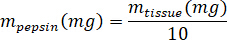
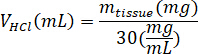
- Weigh the lyophilized kECM and calculate the volume of HCl and mass of pepsin needed to solubilize the kECM to a 3% (30 mg/mL) solution using the following equations, where mpepsin is the mass of pepsin, mtissue is the mass of lyophilized tissue, and VHCl is the volume of 0.01 N HCl:
- In a tissue culture hood, add porcine gastric pepsin, 0.01 N HCl, and the stir bar to the scintillation vial, and leave it on a stir plate at approximately 500 rpm until all the pepsin has dissolved. Transfer the lyophilized kECM to the scintillation vial and leave the solution on a stir plate at approximately 500 rpm for three days.
3. Hydrogel Gelation
- Kidney ECM hydrogel preparation
- Gel the hydrogel by mixing the kECM hydrogel stock solution with 1 N NaOH, 10x Media Supplement (M199), and cell culture media. Keep all the solutions on ice.
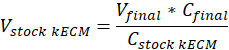
Note: Final gel concentrations of 7.5 mg/mL were used for cell culture. 1 mL of kECM gel was sufficient for cell culture experiments presented. - Determine the volume of workable kECM gel produced and volume of stock kECM hydrogel needed by using the following equation, where Vfinal is the volume of gel created, Vstock kECM is the volume of stock kECM hydrogel needed, Cstock kECM is the concentration of the stock kECM hydrogel, and Cfinal is the concentration of the final gel:
- Determine the volume of neutralizing reagents needed by using the following equations, where VNaOH is the volume of 1 N NaOH, V10X is the volume of M199 10X media supplement, and V1X is the volume of cell culture media:


- In a tissue culture hood, pipette the neutralizing reagents (NaOH, M199, and cell culture media) into a sterile 30 mL self-standing conical tube. Mix the neutralizing reagent solution with a microspatula.
- Use a sterile 1 mL syringe to transfer the appropriate volume of stock kECM hydrogel to the neutralizing reagent solution. Use a microspatula to gently mix the solution until a homogeneous in color hydrogel solution is obtained.
Note: Avoid introducing air bubbles by stirring slowly and gently. - To incorporate cells into the kECM hydrogel, subtract 10 µL of cell culture media (V1X) from the neutralizing solution volume calculations in step 3.1.1.3.
- Suspend cells into 10 µL of cell culture media. Determine the number of cells to be suspended by using the following equation, where #cells implies the number of cells to suspend and Vfinal is the volume of gel created:
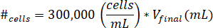
Note: 300,000 cells/mL is the concentration of cells used in the kECM gel. - Pipette the 10 µL of cell suspended solution into the final kECM gel after the kECM stock solution has been mixed with neutralizing reagent solution. Stir the solution with a microspatula until the cells are evenly distributed.
- Suspend cells into 10 µL of cell culture media. Determine the number of cells to be suspended by using the following equation, where #cells implies the number of cells to suspend and Vfinal is the volume of gel created:
- Gel the hydrogel by mixing the kECM hydrogel stock solution with 1 N NaOH, 10x Media Supplement (M199), and cell culture media. Keep all the solutions on ice.
- Use a 1 mL syringe to fill a desired cell culture device with the kECM hydrogel.
- Allow the gel to set at 37 ˚C for 1 h before transferring or plating cells.
The kECM hydrogel provides a matrix for kidney cell culture with similar chemical composition as the native kidney microenvironment. To fabricate the hydrogel, kidney cortex tissue is mechanically isolated from a whole kidney organ and diced (Figure 1). Decellularization with a chemical detergent (Figure 2A.1-A.3) followed by rinses with water to remove detergent particles (Figure 2A.4-A.6) yields isolated kidney cortex ECM. Histological evaluation confirms typical basal laminar proteins such as collagen-IV and laminin and structural proteins such as collagen-I are preserved, with vitronectin as the only noted exception (Figure 2B). Furthermore, protein composition, including preservation of isoforms, in the ECM remains consistent with observed values in native kidney ECM (Figure 3). The ECM (Figure 4A) is mechanically homogenized and lyophilized (Figure 4B) then solubilized to produce the final kECM hydrogel (Figure 4C). The kECM hydrogel appeared opaque with small amounts of visible tissue and was not as viscous as traditional collagen-I hydrogel. Rheological measurement of gelled kECM at a concentration of 15 mg/mL revealed a complex modulus (dynamic elastic modulus) of around 800 Pa over the linear range of strain values, significantly greater than that of 7.5 mg/mL collagen-I (p = 1.602E-14). Human kidney peritubular microvascular endothelial cells (HKMECs) cultured on collagen-I, kECM, and a 1:1 mixture gel showed differences in phenotype, specifically in CD31 expression around cell surfaces and junctions (Figure 5). HKMECs cultured on collagen-I displayed uniform CD31 expression while HKMECs cultured on the two gels containing kECM displayed reduced CD31 expression in uneven distributions. Matrix type did not appear to affect the high PV1 and low VWF expression in the HKMECs.
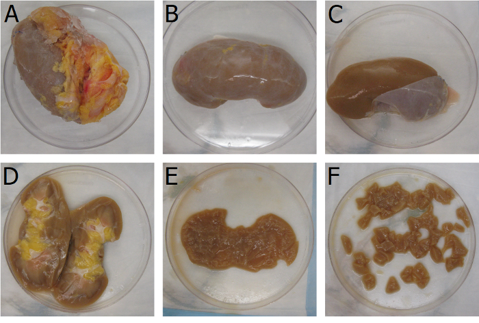
Figure 1: Isolation of kidney cortex tissue. Mechanical processing of a whole kidney organ to isolate the cortex tissue as a base material for the kECM hydrogel. Mechanical isolation begins with (A) the removal of the perirenal adipose tissue. Large pieces of adipose tissue can be torn away from the kidney with hemostat clamps. Remaining pieces of adipose tissue can be removed by running a scalpel against the renal capsule at an angle. (B) The renal capsule is best removed by making a shallow incision along the superior end of the kidney and (C) peeling the renal capsule away from the underlying tissue with hemostat clamps. (D) Bisecting the kidney along the coronal axis allows for the visualization of the cortex and medulla regions. (E) Isolation of the cortex tissue is best done by carving out pieces of the medulla tissue with a scalpel. The color of the cortical region is noticeably darker than that of the medullar region and can be used differentiate the two anatomically distinct tissues. Final processing of the cortex tissue involves (F) dicing the tissue into 0.5 cm3 pieces to aid in subsequent decellularization. Please click here to view a larger version of this figure.
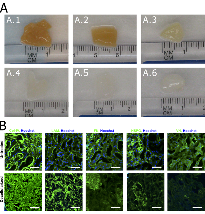
Figure 2: Decellularization of cortex tissue. Visual and histological characterization of decellularized tissue. (A.1) Submerging diced cortex tissue in 1% SDS solution causes lysing of cells and removal of cellular material. After (A.2) 1 h and (A.3) 24 h, the tissue begins to lose color, indicating cellular matter is being removed. By (A.4) 120 h the tissue is blanched and only the ECM remains. Rinses with water at (A.5) 24 h and (A.6) 120 h show no visible changes to the tissue. (B) Immunofluorescence staining of untreated and decellularized cortex tissue reveals near complete removal of cellular matter and preservation of major structural proteins (collagen-IV = Col-IV; laminin = LAM; fibronectin = FN; heparin sulfate proteoglycans = HSPG; and vitronectin = VN). Scale = 100 µm. Please click here to view a larger version of this figure.
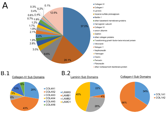
Figure 3: Mass spectroscopy of decellularized tissue. Analysis of decellularized cortex tissue by mass spectrometry to determine ECM protein composition. All ratios are measured as mass percent. (A) Structural and other proteins associated with the basal lamina were present with collagen-IV and -I being the most highly represented. (B.1) Collagen-IV A1 and A2 chains, ubiquitous in all basement membranes, were conserved. Collagen-IV A3 and A5 chains, present only in basement membranes of the glomerulus, were also detected. (B.2) Common isoforms of laminins and (B.3) collagen-I were also detected. This figure was reproduced with permission12. Please click here to view a larger version of this figure.
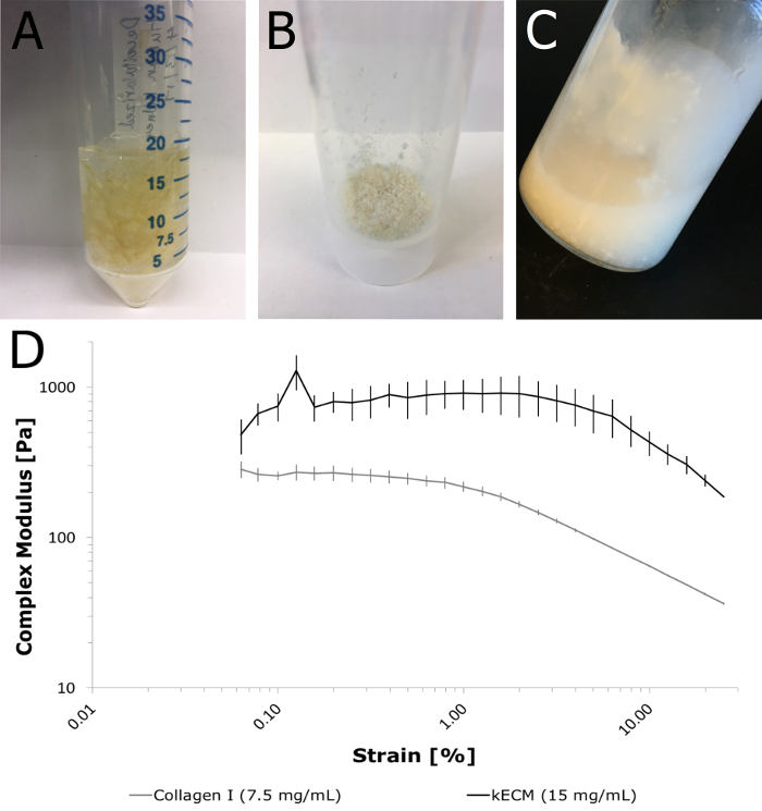
Figure 4: Fabrication of kECM hydrogel. Mechanical and chemical processing of decellularized cortex tissue yields a workable kECM hydrogel with sufficient mechanical properties following gelation with neutralizing reagents. (A) Decellularized cortex tissue is mechanically homogenized with a tissue homogenizer until no visible pieces of ECM remain. (B) A coarse powder was yielded after 3 days under lyophilization. (C) Solubilization in HCl and chemical digestion with pepsin in a scintillation vial resulted in a workable kECM hydrogel. The hydrogel was opaque and of a low viscosity. (D) Physical characterization of the kECM hydrogel following gelation with neutralizing reagents. Rheological experiments were performed with a 30 mm diameter parallel plate system. The sample edges were protected with mineral oil and the loading platform was set at 37 ˚C. The kECM hydrogel was allowed to gel for 1 h prior to testing. Viscoelastic properties of the gel were measured with a strain sweep between 0.01 to 20%. Three samples of both kECM and collagen-I were tested (n = 3). Please click here to view a larger version of this figure.
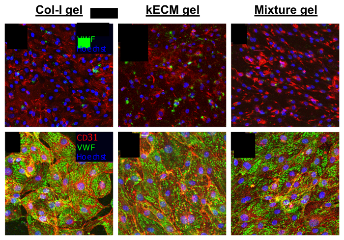
Figure 5: Cell growth characterization. Characterization of morphological differences in HKMECs grown on different matrices. Hydrogels were mixed with neutralizing reagents and set at 37 ˚C for 45 min in open-faced polydimethylsiloxane molds. HKMECs were seeded on the surface of the gels and kept in culture for 72 h before being fixed and stained. Immunofluorescent images of HKMECs cultured in (A and D) 7.5 mg/mL collagen-I gel; (B and E) 7.5 mg/mL kECM gel; and (C and F) 7.5 mg/mL 1:1 mixture gel. (A-C): red = CD31; green = VWF; blue = nuclei; scale bar = 50 µm. (D-F): red = F-actin; green = PV1; blue = nuclei; scale bar = 50 µm. This figure was reproduced with permission12. Please click here to view a larger version of this figure.
