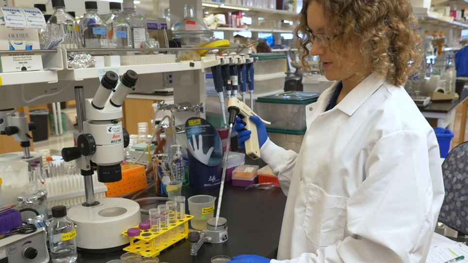
チミジン アナログ エドゥと線虫の生殖における細胞周期解析
Summary
イメージング手法の説明 S 相を識別し、チミジンを使用して線虫の両性生殖における細胞周期ダイナミクスの分析に使用できるアナログ教育。このメソッドは、遺伝子を必要とせず、免疫蛍光染色と互換性のあります。
Abstract
真核生物における細胞周期解析はよく染色体形態、式および/または細胞サイクルのさまざまな段階やヌクレオシド アナログの取り込みに必要な遺伝子産物の局在を利用しています。S 期には、DNA ポリメラーゼはマーキング解析のための細胞の染色体 DNA に EdU など BrdU チミジン類似体を組み込みます。線虫 c. エレガンスのヌクレオシド アナログ エドゥ規則的な文化の中にワームに送られ、蛍光技術と互換性のあります。線虫 c. エレガンスの生殖細胞は透明、遺伝子、減数分裂前期や細胞分化/配偶子形成で発生するので、経路、幹細胞、減数分裂、および細胞周期をシグナル伝達の研究の強力なモデル系、線形アセンブリのようなファッション。これらの機能は、エドゥ サイクリング有糸分裂細胞と生殖細胞の発生の動的側面を勉強する素晴らしいツールを確認します。このプロトコルが正常に野生型に餌をエドゥ細菌を準備する方法をについて説明しますc. の elegansの両性具有、両性の生殖腺を解剖、エドゥの DNA 混入のため様々 な細胞周期を検出する抗体で染色し、発達のマーカーは生殖腺をイメージし、結果を分析します。プロトコルでは、さまざまなメソッドおよび S 相インデックス、M フェーズ インデックス、G2 期間、細胞周期期間、減数分裂のエントリの率および減数分裂前期の進行の率の測定のための分析について説明します。このメソッドは、細胞周期や他の組織、段階、遺伝的背景、および生理的条件でセルの歴史を研究する合わせることができます。
Introduction
動物の開発、数百、数千、百万、十億、または細胞の分裂も兆は、大人の有機体を形成する必要があります。細胞周期細胞イベントのセットから成る G1 (ギャップ)、S (合成)、G2 (ギャップ)、M (有糸分裂) 定義のシリーズと、イベントは各細胞分裂を実行します。細胞周期は、ダイナミックで技術的に難しいことができますリアルタイムで最高評価です。このプロトコルで説明する技法は、段階の測定を行い、静止画像からの細胞周期のタイミングに 1 つを許可します。
線虫(C. elegans) 大人の細胞周期ダイナミクスの研究で S 相を識別するゴールド スタンダードは、ヌクレオシド アナログ 5-エチニル-2′-デオキシウリジン (EdU) など 5-ブロモ-2′-デオキシウリジン (BrdU) 標識両性生殖1,2,3,4,5。彼らはどのような遺伝子構成に依存しないと、エドゥと BrdU をほぼすべての遺伝的背景に使用できます。BrdU を可視化、染色、しばしば共同の追加抗体の汚損によって視覚化他細胞マーカーの評価と互換性がない抗 BrdU 抗体の抗原を公開する過酷な化学治療が必要です。対照的に、エドゥを可視化するクリックケミストリー温和な条件下で発生し、従って抗体染色6,7と互換性のあります。
核のみ S 段階で DNA にチミジン (5-エチニル-2′-デオキシウリジン) 類縁体を組み込むので、ラベルの特異性は、はっきり。可視化は、固定組織で行われます。アジ化物を含む色素までひとりでにエドゥのラベルが表示されていないまたは fluorophore クリックの銅触媒を用いた化学8でエドゥのアルキンと反応して共有結合。エドゥのラベリングの核は、S 期、ラベリングの短いパルスを用いた即時情報を提供できます。エドゥも情報を提供できます動的パルス追跡または連続の分類; を使用してたとえば、パルス追跡実験ラベルは細胞分裂のたびに希釈または開発を経て、非分裂細胞進行を反映します。
線虫の両性生殖は、シグナリング細道、幹細胞、減数分裂、および細胞周期の研究の強力なモデル システムです。大人の生殖は、エントリと減数分裂前期、配偶子形成の段階より近位のコーディネート (図 1) の進行に続いて遠位端見られる幹細胞と偏光の流れ作業です。近位端に卵母細胞は成熟、排卵し受精し、子宮9,10,11の胚発生を開始します。細胞直径 20 〜 長い減数分裂前期のセルではなく、減数分裂期の S 期細胞前駆細胞有糸分裂サイクリングの生殖細胞系幹が含まれています、先端のセル付近と呼びます前駆ゾーン2,4,9,12. 細胞膜遠位の生殖細胞の核間の不完全な分離を提供しますが、前駆細胞のゾーンを経る有糸分裂細胞主として独立していないサイクリングします。若い大人の両性具有の生殖細胞のゾーンの中央の有糸分裂細胞周期期間は ~6.5 h;G1 期は簡単なまたは欠席、静穏化を1,2,13守らないと。生殖細胞系幹細胞の分化本質的に直接分化が発生し、こうしてトランジット増幅部門4を欠いています。Pachytene 段階の分化、約 5 のうち 4 核卵母細胞を形成しないが、アポトーシス、発展途上卵母細胞12,14細胞内容を寄贈ナース細胞として機能を代わりに受ける,15。
ヌクレオシド アナログ S 相の標識細胞に加えて、1 つは有糸分裂と減数分裂抗体の汚損を使用してセルを識別できます。有糸分裂の核は反リン酸化ヒストン H3 に陽性 (Ser10) (pH3 と呼出される) 抗体7,16。減数分裂の核は反彼 3 抗体 (減数分裂期染色体軸蛋白質)17に陽性です。前駆ゾーンの核は、彼 3、核質 REC 818の存在や WAPL 119の存在の有無によって識別できます。WAPL 1 強度、前駆ゾーンで高体生殖腺における最高低初期減数分裂 prophases19の間。プロトコルのいくつかのバリエーションを持ついくつかの細胞周期測定が可能な: 私) S 相の核を識別し、S 期のインデックスを測定II) M 段階で核を識別し、M フェーズ インデックスを測定III) 原子核は、有糸分裂または減数分裂 S 相; かどうかを決定します。IV) G2; 時間を測定します。V) G2 + M + G1 フェーズの duartion を測定します。VI) 減数分裂の入力の速度を測定します。VII) 減数分裂の進行の率を推定します。
1 つは、ウェット実験の種類から複数の細胞周期測定を行うことができます。以下のプロトコルは、エドゥ アンチ pH3 抗体と前駆ゾーン細胞アンチ WAPL 1 抗体の染色による染色による細菌や共同ラベリング M 期の細胞をラベルにc. の elegans大人の両性具有を供給することにより 30 分パルス ラベルについて説明します。エドゥのフィード (ステップ 2.5)、抗体の種類の期間の変更の採用 (手順 5)、追加の計測に必要な解析 (ステップ 8.3)。
Protocol
Representative Results
Discussion
エドゥ ラベル細菌 (ステップ 1) の準備は、このプロトコルのトラブルシューティングの最初の点が重要です。4 h エドゥ パルス、エドゥ ラベル細菌の新しいバッチごとに便利なコントロールを作ってこれに非常に確実に野生型若い大人の両性具有のラベルです。さらに、(より古い動物または特定の咽頭/グラインダー欠陥変異) 腸を入力そのままのエドゥ ラベル細菌はクリックケミストリー?…
Disclosures
The authors have nothing to disclose.
Acknowledgements
MG1693; のエシェリヒア属大腸菌のストック センターに感謝しておりますWormbase;国家機関の健康オフィスの研究インフラストラクチャのプログラム (P40OD010440) 系統; によって資金が供給される線虫の遺伝学センターザック ピンカス統計的アドバイス;愛平公使風水の試薬;ルーク ・ シュナイダー、アンドレア ・ シャーフ、サンディープ クマー、トレーニング、アドバイス、サポート、および有用な議論; のジョン ・フィードバックのために本稿では Kornfeld と Schedl ラボ。この作品は一部で健康の国民の協会によって支えられた [R01 AG02656106A1 株式会社、TS に R01 GM100756 へ] と全米科学財団を経て交わり [DGE 1143954 と ZK を DGE 1745038]。健康の国民の協会、全米科学財団、研究、収集、分析、およびデータの解釈のデザインにも原稿を書くに任意の役割を持っていた。
Materials
| E. coli MG1693 | Coli Genetic Stock Center | 6411 | grows fine in standard unsupplemented LB |
| E. coli OP50 | Caenorhabditis Genetics Center | OP50 | |
| Click-iT EdU Alexa Fluor 488 Imaging Kit | Thermo Fisher Scientific | C10337 | |
| 5-Ethynyl-2′-deoxyuridine | Sigma | 900584-50MG | or use EdU provided in kit |
| Glucose | Sigma | D9434-500G | D-(+)-Dextrose |
| Thiamine (Vitamin B1) | Sigma | T4625-5G | Reagent Grade |
| Thymidine | Sigma | T1895-1G | BioReagent |
| Magnesium sulfate heptahydrate | Sigma | M1880-1KG | MgSO4, Reagent Grade |
| Sodium Phosphate, dibasic, anhydrous | Fisher | BP332-500G | Na2HPO4 |
| Potassium Phosphate, monobasic | Sigma | P5379-500G | KH2PO4 |
| Ammonium Chloride | Sigma | A4514-500G | NH4Cl, Reagent Plus |
| Bacteriological Agar | US Biological | C13071058 | |
| Calcium Chloride dihydrate | Sigma | C3881-500G | CaCl |
| LB Broth (Miller) | Sigma | L3522-1KG | Used at 25g/L |
| Levamisole | Sigma | L9756-5G | 0.241g/10ml |
| Phosphate buffered saline | Calbiochem Omnipur | 6506 | homemade PBS works just as well |
| Tween-20 | Sigma | P1379-500ML | |
| 16% Paraformaldehyde, EM-grade ampules | Electron Microscopy Sciences | 15710 | 10ml ampules |
| 100% methanol | Thermo Fisher Scientific | A454-1L | Gold-label methanol is critical for proper morphology with certain antibodies |
| Goat Serum | Gibco | 16210-072 | Lot 1671330 |
| rabbit-anti-WAPL-1 | Novus biologicals | 49300002 | Lot G3048-179A02, used at 1:2000 |
| mouse-anti-pH3 clone 3H10 | Millipore | 05-806 | Lot#2680533, used at 1:500 |
| goat-anti-rabbit IgG-conjugated Alexa Fluor 594 | Invitrogen | A11012 | Lot 1256147, used at 1:400 |
| goat-anti-mouse IgG-conjugated Alexa Fluor 647 | Invitrogen | A21236 | Lot 1511347, used at 1:400 |
| Vectashield antifade mounting medium containing 4',6-Diamidino-2-Phenylindole Dihydrochloride (DAPI) | Vector Laboratories | H-1200 | mounting medium without DAPI can be used instead, following a separate DAPI incubation |
| nail polish | Wet n Wild | DTC450B | any clear nail polish should work |
| S-medium | various | see wormbook.org for protocol | |
| M9 buffer | various | see wormbook.org for protocol | |
| M9 agar | various | same recipe as M9 buffer, but add 1.7% agar | |
| Nematode Growth Medium | various | see wormbook.org for protocol | |
| dissecting watch glass | Carolina Biological | 42300 | |
| Parafilm laboratory film | Pechiney Plastic Packaging | PM-996 | 4 inch wide laboratory film |
| petri dishes | 60 mm diameter | ||
| Long glass Pasteur pipettes | |||
| 1ml centrifuge tubes | MidSci Avant | 2926 | |
| Tips | |||
| Serological pipettes | |||
| 500 mL Erlenmyer flask | |||
| Aluminum foil | |||
| 25G 5/8” needles | BD PrecisionGlide | 305122 | |
| 5ml glass centrifuge tube | Pyrex | ||
| Borosilicate glass tubes 1ml | |||
| glass slides | |||
| no 1 coverslips 22 x 40 mm | no 1.5 may work, also | ||
| 37 °C Shaker incubator | |||
| Tabletop Centrifuge | |||
| Clinical Centrifuge | IEC | 428 | with 6 swinging bucket rotor |
| Mini Centrifuge | |||
| 20 °C incubator | |||
| 4 °C refrigerator | |||
| -20 °C freezer | |||
| Observer Z1 microscope | Zeiss | ||
| Plan Apo 63X 1.4 oil-immersion objective lens | Zeiss | ||
| Ultraview Vox spinning disc confocal system | PerkinElmer | Nikon spinning disc confocal system works very well, also, as described here: http://wucci.wustl.edu/Facilities/Light-Microscopy |
References
- Fox, P. M., Vought, V. E., Hanazawa, M., Lee, M. -. H., Maine, E. M., Schedl, T. Cyclin E and CDK-2 regulate proliferative cell fate and cell cycle progression in the C. elegans germline. Development. 138 (11), 2223-2234 (2011).
- Crittenden, S. L., Leonhard, K. A., Byrd, D. T., Kimble, J. Cellular analyses of the mitotic region in the Caenorhabditis elegans adult germ line. Molecular biology of the cell. 17 (7), 3051-3061 (2006).
- Seidel, H. S., Kimble, J. Cell-cycle quiescence maintains Caenorhabditis elegans germline stem cells independent of GLP-1/Notch. eLife. 4, (2015).
- Fox, P. M., Schedl, T. Analysis of Germline Stem Cell Differentiation Following Loss of GLP-1 Notch Activity in Caenorhabditis elegans. 유전학. 201 (9), 167-184 (2015).
- Kocsisova, Z., Kornfeld, K., Schedl, T. Cell cycle accumulation of the proliferating cell nuclear antigen PCN-1 transitions from continuous in the adult germline to intermittent in the early embryo of C. elegans. BMC Developmental Biology. 18 (1), (2018).
- Salic, A., Mitchison, T. J. A chemical method for fast and sensitive detection of DNA synthesis in vivo. Proceedings of the National Academy of Sciences. 105 (7), 2415-2420 (2008).
- vanden Heuvel, S., Kipreos, E. T. C. elegans Cell Cycle Analysis. Methods in Cell Biology. , 265-294 (2012).
- ThermoFisher. . Click-iT EdU Imaging Kits. , (2011).
- Pazdernik, N., Schedl, T. . Germ Cell Development in C. elegans. , 1-16 (2013).
- Hirsh, D., Oppenheim, D., Klass, M. Development of the reproductive system of Caenorhabditis elegans. 발생학. 49 (1), 200-219 (1976).
- Brenner, S. The genetics of Caenorhabditis elegans. 유전학. 77 (1), 71-94 (1974).
- Hansen, D., Schedl, T. Stem cell proliferation versus meiotic fate decision in Caenorhabditis elegans. Advances in Experimental Medicine and Biology. 757, 71-99 (2013).
- Jaramillo-Lambert, A., Ellefson, M., Villeneuve, A. M., Engebrecht, J. Differential timing of S phases, X chromosome replication, and meiotic prophase in the C. elegans germ line. 발생학. 308 (1), 206-221 (2007).
- Agarwal, I., Farnow, C., et al. HOP-1 presenilin deficiency causes a late-onset notch signaling phenotype that affects adult germline function in Caenorhabditis elegans. 유전학. 208 (2), 745-762 (2018).
- Gumienny, T. L., Lambie, E., Hartwieg, E., Horvitz, H. R., Hengartner, M. O. Genetic control of programmed cell death in the Caenorhabditis elegans hermaphrodite germline. Development. 126 (5), 1011-1022 (1999).
- Hendzel, M. J., Wei, Y., et al. Mitosis-specific phosphorylation of histone H3 initiates primarily within pericentromeric heterochromatin during G2 and spreads in an ordered fashion coincident with mitotic chromosome condensation. Chromosoma. 106 (6), 348-360 (1997).
- Zetka, M. C., Kawasaki, I., Strome, S., Mü Ller, F. Synapsis and chiasma formation in Caenorhabditis elegans require HIM-3, a meiotic chromosome core component that functions in chromosome segregation. Genes & development. 13 (17), 2258-2270 (1999).
- Hansen, D., Hubbard, E. J. A., Schedl, T. Multi-pathway control of the proliferation versus meiotic development decision in the Caenorhabditis elegans germline. 발생학. 268 (2), 342-357 (2004).
- Crawley, O., Barroso, C., et al. Cohesin-interacting protein WAPL-1 regulates meiotic chromosome structure and cohesion by antagonizing specific cohesin complexes. eLife. 5 (2), 1-26 (2016).
- Zhao, H., Halicka, H. D., et al. DNA damage signaling, impairment of cell cycle progression, and apoptosis triggered by 5-ethynyl-2′-deoxyuridine incorporated into DNA. Cytometry Part A. 83 (11), 979-988 (2013).
- Stiernagle, T. Maintenance of C. elegans. WormBook. , 1-11 (2006).
- Gervaise, A. L., Arur, S. Spatial and Temporal Analysis of Active ERK. in the C. elegans Germline. Journal of Visualized Experiments. 117 (117), e54901-e54901 (2016).
- Vos, K. . De Cell Counter Plugin. , (2015).
- Schindelin, J., Arganda-Carreras, I., et al. Fiji: an open-source platform for biological-image analysis. Nature Methods. 9 (7), 676-682 (2012).
- Rasband, W. . ImageJ. , (2016).
- Michaelson, D., Korta, D. Z., Capua, Y., Hubbard, E. J. A. Insulin signaling promotes germline proliferation in C. elegans. Development. 137 (4), 671-680 (2010).
- Qin, Z., Jane, E., Hubbard, A., Hubbard, E. J. A. Non-autonomous DAF-16/FOXO activity antagonizes age-related loss of C. elegans germline stem/progenitor cells. Nature communications. 6 (5), 7107 (2015).
- Luo, S., Kleemann, G. A., Ashraf, J. M., Shaw, W. M., Murphy, C. T. TGFB and Insulin Signaling Regulate Reproductive Aging via Oocyte and Germline Quality Maintenance. Cell. 143 (2), 299-312 (2010).
- Narbonne, P., Maddox, P. S., Labbe, J. -. C. daf-18/PTEN locally antagonizes insulin signalling to couple germline stem cell proliferation to oocyte needs in C. elegans. Development. , 4230-4241 (2015).
- Cinquin, A., Chiang, M., et al. Intermittent Stem Cell Cycling Balances Self-Renewal and. Senescence of the C. elegans Germ Line. PLoS Genetics. 12 (4), 1005985 (2016).
- . . Invitrogen EdU (5-ethynyl-2’-deoxyuridine). , 1-7 (2010).
- Tuttle, A. H., Rankin, M. M., et al. Immunofluorescent Detection of Two Thymidine Analogues (CldU and IdU) in Primary Tissue. Journal of Visualized Experiments. 46 (46), e2166-e2166 (2010).

