All animal procedures were performed in accordance with an approved institutional animal care and use committee (IACUC) protocol of the University of Colorado Denver (protocol # 00250).
1. Tumor Cell Culture
NOTE: B4B8 and LY2 cell lines were used to generate orthotopic HNSCC tumors: B4B8 tumor cells were derived from carcinogen-transformed mucosal keratinocytes (from BALB/C mice)11. LY2 tumor cells were derived from lymph node metastases of a spontaneously transformed BALB/C keratinocyte line (Pam 212)12,13. Both cell lines were kindly provided by Dr. Nadarajah Vigneswaran (UTHealth, Houston, TX, USA).
- Use Dulbecco’s modified Eagle’s medium (DMEM) F/12 supplemented with 10% fetal bovine serum (FBS) and 1% antimicrobial reagent for the cell line maintenance. Maintain these cell lines in a sterile incubator at 37 °C and 5% CO2. Cells should be used for inoculation before exceeding 15 passages.
- Plate 2 x 10⁶ to 4 x 10⁶ cells in 175 cm2 cell culture flasks.
NOTE: Ensure the cells are less than 90% confluent to avoid inducing a stress response. - When the cells are 70% confluent (~48 h), remove the flask from the incubator and wash the cells 3x with cold phosphate-buffered saline (PBS).
- Detach the cells from the flask using 0.25% trypsin, enough to cover the surface of the plate.
- To do this, dispense 4 mL of trypsin in a 175 cm2 flask containing 70% confluent cells. Incubate the cells with trypsin for 3–4 min in the cell culture incubator at 37 °C and 5% CO2.
- Visualize the cells under a microscope to ensure cell detachment.
- Add 12 mL of DMEM F/12 media containing FBS to neutralize trypsin activity.
- Aspirate the cell suspension and place it into a 50 mL conical tube.
- Shake the tube containing the cells by inverting it 3x–4x.
- Optionally, mix a 10 µL aliquot of cell suspension with 10 µL of Trypan blue in a microcentrifuge tube and count the cells using a hemocytometer. Determine cell viability by subtracting the number of Trypan-blue-positive cells from the total number of cells and divide by the total number of cells.
- Centrifuge the cell suspension at 300 x g for 5 min at 4 °C.
- Resuspend the cells in serum-free and antibiotic-free DMEM at an appropriate volume so that 1 x 10⁶ cells are present in 50 µL of media.
NOTE: Based on the in vivo cell line aggressiveness (determined empirically), 1 x 10⁶ cells per injection site per mouse was deemed appropriate for B4B8 and LY2 cells. - Place the vial containing the cell suspension on ice.
- Place the pre-thawed basement membrane matrix on ice.
NOTE: The basement membrane matrix was thawed overnight at 4 °C.
2. Cell Injection into Mice
- Prepare a 1:1 mixture of cells:basement membrane matrix (50 µL each).
- Add the cells first; then, gradually pipette the basement membrane matrix. Avoid introducing air bubbles. Ensure that the mixture is made immediately before the animal injection. Adding cells to basement membrane matrix for an extended period of time can result in cell settling in the matrix mixture, which makes the mixture difficult to shake rigorously. This will cause a considerable variability in tumor size between mice.
- Mix gently. Ensure all steps involving matrix are performed on ice. Basement membrane matrix will polymerize at room temperature.
- Prepare syringes for inoculation.
- Load 0.5 mL insulin syringes (23 G) with 100 µL of the cell/basement membrane matrix solution.
- Keep the syringes on ice to avoid basement membrane matrix polymerization.
- Anesthetize mice by placing them in a chamber with isoflurane and oxygen (2.5%).
- Ensure the mice are deeply anesthetized before performing the injection (by ensuring a lack of response to a toe pinch).
- Insert the needle into the right or left buccal region. This is performed through the available open space on either side of the mouth.
- Ensure that the mouse’s tongue is not in the way.
NOTE: It is easy to poke the tongue, which will result in tongue tumors. Move the tongue to the opposite side if necessary. - Keep the syringe parallel to the buccal region while inside the oral cavity.
- When ready to inject, pull the syringe back and slowly insert the syringe at a 10° angle.
- Inject 100 µL of the cell/basement membrane matrix suspension over a period of 5 s.
- Hold the syringe in place for an additional 5 s to ensure all material is injected.
NOTE: For control nontumor-bearing mice, inject a mixture of serum-free media and matrix (as described above) without the tumor cells. - Withdraw the syringe gently.
- Continue the above procedure with the remaining mice.
- Allow for 1 week until tumors begin to appear grossly (50–200 mm3 for B4B8 and LY2 cells).
3. Mouse Monitoring
- Perform the first measurement using calipers at 1 week after the tumor cell injection. In order to calculate the tumor volume using external calipers, determine the greatest longitudinal diameter (length) and the greatest transverse diameter (width) and use the modified ellipsoidal formula14,15.
- Continue to perform regular caliper measurements for tumor volume (1x–2x per week at regular intervals).
- Measure animal weight to assess the effects of tumor growth on feeding.
4. Harvesting Tumors
- When the experimental endpoint is reached, euthanize the animals using appropriate measures (e.g., CO2 asphyxiation, decapitation, or cervical dislocation).
NOTE: In this study, the endpoint was reached if the mice became moribund (with a weight loss of >15% of their initial weight, a lack of grooming, cachexia) and/or if the tumor size reached 1,000 mm3. - Begin dissecting the animal by creating a long incision through the midline in the neck region.
- Use blunt forceps to grab the skin and sharp scissors to cut the skin.
- Insert scissors gently under the skin covering the tumor and create air pockets by pushing the scissors across and into the skin.
- Once the skin is sufficiently detached from the tumor, identify the draining lymph nodes (DLNs) and excise them to avoid having the tumor tissue confounded by the presence of lymph nodes.
NOTE: Large tumors may reach and/or cover the DLN. Depending on the type of assay being performed, the isolation of intact lymph nodes is essential to avoid spillage of immune cells into the tumor, which will result in skewing the assay results. - Cut through the borders of the tumor until the entire volume is detached.
5. Tumor Processing for Downstream Applications
- For the downstream histologic examination, place tumors in 10% formalin at room temperature. To avoid overfixation, replace the formalin with 70% ethanol. For flow cytometry analysis, process the tumors as described below.
NOTE: The tissue can be kept in formalin for 72 h before overfixation can become problematic for certain types of staining. - Cut the tumor into 1–2 mm-sized pieces using a sterile razor blade or sharp scissors.
- Place the cut tumor pieces in a 50 mL conical tube with collagenase III (4,250 units per sample), DNase I (0.1 mg per sample), and trypsin inhibitor (1 mg per sample).
- Incubate at 37 °C for 30 min with shaking every 10 min.
NOTE: The samples can be placed in a resealable bag, sterilized with 90% ethanol, and placed in a cell culture incubator. - After 30 min, add 20 mL of Hank’s balanced salt solution (HBSS) and spin at 300 x g for 5 min.
NOTE: HBSS is comprised of potassium chloride, sodium chloride, sodium bicarbonate, sodium phosphate dibasic, sodium phosphate monobasic and glucose. - Discard the supernatant and resuspend the pellet in 2–3 mL of red blood cell (RBC) lysis buffer. Pipette rigorously.
NOTE: RBC lysis buffer is comprised of ammonium chloride, sodium bicarbonate, and disodium. - Incubate for 2 min at room temperature.
NOTE: Longer incubation can be toxic to other cell populations. - Add 20 mL of HBSS to neutralize the effect of lysis buffer.
- Centrifuge at 300 x g for 5 min and discard the supernatant.
- Resuspend the pellet in 10 mL of HBSS.
- Pass the solution through a 70 µm nylon restrainer and centrifuge at 300 x g for 5 min. Discard the supernatant.
- Repeat steps 5.8–5.10 and pass the cell suspension through a 40 µm nylon restrainer to ensure any additional debris is removed from the suspension.
6. Cell Staining and Data Acquisition
- Resuspend the cell pellet in 1 mL of HBSS.
- Count the cells using a hemocytometer or an automated cell counter, as described in step 1.7
- Determine the total cell number and appropriate concentration for staining.
NOTE: The ideal cell concentration for flow cytometry staining is 1–2 million cells. For example, if staining is performed in a 96-well plate and there are 10 million cells, resuspend the sample in 1 mL and plate 100 µL for 1 million cells per well. - Add Fc block (CD16/CD32) at a concentration of 1:100 in order to prevent nonantigen-specific binding of immunoglobulins to the FcγIII and FcγII receptors. Incubate for 5 min at room temperature.
- Centrifuge and resuspend the cell pellet in 100 µL of flow cytometry cell staining buffer (comprised of saline solution containing 5% FBS, 2% EDTA, and 1% HEPES).
- Perform cell surface staining according to supplier-provided instructions (adding each antibody at the appropriate dilution). Incubate for 60 min at room temperature.
- Centrifuge and resuspend the cell pellet in 100 µL of HBSS. Repeat 2x to get rid of excess antibody.
- Run samples on an appropriate flow cytometer.
- If analyzing intracellular markers (e.g., foxp3), perform cell permeabilization and staining according to the supplier’s instructions.
The in vitro assessment of LY2 and B4B8 cell proliferation showed that both cell lines have similar doubling times (21 h and 23 h, respectively). In vivo, both cell lines formed a single, visible, and palpable mass within 1 week of inoculation (Figure 1A). In mice bearing LY2 tumors, the jaw was displaced by 3 weeks due to tumor burden (Figure 1A). Control mice that did not receive tumor cells did not develop tumors as anticipated. LY2 tumors grew at a higher rate compared to B4B8 tumors. The average tumor volume on day 21 in LY2 tumor-bearing mice was 632 ± 10 mm3 compared to 162 ± 4 mm3 in B4B8 tumors (Figure 1B). Mice exhibiting jaw displacement rapidly developed weight loss due to dysphagia (Figure 1C). Mice exhibiting significant weight loss defined as >15% of their initial weight and/or having a tumor volume of >1 cm3 were euthanized within 1 day of the noted observation. The median survival in LY2 mice was 22.5 days, compared to 52.0 days in B4B8 tumor-bearing mice (Figure 1D).
Magnetic resonance imaging (MRI) of tumor-bearing mice showed well-demarcated tumors extending into the inner layer of the buccal mucosa (Figure 2A). Tumors did not invade into the tongue or other nearby organs (esophagus, bronchus, thymus) as shown with histological assessment. Signal heterogeneity on MR images was representative of the presence of vascularized hyperintense regions (denoted with V) and necrotic hypointense regions (denoted with N) (Figure 2A). A gross pathological examination showed enlarged DLNs (Figure 2B). We further assessed tumor volume with computed tomography (CT) to determine the reliability of the caliper measurements. Tumors were delineated using a digital imaging and communications in medicine (DICOM) image analysis software16. Assessment of LY2 tumor volume by calipers and CT imaging showed an excellent correlation between the two methods (R2 = 0.8493; Figure 2C). This indicates that tumor growth is primarily exophytic and caliper measurement is a reliable method for the assessment of tumor growth. Histologic examination showed that all LY2 tumor-bearing mice developed poorly differentiated squamous cell carcinoma (Figure 2D). Nine out of nine LY2 tumor-bearing mice developed metastases to the first and second echelon lymph nodes. Nodal metastases were primarily subcapsular (with seven out of nine mice) and within sinuses (with five out of nine mice), with two out of nine mice demonstrating intracapsular invasion. In contrast, mice bearing B4B8 tumors developed moderately differentiated squamous cell carcinoma (Figure 2E) and did not metastasize to regional or distant sites, including DLNs. Necrosis was assessed based on a previously established three-grade scale: 0 = no visible necrosis, 1 = scant, 2 = moderate, and 3 = severe17. All LY2 tumor-bearing mice had histologically confirmed necrosis with the majority (seven out of ten) demonstrating moderate to severe necrosis (Figure 2F). No evidence of necrosis was observed in B4B8 tumors.
We focused on the LY2 tumor model for further characterization of the tumor-immune microenvironment. Tumors were harvested at 3 weeks after the tumor inoculation and digested using collagenase III, followed by the staining of the cell surface and intracellular markers for flow/mass cytometry (Figure 3A). Samples were processed, using a mass cytometer, at the University of Colorado Denver Flow Cytometry Shared Resource. Gating and data analysis were performed using flow cytometry commercial software. The data obtained showed the presence of numerous immune cell populations to varying degrees. Total immune cells (CD45+) represented 7.3% of the total tumor (Figure 3B). In absolute numbers, we observed, on average, 26 CD45+ cells (SD = 0.81) per milligram of tumor tissue (Figure 3B). Myeloid cells (CD11b+) represented 37.8% of all CD45+ cells (Figure 3C). Myeloid cells were largely comprised of macrophages (F4/80+, 63.0%), followed by myeloid-derived suppressor cells (MDSCs) (Gr1+, 8.51%) and neutrophils (Ly6G+, 5.87%). Total T cells comprised 15.9% of CD45+ cells with CD4+ T cells comprising the majority of all T cells (79.4%), of which 53.4% were regulatory T cells (Tregs) (CD4+FoxP3+) (Figure 3D). Natural killer (NK) cells comprised 1.75% of all CD45+ cells. These data highlight the presence of various immune infiltrates in LY2 orthotopic HNSCC tumors which likely play a role in mediating tumor progression and immune evasion.
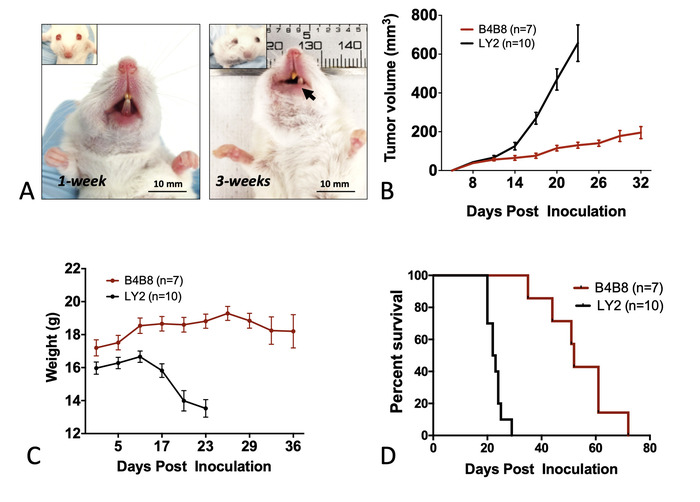
Figure 1: Monitoring mice bearing orthotopic HNSCC tumors. (A) Representative images of tumor-bearing mice. The inoculated buccal develops a tumor within 1 week of inoculation. At 3 weeks, displacement of the jaw is observed. (B) Tumor growth rate in B4B8 and LY2 tumor-bearing mice as determined by caliper measurements. (C) Changes in the body weight of mice bearing B4B8 or LY2 tumors. (D) Survival of B4B8 and LY2 tumor-bearing mice. The bars represent the standard error of the mean (SEM) of 7–10 mice per group. Please click here to view a larger version of this figure.
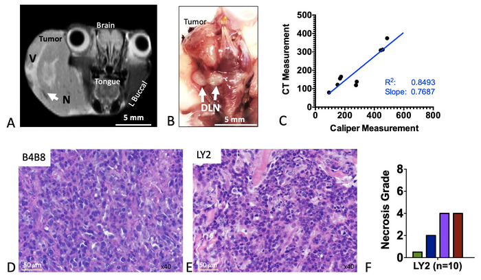
Figure 2: Imaging and histologic features of murine HNSCC tumors. (A) Representative coronal MR image of an LY2 tumor-bearing mouse. Anatomic sites are labeled. Areas of signal hyperintensity represent vascularized regions (denoted V). Regions of signal hypointensity represent necrotic areas (denote N). (B) Representative gross image of a dissected tumor-containing region. White arrows point to enlarged draining lymph nodes (DLNs). (C) Correlation of tumor volume as assessed by caliper measurement versus computed tomography (CT)-based assessment. Slope, correlation coefficient (R2), and line of best fit are shown. (D) Representative hematoxylin and eosin (H&E) image of LY2 tumor. (E) Representative H&E image of B4B8 tumor. (F) Quantification of necrosis in LY2 tumors, based on an H&E four-grade scoring system. Please click here to view a larger version of this figure.
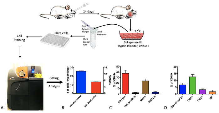
Figure 3: Analysis of baseline intratumoral immune populations using cytometry by time-of-flight (CyTOF). Tumors were processed into a single-cell suspension using collagenase-based enzymatic digestion and, then, stained with cell surface and intracellular markers. (A) Mass cytometry was performed using the CyTOF platform. (B) Absolute and relative quantitative assessment of total immune cells (CD45+). (C) Quantification of myeloid-immune subpopulations. (D) Quantification of lymphoid immune subpopulations. Please click here to view a larger version of this figure.
