The goal of this protocol is to test the ability of progenitor cells derived from human perivascular adipose tissue to differentiate into multiple cell lineages. Differentiation was compared to mesenchymal stem cells derived from human bone marrow, which is known to differentiate into adipocyte, osteocyte, and chondrocyte lineages.
Adipose tissue is a rich source of multi-potent mesenchymal stem cells (MSC) capable of differentiating into osteogenic, adipogenic, and chondrogenic lineages. Adipogenic differentiation of progenitor cells is a major mechanism driving adipose tissue expansion and dysfunction in response to obesity. Understanding changes to perivascular adipose tissue (PVAT) is thus clinically relevant in metabolic disease. However, previous studies have been predominately performed in the mouse and other animal models. This protocol uses human thoracic PVAT samples collected from patients undergoing coronary artery bypass graft surgery. Adipose tissue from the ascending aorta was collected and used for explantation of the stromal vascular fraction. We previously confirmed the presence of adipose progenitor cells in human PVAT with the capacity to differentiate into lipid-containing adipocytes. In this study, we further analyzed the differentiation potential of cells from the stromal vascular fraction, presumably containing multi-potent progenitor cells. We compared PVAT-derived cells to human bone marrow MSC for differentiation into adipogenic, osteogenic, and chondrogenic lineages. Following 14 days of differentiation, specific stains were utilized to detect lipid accumulation in adipocytes (Oil red O), calcific deposits in osteogenic cells (Alizarin Red), or glycosaminoglycans and collagen in chondrogenic cells (Masson’s Trichrome). While bone marrow MSC efficiently differentiated into all three lineages, PVAT-derived cells had adipogenic and chondrogenic potential, but lacked robust osteogenic potential.
Adipose tissue is a rich source of multi-potent mesenchymal stem cells (MSC) capable of differentiating into osteogenic, adipogenic, and chondrogenic lineages1. This tissue expands through hypertrophy of mature adipocytes and de novo differentiation of resident MSC to adipocytes. Perivascular adipose tissue (PVAT) surrounds blood vessels and regulates vascular function2,3. Obesity-induced PVAT expansion exacerbates cardiovascular pathologies. While the multipotent potential of MSC from human subcutaneous adipose depots have been well studied4,5, no studies have explanted and evaluated the differentiation capacity of human PVAT-derived progenitor cells, likely due to the invasiveness of procurement. Thus, the goal of this work is to provide a methodology to explant and propagate progenitor cells from human aortic PVAT from patients with cardiovascular disease and to test their propensity to differentiate to osteogenic, chondrogenic, and adipogenic lineages. Our source of PVAT is from the site of anastomosis of the bypass graft on ascending aorta of obese patients undergoing coronary artery bypass graft surgery. Freshly-isolated PVAT is enzymatically-dissociated and the stromal vascular fraction is isolated and propagated in vitro, enabling us to test for the first time the differentiation capacity of human PVAT-derived progenitor cells.
Using primary cultured human PVAT stromal vascular fraction, we tested three assays designed to induce stem/progenitor cells to differentiate toward adipogenic, osteogenic, or chondrogenic lineages. Our prior study identified a population of CD73+, CD105+, and PDGFRa+ (CD140a) cells that can robustly differentiate into adipocytes6, although their multipotency was not tested. PVAT directly regulates vascular tone and inflammation7. The rationale for testing the differentiation potential of this novel cell population is to begin to understand the specialized influence of PVAT on vascular function, and mechanisms of PVAT expansion during obesity. This methodology enhances our understanding of the functions of adipose-tissue derived progenitor cells and enables us to identify and compare similarities and differences of progenitor cells from different tissue sources. We build upon established and validated approaches for isolating and differentiating MSC towards different lineages and optimize procedures to maximize the viability of human PVAT-derived progenitor cells. These techniques have broad applications in the fields of stem and progenitor cell research and adipose tissue development.
The use of human tissues in this study was evaluated and approved by the Institutional Review Board of Maine Medical Center, and all personnel received appropriate training prior to experimentation.
1. Preparations
2. Protocol 1: Culture Human PVAT Cells from the Stromal Vascular Fraction
NOTE: PVAT is resected from the site of graft anastomosis on the ascending aorta of anesthetized patients undergoing coronary artery bypass graft procedures. Aortic PVAT is placed in a 15 mL conical containing 10 mL ice cold high glucose DMEM F12 and transferred from the operating room to the laboratory within 2 h of resection. Aortic PVAT is discarded tissue during bypass procedure and has been deemed as non-human subjects research by Maine Medical Center’s Internal Review Board.
3. Protocol 2: Culture Human Bone Marrow MSC Colonies
NOTE: Human bone marrow MSC are isolated as described8 and stored as early passage frozen stocks in freeze media (70% FBS 20% basal DMEM and 10% DMSO) at ~100,000 cell/mL in liquid N2.
4. Protocol 3: Plate and Induce Adipogenic, Osteogenic, and Chondrogenic Lineages
5. Protocol 4: Culture Adipogenic, Osteogenic, and Chondrogenic Lineages for 14 Days
6. Protocol 5: Staining Adipogenic Condition with Oil Red O
7. Protocol 6: Staining Osteogenic Condition with Alizarin Red
8. Protocol 7: Staining Chondrogenic Condition with Masson’s Trichrome
Isolation of stromal vascular fraction from human PVAT
Figure 1A shows a schematic of the anatomical region where the PVAT overlying the ascending aorta was obtained. We previously described the patient populations undergoing coronary artery bypass grafting from which these samples were derived6. Figure 1B shows an example of the human PVAT obtained following surgery. Figure 1C shows a representative PVAT tissue section stained with Masson’s Trichrome stain. Figure 1D is a phase contract micrograph showing a population of PVAT-derived stromal cells during the expansion phase prior to differentiation. Cells can be expanded and frozen for future use. Cells are typically frozen at a density of 2.5 x 10⁵ cells/mL in a media comprised of 70% FBS, 20% basal (antibiotic free) DMEM F12 and 10% DMSO in an isopropanol cryochamber at -80 °C for 24 h and moved to the liquid phase of a liquid N2 freezer for long-term storage.
Adipogenic differentiation
Studies were performed in parallel with human bone marrow-derived MSC (Figure 2A,B) and PVAT-derived progenitor cells (Figure 2C,D). The left panels of Figure 2 show the non-induced condition, where no lipid accumulation is evident. The right panels show cells following adipocyte differentiation and staining of neutral lipids with Oil Red O. While the degree of differentiation in the human aortic PVAT-derived cells is more robust, both human cell sources exhibited the ability to differentiate towards the adipogenic lineage.
Osteogenic differentiation
The osteogenic differentiation protocol was used for human bone marrow-derived MSC (Figure 3A–C) and PVAT-derived cells (Figure 3D–F). Non-induced cells (Figure 3A,D) did not stain with Alizarin Red. After the osteogenic differentiation protocol, the human MSC developed calcified nodules that stained with Alizarin Red (Figure 3B–C), while human aortic PVAT-derived cells did not (Figure 3E–F). These data indicate that our preparation of cells from the stromal vascular fraction of human PVAT lack significant number of progenitors with the ability to undergo osteogenesis. Depending on the study and time course of differentiation, it is advisable to follow up standard stains with detection of molecular markers that define osteogenic lineage commitment (e.g. RUNX2, osterix, alkaline phosphatase) or osteoblasts (osteopontin, osteocalcin, alkaline phosphatase, BAP1).
Chondrogenic differentiation
Cells derived from both human bone marrow MSC (Figure 4A) and human PVAT (Figure 4B) display features characteristic of chondrogenic differentiation, with abundant collagen accumulation in the micromass. Micromasses formed from human bone marrow MSC and aortic PVAT-derived cells also exhibited abundant accumulation of glycosaminoglycans (blue) as indicated by Alcian blue staining (Figure 4C–D, respectively). Morphologically, structures similar to lacunae were detected with cells sitting in cavities surrounding by collagen deposition (Figure 4, arrows). Depending on the study and time course of differentiation, the detection of specific chondrogenic markers (aggrecan, collagen type II, osteonectin, Sox9) is useful.
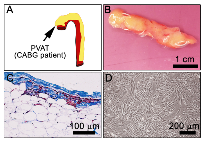
Figure 1: Morphological characteristics of human PVAT. (A) Cartoon depiction of the human aorta (red) with surrounding PVAT (yellow). Arrow indicates ascending aorta. (B) A 480 mg piece of human aortic PVAT from a CABG patient in DMEM prior to dissociation. (C) Masson’s trichrome staining of formalin-fixed paraffin-embedded human PVAT (dark brown/black = nuclei, blue/purple = connective tissue, pink = cytoplasm; Note, RBC appear reddish-pink. (D) Representative image of explanted PVAT-stromal cells at 7 days in culture. Please click here to view a larger version of this figure.
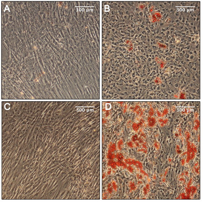
Figure 2: Adipogenic differentiation. (A) Phase microscopy of the human bone marrow MSC in the non-induced condition shows no evidence of differentiation. (B) Phase microscopy of the human bone marrow MSC in the induced adipogenic condition shows some differentiation and immobilization of neutral lipids, stained with Oil Red O after 14 days. (C) Phase microscopy of the human aortic PVAT-derived cells in the non-induced adipogenic condition shows no evidence of differentiation. (D) Phase microscopy of the human aortic PVAT-derived cells in the induced adipogenic condition shows robust differentiation and immobilization of neutral lipids, stained with Oil Red O after 14 days. Please click here to view a larger version of this figure.
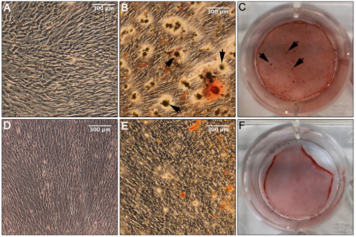
Figure 3: Osteogenic differentiation. (A) Phase microscopy of the human bone marrow MSC in the non-induced osteogenic condition shows no evidence of differentiation toward an osteogenic lineage. (B) Phase microscopy of the human bone marrow MSC in the induced condition after 14 days in culture shows evidence of differentiation and formation of calcium deposits stained with Alizarin Red (arrows). (C) An image of the well containing the human bone marrow MSC in the induced osteogenic condition following staining with Alizarin Red indicates abundant calcium deposition, suggesting successful differentiation toward an osteogenic lineage (arrows, calcium deposition). (D) Phase microscopy of the aortic human PVAT-derived cells in the non-induced osteogenic condition exhibits no indication of differentiation toward an osteogenic lineage. (E) Phase microscopy of the aortic human PVAT-derived cells in the induced osteogenic condition after 14 days in culture shows no evidence of differentiation toward an osteogenic lineage or deposition of calcium when stained with Alizarin Red, only non-specific staining. (F) An image of the well containing the human aortic PVAT-derived cells in the induced osteogenic condition following staining with Alizarin Red shows only non-specific staining. Please click here to view a larger version of this figure.
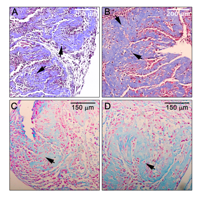
Figure 4: Chondrogenic differentiation. (A,B) Light microscopy of a section of the micromass formed by human bone marrow MSC (A) or human aortic PVAT progenitor cells (B) in the induced chondrogenic condition after 14 days in culture and subsequently stained with Masson’s Trichrome, which indicates significant deposition of collagen (blue) and suggests successful differentiation toward a chondrogenic lineage. (C,D) Light microscopy of a section of the micromass formed by human bone marrow MSC (C) or human aortic PVAT progenitor cells (D) in the induced chondrogenic condition after 14 days in culture and subsequently stained with Alcian blue, which indicates deposition of acidic proteoglycans (e.g. glycosaminoglycans) as are typically found in cartilage. Counterstain, nuclear fast red; arrows, lacunae-like structures. Please click here to view a larger version of this figure.
Adipose progenitor cells from different depots vary widely in phenotype and differentiation potential9. Culturing PVAT-derived progenitors from a single patient donor in simultaneous induction down three different lineages, adipogenic, osteogenic, and chondrogenic, allows for a well-controlled investigation of the pluripotent capacity of this novel population of progenitor cells. The methodology described in this report can be used to test the differentiation capacity of progenitor cells from human PVAT and to understand their function in regulating PVAT pathologies and vascular tone. Some benefits of this technique are its simplicity and use of primary human tissue from coronary artery bypass patients, thus enabling the investigation of PVAT progenitor cell function from patients with severe cardiovascular disease. However, explants from a 500 mg piece of tissue typically yield <1000 adherent cells and thus require 5–7 passages to obtain adequate numbers, highlighting one important caveat to this approach. Critical for success of the tri-lineage differentiation experiments is the quality of the initial cell explant from donor PVAT. It is essential to finely mince the tissue thoroughly prior to enzymatic dissociation. Additionally, unlike other explant protocols, we found a dramatic loss of cell number, viability and overall quality when red blood cell lysis buffer was used prior to plating. Instead, it is strongly recommended to plate the entire stromal vascular fraction (including red blood cells). After 24 h, adherent cells will have attached and non-lysed red blood cells can be removed through repeated washes with HBSS. Passaging of the PVAT-derived stromal cells should be no greater than 1:2 splits. We observed a reduction in growth rate when the cells were split 1:3 or greater.
The most difficult component of the trilineage differentiation assay is the chondrogenic condition, as it requires a less common method of culturing cells in a “micromass” and histological embedding and sectioning. Care must be taken to properly plate the 10 µL droplet of 100,000 cells and not disturb the mass on adding culture medium. We found variation between assays as to whether or not the micromass remained adhered to the plate or suspended in the medium. Even when the mass did not remain adhered, the micromass structure and cell viability remained consistent.
Stem or progenitor cells within the vascular microenvironment are responsible for tissue repair in response to injury or disease. Most well characterized are populations that derive from pericyte/adventitial compartments, although there is still controversy about whether there is a standardized molecular identification of these populations in humans10,11. In this protocol, we study a progenitor cell population specifically derived from human PVAT to test their propensity to differentiate to an osteogenic, chondrogenic or adipogenic lineage. Using staining techniques to define cellular commitment to each lineage, we demonstrate that unlike MSC from human bone marrow, progenitor cells from human PVAT were not able to differentiate towards an osteogenic lineage (Figure 3) but exhibit robust adipogenic differentiation. The chondrogenic differentiation capacity is comparable between bone marrow and PVAT-derived cells. This suggests that PVAT-derived progenitor cells might be more lineage restricted than bone marrow MSC or that the PVAT progenitors do not readily differentiate into osteogenic lineages. Future studies should include an investigation of gene and protein expression of markers for each lineage to more quantitatively evaluate the differentiation capacity of PVAT-derived cells.
Adipose-derived stem cells have been a focus for regenerative medicine applications, due to easy access and the high number of cells that can be derived from adipose tissue12,13. Within the stromal vascular fraction of adipose tissue, there are vascular cells, inflammatory cells, fibroblasts, preadipocytes, and adipose-derived stem cells. There are differences in the stem cell population based on anatomical location of the adipose depot and adipocyte characteristics14. Most importantly, the adipose-derived stem cell appears to have the capacity not only to differentiate into mesenchymal lineages15, but also potential to adopt neuronal16,17 and epidermal18 fates.
Numerous studies have focused on stem/progenitor cells derived from human subcutaneous white adipose tissues, but very few studies have addressed characteristics of the progenitor populations in PVAT. Recent studies have isolated adipocyte progenitor cells from PVAT surrounding mesenteric vessels or thoracic aorta of the rat. These CD34+/CD140a+ populations differentiated into adipocytes, although the capacity to derive other lineages was not tested19.
We recently characterized adipose progenitor cells derived from the stromal vascular fraction of human PVAT from patients with advanced coronary artery disease6. These adipose progenitor cells were CD73+, CD105+, and CD140a+, and efficiently differentiated into adipocytes with thermogenic characteristics (UCP1 expression)6. In the current work, we found a limited lineage potency of the PVAT-derived cells compared to bone marrow-derived MSC, suggesting either a lack or minimal population of adipose-derived stem cells that have been characterized from other human adipose tissues. A consideration for this protocol is the expected variability between human specimens, which may affect the number of stem/progenitor cells within the PVAT sample and their capacity to undergo differentiation to distinct lineages. Age, gender, BMI, medications and other clinical parameters likely affect the phenotype of progenitor cells within PVAT. Thus, expected future modifications of this protocol await a more clearly defined progenitor population within human PVAT, and possibly using cell sorting to isolate specific sub-populations for lineage studies.
The authors have nothing to disclose.
We acknowledge the assistance of Research Navigation at Maine Medical Center for assisting with the procurement of clinical tissue, and the Histopathology and Histomorphometry Core (supported by 1P20GM121301, L. Liaw PI) at Maine Medical Center Research Institute for sectioning and staining. This work was supported by NIH grant R01 HL141149 (L. Liaw).
animal-free collagenase/dispase blend I | Millipore-Sigma | SCR139 | 50mg |
Alcian Blue | NewComerSupply | 1003A | 1% Aqueous solution pH 2.5 |
Alizarin Red | Amresco | 9436-25G | |
alpha-MEM | ThermoFisher | 12561056 | |
Aniline Blue | NewComerSupply | 10073C | |
antibiotic/antimycotic | ThermoFisher | 15240062 | |
Beibrich's scarlet acid fuchsin | Millipore-Sigma | A3908-25G | |
b-glycerophosphate | Millipore-Sigma | G9422-10G | |
Biebrich Scarlet | EKI | 2248-25G | |
biotin | Millipore-Sigma | B4501-100MG | |
Bouin's fixative | NewComerSupply | 1020A | |
bovine serum albumin | Calbiochem | 12659 | stored at 4C |
Cell detachment solution | Accutase | AT104 | |
cell strainer (70mm) | Corning | 352350 | |
dexamethasone | Millipore-Sigma | D4902-100MG | |
DMEM | Corning | 10-013-CV | 4.5g/L glucose, L-glut and pyruvate |
DMEM/F12 medium | ThermoFisher | 10565-042 | high glucose, glutamax, sodium bicarbinate |
DMSO | Millipore-Sigma | D2650 | |
fetal bovine serum | Atlanta Biologicals | S11550 | |
FGF2 | Peprotech | 100-18B | |
formalin | NewComerSupply | 1090 | |
gelatin, bovine skin | Millipore-Sigma | G9391-500G | |
glutamax | ThermoFisher | 35050061 | glutamine supplement |
HBSS | Lonza | 10-547F | |
IBMX | Millipore-Sigma | I5879-250MG | |
insulin solution | Millipore-Sigma | I9278-5ML | |
Oil red O | Millipore-Sigma | O0625-100G | |
pantothenic acid | Millipore-Sigma | P5155-100G | |
penicillin-streptomycin solution | ThermoFisher | 15240062 | 100ml |
permount | Fisher | SP15-500 | |
phosphotungstic/phosphomoybdic acid solution | Millipore-Sigma | P4006-100G/221856-100G | |
primocin | Invivogen | ant-pm-1 | Antimicrobial reagent for culture media. |
rosiglitazone | Millipore-Sigma | R2408-10MG | |
TGFb1 | Peprotech | 100-21 | |
Weigert's hematoxylin | EKI | 4880-100G |