Science Education
>

An In Vitro Hemodynamic Loop Model to Investigate the Hemocytocompatibility and Host Cell Activation of Vascular Medical Devices

Instructor Prep
concepts
Student Protocol
JoVE 신문
의학
Author Produced
This content is Free Access.
JoVE 신문 의학
An In Vitro Hemodynamic Loop Model to Investigate the Hemocytocompatibility and Host Cell Activation of Vascular Medical Devices

This study was approved by the Ethics Committee of the medical faculty of the University Hospital Magdeburg (application number 88/18) and the subjects provided written informed consent prior to the blood drawing procedure.

1. Heparin stock preparation and blood sampling

  1. Calculate the amount of blood needed for the entire experiments.
    NOTE: Almost, 5 mL of heparinized blood is required for each vascular device in duplicates (loops). Likewise, 5 mL of blood is required for each baseline and static condition which is kept aside at room temperature.
  2. Prepare a heparin stock solution at the concentration of 100 IU/mL by diluting the unfractionated heparin (e.g., Rotexmedia) in deionized water. Use 150 µL of this solution for the heparinization of 10 mL fresh blood.
  3. Calculate the amount of blood as well as plasma that are needed for whole blood cell count, ELISA and FACS assays.
    NOTE: The measurements represented in this study are obtained from two loops running in parallel (one as duplicate) with a total blood volume of 10 mL.
  4. Based on the required amount of blood, fill 10 mL syringes with 150 µL of heparin stock solution to prevent blood coagulation with a final heparin concentration of 1.5 IU/mL blood.
  5. Draw blood using a butterfly (size: 21 G). Fill the syringes very gently to avoid hemolysis or cell activation due to excessive vacuum.
    NOTE: Permission from the local ethical committee should be obtained before the commencement of the experiments. Obtain informed consent from each human blood donor. Ensure that the blood donor is healthy and does not take any medication, especially no antiplatelet agents or non-steroidal anti-inflammatory drugs for at least 10 days prior to the experiments. Of note, same donor is preferred when comparing different types of vascular devices for the first time as presented in this manuscript. To further evaluate the inter-individual differences, the protocol can be repeated with different donors.
  6. Collect blood in a glass beaker, avoid excessive agitation.

2. In vitro hemodynamic loop assembly

  1. Fill the water bath until the water level reaches up to the center of the rotation unit. Set water temperature to 37 °C.
  2. Loop assembly for testing different tubing materials (polyPVC, hepPVC, PVC and latex)
    1. Cut two 50 cm long pieces of each tube material (inner diameter 5 mm) with the tube cutter. Ensure that the cutting surface is flat, as this is particularly important for perfect closure for the loops with small diameters so that blood flows without any distortion on the fitting edge.
      NOTE: This entire protocol utilizes the tubes with an inner diameter of 5 mm.
    2. To ease the handling and to avoid post-sample processing delay after rotation, run only four loops (2 materials in duplicates) in parallel.
    3. To generate a loop shape, plug the open endings of the tubes into a short piece of silicon tube fitting the outer diameter of the investigative tube.
    4. Use the polycarbonate tension bands to ensure proper closing of the loops. When used for the first time, adjust the length of the bands to fit the outer diameter of the loop by cutting the tension band with scissors in required lengths and secure it with a 3 mm torque wrench.
    5. Carefully tighten the locking screw of the tension band connector under inspection of the tube endings. Adjust the closing force so that no gap remains between the tube endings. If the locking screw is totally tightened and the tension of the polycarbonate band seems too low to close the gap between the tube endings, open the locking the system and cut a few mm of the tension band. Repeat this, until accurate loop closure is achieved (Figure 1A).
    6. If the tubing material is very soft and tends to slip inside the tension bands, fix the loop with electrical tape to the tension band (Figure 1B,C).
    7. Prepare an additional loop of PVC for temperature control with the same dimensions as the test loops.
    8. Secure the loops in the loop cradle of the rotation unit outside the water bath. Thereafter, attach the loop cradle to the rotation unit inside the water bath (Figure 1E).
    9. Dismantle the tension band partly from the loops and unplug one end of each tube to open the loop.
    10. Gently fill each loop with 5 mL of blood with a 5 mL serological pipette. Mix blood gently twice in the glass beaker by slowly pipetting up and down before loading into the loops.
    11. Take the disposable thermometer and place it inside the temperature control loop. If the thermometer is too large, cut it with scissors to fit smaller tubes. Fill the loop with 5 mL of deionized water at room temperature.
    12. Close the loops and ensure whether the loops, the tension bands and the rack are properly fitted.
    13. Set rotation speed to 30 rounds per minute (rpm) and rotate for 3 h.
  3. Loop assembly for testing the impact of improper loop closure (gap)
    1. Prepare four loops (polyPVC) as described in 2.2.
    2. Close two of the loops properly with the tension bands and avoid any gap between the tube endings.
    3. For the other two loops plug the open endings into the bigger tube as described in 2.2.3, but leave a gap of 1-2 mm in between the loop endings. Do not use the tension bands for these loops (Figure 1D).
    4. Prepare one temperature control loop as described in 2.2.7 and 2.2.11.
    5. Fill all loops with blood as described in 2.2.10.
    6. Set the rotation speed to 30 rpm and rotate for 3 h.
  4. Loop assembly for stent testing
    1. Prepare four loops as described in 2.2 and following.
      NOTE: To assess the blood biocompatibility of stents, the tubing material itself should be tested to be biocompatible in order to prevent masking of cell activation. If no data exists, the tube material itself can be tested as described in 2.2. Furthermore, the diameter range within the stent can be applied (see manufacturer’s instructions) and should fit the inner diameter of the tube material.
    2. Open two of the loops and take the tube out of the tension band system.
    3. Insert the stent into the middle of the tube as per the manufacturer’s instructions.
    4. Use the other two loops without stent as control. Fill all loops with blood as described in 2.2.10.
    5. Set rotation speed to 30 rpm and rotate for 3 h.
  5. Temperature control
    1. At any time during duration and when rotation is stopped, the temperature of the blood inside the loops is indicated by the thermometer inside the temperature control loop. To read off the temperature, stop the rotation and immediately read off the temperature indicated by the thermometer.

3. Blood sample processing

  1. After rotation, let the loops stand in the rack for 2 min in an upward position to let the blood accumulate at the bottom of the loops, avoiding any spilling while opening the loops.
  2. Inspect the blood left for static conditions: If this blood is coagulated, an improper heparinization is ultimately suspected. In this case, repeat the experiment preferably also with another blood donor.
  3. Carefully take the loops out of the rack. Open the connectors and let the blood flow into a 10 mL glass beaker.
  4. Pool the blood from the tubes that are run in duplicates.
  5. Draw fresh blood for baseline analysis from the same donor as described in 1.5 and 1.6.
  6. Collection of sodium citrate blood
    1. For the collection of sodium citrate blood, fill four 1.5 mL tubes, each with 111 µL sodium citrate solution collected from sodium citrate tubes, to generate a final concentration of 3.2% of sodium citrate.
    2. Fill each tube with 1 mL of blood from the glass beakers as described in 1.6 and 3.3.
    3. Keep the blood at room temperature and use the this blood for FACS analyses as described in 12.
      NOTE: To change the length of the loops for different experimental setups, properly plan aliquots based on the amount of measurements to be made. It may be necessary to establish all required measurements with fresh blood prior to the real experiments to ensure the volume of blood in the loops is enough for all measurements.
  7. Collection of ethylenediaminetetraacetic acid (EDTA) blood and plasma samples.
    1. From each glass beaker with blood (see 1.6 and 3.3) transfer 4.5 ml of blood into one 5 ml EDTA tube. Fill two EDTA tubes from each 10 ml glass beaker with blood.
    2. Gently mix the blood and keep it on ice.
    3. Transfer 2 mL of blood into a 2 mL locking centrifugation tube and use this blood for blood cell count and free hemoglobin measurement as described in 5. And 6.
    4. Centrifuge the remaining blood at 3,500 x g for 20 min.
    5. Carefully collect the plasma in the supernatant in 500 µL aliquots and immediately freeze at -80 °C.

4. Scanning electron microscopy and µCT images

  1. After blood sample processing, rinse the emptied loops prepared as described in 2.3 with 10 mL of phosphate-buffered saline (PBS) each.
  2. Carefully cut off a 1 cm long sample from each end of each tube with a scalpel.
  3. Incubate sample overnight at 4 °C in a 2% glutaraldehyde solution.
    CAUTION: Inhalation of aldehyde vapors can cause nasal symptoms such as a runny nose ore persistent stuffiness and airway irritation, and contact with skin causes dermatitis. Aldehydes should be handled in a fume-hood while wearing gloves, a protective gown and safety goggles.
  4. Rinse the sample (3 times) with PBS.
  5. Prepare a 1% solution of osmium tetroxide (OsO4) in deionized water and incubate sample for 15 min at room temperature.
    CAUTION: OsO4 is a strong oxidizing agent, it can be reduced by exposure to light. To avoid the reduction during preparation, store OsO4 in a brown glass bottle. OsO4 is extremely volatile, its fumes are toxic to eyes, nose and throat. Always work under a fume-hood and use gloves and protecting clothing, ensuring that no part of the body is exposed to OsO4. Contact your institution’s guidelines regarding handling of waste disposal and storage. In general, OsO4 is storable for several months, but it needs special glass bottles with Teflon liner and a desiccator, because OsO4 can discolor internal surfaces and contents of the refrigerator in the presence of leaking fumes.
  6. Take out samples, transfer them in a new 15 mL centrifuge tubes and rinse samples 3 times with PBS.
  7. Prepare a series of ethanol with varying concentrations (25%, 50%, 75%, 95%, 100%)
  8. Dehydrate samples in ethanol: incubate for 20 min each in 25%, 50%, 75% and 90% and 30 min in 100%.
  9. Take samples out of 100% ethanol and let it air dry overnight at room temperature.
  10. Examine samples in the scanning electron microscope at an acceleration voltage of 5 kV and with the X-ray µCT scanner.

5. Blood cell count

  1. Take 2 mL of the EDTA blood obtained as described in 3.7.3
  2. Insert the tube into the automated hematology analyzer and follow the manufacturer’s instructions.

6. Measurement of free hemoglobin (fHb) in plasma

  1. Thaw one plasma sample from each condition obtained as described in 3.7.5. Store on ice after thawing.
    NOTE: Always thaw frozen plasma samples in a water bath at 37 °C and immediately transfer to ice, containing some water, to lower the temperature. This is important to avoid activation of blood components during thawing.
  2. Use the fHb reagent and follow the manufacturer’s instructions. Avoid contamination after opening and protect the reagent from direct light (sun, UV light).
  3. Use a pipetting scheme (see manufacturer’s instructions) with 1:5 dilution.
  4. Add 1000 µL of the hemoglobin reagent in a 1.6 mL semi-micro cuvette. Use this cuvette to determine the blank value.
  5. Add 1000 µL of the hemoglobin reagent and 250 µL of the plasma sample in another semi-micro cuvette.
  6. Mix the contents in both cuvettes by flushing the pipette thoroughly by repeatedly filling with reaction mixture, and incubate at least 3 min at room temperature.
  7. Determine the extinction (E) of the sample against fHb reagent as blank reagent. Calculate the fHb-concentration (mmol/l) of the sample: E540-E680 x 0.452.

7. Measurement of FPA

  1. Thaw a plasma sample from each condition obtained as described in 3.7.5 and store on ice after thawing.
  2. Use the FPA Elisa kit and follow the manufacturer’s instructions.

8. Measurement of sC5b9

  1. Thaw a plasma sample from each condition obtained as described in 3.7.5 and store on ice after thawing.
  2. Use the sC5b-9 ELISA kit and follow the manufacturer’s instructions.

9. Measurement of PMN

  1. Thaw a plasma sample from each condition obtained as described in 3.7.5 and store on ice after thawing.
  2. Use the PMN-Elastase ELISA kit and follow the manufacturer’s instructions.

10. Measurement of TNF

  1. Microplates must be coated one day before running the ELISA. For coating, add 100 µL of capture antibody solution to all wells, seal plate and incubate overnight at 2 °C-8 °C.
  2. Thaw one plasma sample from each condition obtained as described in 3.7.5. Store on ice after thawing.
  3. Use the TNF ELISA kit and follow the manufacturer’s instructions.

11. Measurement of IL-6

  1. Microplates must be coated with capture antibodies one day prior to the experiment. For coating, add 100 µL of capture antibody solution to all wells of the microplate provided with the set, seal plate and incubate overnight at 4 °C
  2. Thaw a plasma sample from each condition obtained as described in 3.7.5. and store on ice after thawing.
  3. Use the IL-6 ELISA kit and follow the manufacturer’s instructions..

12. FACS analyses

  1. Prepare 500 mL of FACS buffer by adding 10 mL of fetal calf serum and 2 mL of EDTA solution (0.5 M) to 488 mL of 1x PBS. The FACS buffer can be stored for 4 weeks at 4°C.
  2. Use 100 μL of the sodium citrate blood (see 3.6.3) for each staining procedure (monocyte platelet aggregates (MPA), platelet activation (PA), leukocyte integrins (LI)).
  3. Pipette 100 μL of blood from each sample into a 5 mL FACS tube. From each sample, prepare 3 tubes for antibody staining and label with MPA (monocyte platelet aggregates) panel, PA (platelet aggregates) panel and LI (leukocyte integrin) panel.
  4. Mix the rest of the 4 samples into one 5 mL FACS tube. Use this mix for unstained and fluorescence minus one (FMO) controls.
  5. Prepare 6 FACS tubes by pipetting 100 µL of the mixed sample blood into each tube. Label 3 tubes as unstained and 3 tubes as FMO-CD41, FMO-CD62P and FMO-CD162.
  6. Add 100 µL of 4% paraformaldehyde solution and incubate for 15 min in the dark at room temperature.
    CAUTION: Paraformaldehyde is toxic to skin and eyes. It can cause serious pulmonary irritation when inhaled and can lead to lung damage after prolonged exposure. Furthermore, it is classified as a carcinogen and a reproductive toxin. Always wear gloves and safety glasses and work under the fume hood.
  7. Add 1 mL of wash buffer to each tube and centrifuge at 287 x g for 5 min. Discard the supernatant and repeat wash step two times.
  8. For red blood cell (RBC) lysis, add 1 mL of 1x buffer RBC lysis buffer (dilute 10x RBC lysis buffer in deionized water), mix by slowly pipetting up and down and incubate for 5 min at RT in the dark.
  9. Prepare compensation beads for single stains. To 1 mL of FACS buffer add 4 drops of each positive and negative beads. Vortex thorough and add 100 µL of beads solution to one 5 mL FACS tube.
  10. Prepare 4 tubes with beads and label as CD14-FITC, CD41-BV421, CD45-APC and CD62P-PE. Add 1 mL of FACS buffer to each tube and centrifuge at 287 x g for 5 min. Discard supernatant and keep on ice.
  11. After RBC lysis, add 1 mL of FACS buffer to each tube and wash as described in 12.7. Discard supernatant.
  12. Prepare antibody cocktails:
    1. MPA panel: Add 4 µL of CD45-APC, 4 µL of CD14-FITC and 4 µL of CD41-BV421 to 388 µL of FACS buffer.
    2. PA panel: Add 1.6 µL of CD41-BV421 and 12 µL of CD62P-PE to 386.4 µL of FACS buffer.
    3. LI panel: Add 4 µL of CD45-APC and 8 µL of CD162-BV421 to 388 µL of FACS buffer. Keep on ice.
  13. Prepare single stains: Add 0.5 µL to 499.5 µL of FACS buffer each for CD14-FITC, CD41-BV421 and CD45-APC. For CD62P-PE add 0.3 µL to 499.7 µL of FACS buffer. Keep on ice.
  14. Prepare FMO controls: (i) MPA panel (FMO-CD41): Add 1 µL CD14-FITC antibody and 1 µL of CD45-APC antibody to 98 µL FACS buffer. (ii) PA panel (FMO-CD62P): Add 0.4 µL of CD41-BV421 to 99.6 µL of FACS buffer. (iii) LI panel (FMO-CD162): Add 1 µL of CD45-APC to 99 µL of FACS buffer. Keep FMO controls on ice.
  15. Vortex antibody cocktails and add 100 µL of each antibody cocktail as prepared in 12.12 to each of the respective labeled tubes (MPA, PA and LI panel) as described in 12.3 and mix gently by pipetting up and down. Keep on RT in the dark.
  16. Vortex single stain antibody dilutions and add 100 µL of the single stain antibody dilutions as prepared in 12.13. to each of the respective labeled tubes with beads as described in 12.7 and mix gently by pipetting up and down. Keep on RT in the dark.
  17. Vortex FMO antibody cocktails and add 100 µL of each FMO-1 antibody cocktail as prepared in 12.14. to each of the respective labeled tubes (FMO controls) as described in 12.5. and mix gently by pipetting up and down. Keep at RT in the dark.
  18. Incubate all tubes with antibody staining for 30 min at RT in the dark.
  19. Take the three tubes for unstained control (see 12.4) and resuspend pellet in 250 µL of FACS buffer. Keep on ice.
  20. After incubation time, wash all tubes except unstained controls one time with FACS buffer as described in 12.7. Resuspend pellet in in 250 µL of FACS buffer and acquire data on the flow cytometer.
  21. Use unstained cells for negative settings and compensation mouse beads for positive settings in FACS software. Further, use FMO to control the gating strategy, where FMO-CD41 is used in MPA panel, FMO-CD62P is used in PA panel and FMO-CD162 is used in LI panel.
  22. Acquire almost 0.5 x 10⁶ – 0.25 x 10⁶ events per tube for FACS. Save data and analyze with an analysis software (version 9.9.6).

An In Vitro Hemodynamic Loop Model to Investigate the Hemocytocompatibility and Host Cell Activation of Vascular Medical Devices

Learning Objectives

All presented data, except FACS plots, were analyzed with a statistics software. The FACS plots were analyzed using flow cytometry software.

The analysis of whole blood cell count did not show any significant differences with respect to erythrocytes between all tested conditions (Figure 2). But, platelets and leukocytes were drastically reduced in the latex group, indicating a very poor biocompatibility of latex. This is further underlined by increased levels of free hemoglobin in the latex group, indicating the fact that except for the latex group, none of the other vascular devices or conditions led to extensive hemolysis (Figure 2). Further, the coated PVC tubes, polyPVC and hepPVC, as well as the tested stent did not lead to thrombosis by means of platelet and leukocyte loss, while latex exhibited the highest platelet and leukocyte loss, followed by uncoated PVC tubes that showed a decreased trend.

While all the tested vascular devices led to increased activation of the coagulation system (FPA) and complement component (sC5b-9), the hepPVC loops exhibited a trend for decreased levels of FPA and sC5b-9 when compared specifically to polyPVC loops (Figure 3). Interestingly, uncoated PVC and Gap loops showed lower levels of FPA compared to polyPVC, though not reaching the level of statistical significance. Nevertheless, latex loops exhibited significantly increased levels of FPA when compared to baseline and static conditions.

In accordance with the whole blood cell counts, latex loops exhibited highest levels of TNF, IL-6 and PMN elastase (Figure 4), reaching the level of statistical significance when compared to rest of the groups in terms of TNF and IL-6 (Figure 4A,B), whereas to static and baseline conditions in terms of PMN elastase (Figure 4C). These results indicate the potent activation of leukocytes by latex. The baseline levels of activation markers were always comparable to static conditions, indicating a proper heparinization of the blood.

Interestingly, it was shown that platelet and leukocyte counts for gap induced loops were only slightly reduced with moderate activation of the coagulatory system (FPA) and leukocytes (PMN elastase), though improper loop closure with resulting flow turbulences and blood contact to the uncoated, rough cutting surface led to macroscopically visible clots at the splice (Figure 1F). The clots and its distribution over the whole splice surface were evident with µCT and SEM images, while no clot was found when the loops were closed with the external closing device leaving no gap between the loop endings (Figure 5).

Flow cytometric analysis of host blood cells that were stained with platelet specific markers, CD41 and platelet activation marker CD62P, are shown in Figure 6A,B. Here, the latex tubes exhibited exceedingly high median fluorescence intensity (MFI) for CD62P on blood platelets, followed by stent, whereas heparin coated polyPVC tubes exhibited minimal activation of platelets depicting anti-thrombogenic property of polyPVC tubes. Furthermore, leukocytes were classified based on the CD45 and SSC (side scatter) based granularity into (i) granulocytes; (ii) monocytes and (iii) lymphocytes (Figure 7), and the expression of CD162+ integrin was detected on each subpopulation of leukocytes that are known to interact with the CD62P on platelets24. It was noticed that the integrin expressions were drastically reduced on granulocytes and lymphocytes in latex loops. This result was in line with lowered levels of total frequencies of leukocytes in the latex loops (Figure 2). In general, the integrin levels were higher among monocytes when compared to granulocytes and lymphocytes, indicating the likelihood for the monocyte interaction with activated platelets. In this regard, monocyte platelet aggregates were also evaluated by staining the blood cells with CD14 (as monocyte marker) and CD41 (as platelet marker) and ultimately to identify double positive cells i.e. CD14+CD41+MPA (Figure 8). Here, we noticed that the stent group exhibited the highest levels of CD41 expression on the MPA, followed by the latex group, indicating an increased tendency to form MPA, despite the reduced frequency of monocyte (<1 %) in the latex loops.

Figure 1
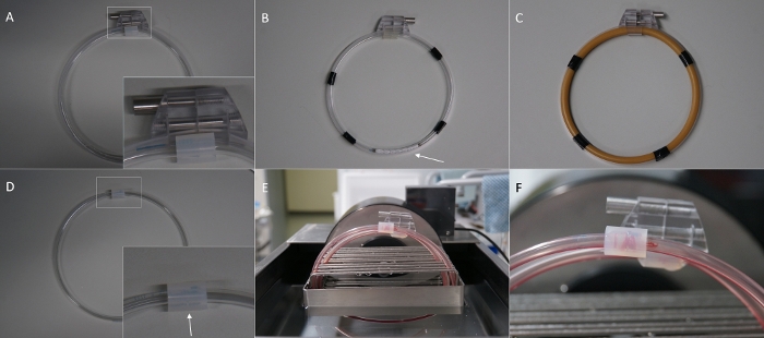
Figure 1: Overview of the in vitro hemodynamic loop model and its modifications. (A) Loop for the gap experiment with external loop closing system, leaving no gap at the splice. (B) Loop made of polyPVC coated PVC tube and stent inside (arrow). (C) Loop made of latex tube. (D) Loop for the gap experiment without the external loop closing system leaving a gap between the tube endings (arrow). (E) Loops placed in the loop cradle inside the water bath and filled with blood. (F) Thrombus resulting in a gap at the splice (arrow) after rotation. Please click here to view a larger version of this figure.

Figure 2
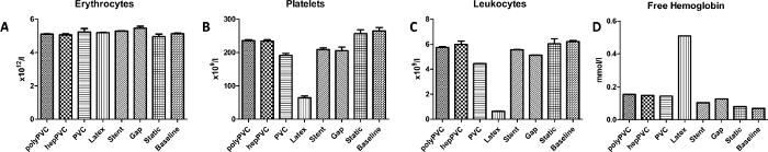
Figure 2: Results for blood cell count and plasma hemoglobin. (A) Erythrocytes count. (B) Platelets count. (F) Leukocytes count. (D) Free plasma hemoglobin. The results indicate the poor biocompatibility of latex, leading to excessive hemolysis. Data are presented as mean value; error bars indicate SEM. n=1. Please click here to view a larger version of this figure.

Figure 3
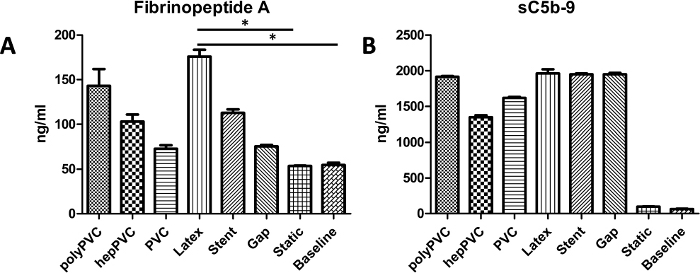
Figure 3: Results for activation of the coagulation and complement system. (A) Coagulation system activation, measured by levels of Fibrinopeptide A (FPA) (B) Complement system activation, measured by levels of sC5b-9. While latex tubes evoked significant elevated levels of the FPA, the complement activation was strong for all tested materials. Data are presented as mean value, error bars indicate SEM. *p<0.05, n=1. Please click here to view a larger version of this figure.

Figure 4
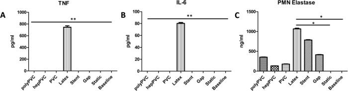
Figure 4: Leukocyte activation markers. (A) Tumor necrosis factor alpha (TNF). (B) Interleukin 6 (IL-6) (C) PMN Elastase. The results indicate increased activation of leukocytes due to elevated levels of the analyzed markers, followed by stent loops, that only led to increased levels for PMN Elastase but not TNF or IL-6. Data are presented as mean value, error bars indicate SEM. *p<0.5; **p<0.01, n=1. Please click here to view a larger version of this figure.

Figure 5
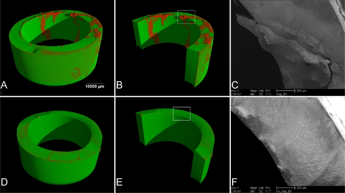
Figure 5: Imaging of the splice of the loops. (A) µ-computer tomography (µCT) of loops with improper closing (gap). The red areas indicate thrombus material. (B) Rendering of the luminal side of the tube. The rectangular selection indicates the area for scanning electron microscopy (SEM) (C). (D) µCT of loops with external loop closing device and no gap at the splice, and (E) rendering and view of the luminal surface. No thrombus material was found. (F) SEM image of the rectangular selection in (E). No thrombus material was found on the cutting surface. Please click here to view a larger version of this figure.

Figure 6
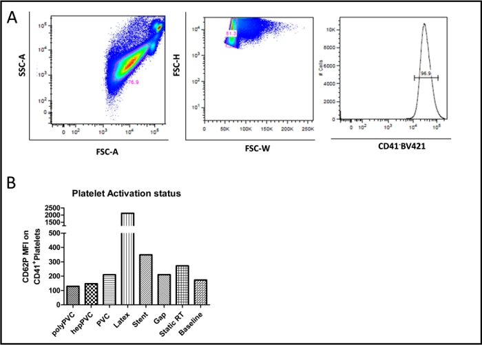
Figure 6: FACS plot for platelet activation (CD62P). (A) Representative FACS plot (basic condition) showing the blood CD41+ platelets. (B) Graph showing the platelet activation status reflected by the mean fluorescence intensity (MFI) of the different types of vascular devices in comparison to the static RT and baseline conditions. The data bars present data from single measurements. Please click here to view a larger version of this figure.

Figure 7
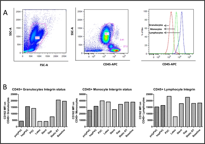
Figure 7: FACS plot for leukocyte integrin (CD162). (A) Representative FACS plot (basic condition) showing the blood CD45+ leukocytes and subgroups (B) Graph showing the leukocyte CD162+ integrin mean fluorescence intensity (MFI) of the different types of vascular devices in comparison to the static and baseline conditions. The data bars present data from single measurements. Please click here to view a larger version of this figure.

Figure 8
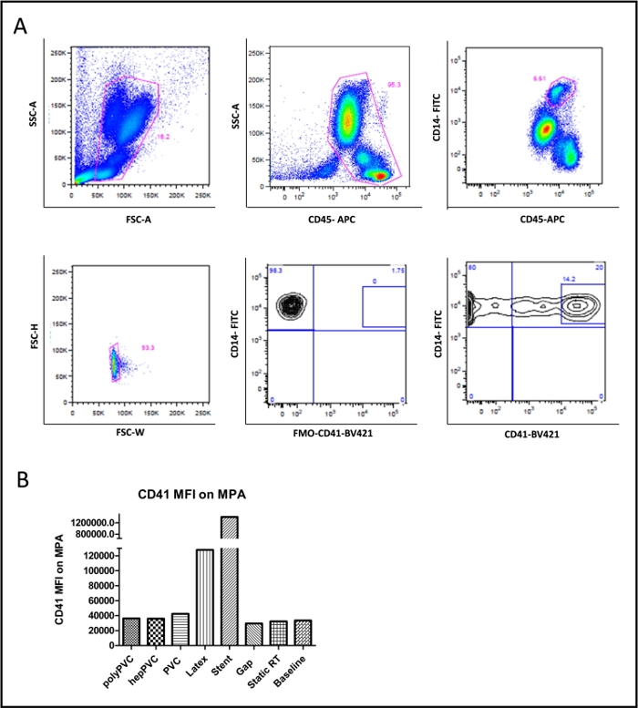
Figure 8: FACS plot for platelet monocyte aggregates (CD41/CD14). (A) Representative FACS plot (basic condition) showing the gating for blood monocytes (CD45+/CD14+), platelets (CD41+) and monocyte platelet aggregates (CD41+/CD14+) (B) Graph showing the CD41+ mean fluorescence intensity (MFI) on monocyte platelet aggregates for the various vascular devices compared to the static and baseline conditions. The data bars present data from single measurements. Please click here to view a larger version of this figure.

List of Materials

5 ml tube, K3 EDTA Sarstedt 32332
Anti-Mouse Ig, κ/Negative Control Compensation Particles Set Becton Dickinson BioSciences 552843
APC anti-human CD45 Antibody BioLegend 368512
BD LSR Fortessa II cell analyzer Becton Dickinson 647465
BD Vacutainer Citrate Tubes Becton Dickinson 369714
BD Vacutainer one-use holder Becton Dickinson 364815
BD Vacutainer Safety-Lok butterfly canula 21 G Becton Dickinson 367282
Beaker glass ROTILABO short 10 ml Carl Roth GmbH + Co. KG X686.1
Beaker glass ROTILABO short 50 ml Carl Roth GmbH + Co. KG X688.1
Brilliant Violet 421 anti-human CD162 Antibody BioLegend 328808
Brilliant Violet 421 anti-human CD41 Antibody BioLegend 303730
Centrifuge ROTINA 420 | 420 R Hettich Zentrifugen 4701 | 4706
Centrifuge tubes, 50 ml Greiner Bio-One GmbH 227261
CHC Super modified, 5mm PVC tubing Corline Sweden 1807-148 Referred to as hepPVC tube
Circular Precision Cutter ebo kunze industriedesign, Neuffen, Germany CLS 007-20
Closing Unit (complete with tension bands) ebo kunze industriedesign, Neuffen, Germany CLS 008-20
Electric tape Scotch Super 33+ VWR MMMA331933
ELISA MAX Deluxe Set Human IL-6 BioLegend 430504
ELISA MAX Deluxe Set Human TNF-a BioLegend 430204
Eppendorf Pipette Research plus, single channel, inkl. epT.I.P.S. box, 0,1 – 2,5 µL, gray Eppendorf AG 3123000012
Eppendorf Pipette Research plus, single channel, inkl. epT.I.P.S. box, 0,5 – 10 µL, gray Eppendorf AG 3123000020
Eppendorf Pipette Research plus, single channel, inkl. epT.I.P.S. box, 10 – 100 µL, yellow Eppendorf AG 3123000047
Eppendorf Pipette Research plus, single channel, inkl. epT.I.P.S. box, 100 – 1,000 µL, blue Eppendorf AG 3123000063
Eppendorf Pipette Research plus, single channel, inkl. epT.I.P.S. box, 20 – 200 µL, yellow Eppendorf AG 3123000055
Eppendorf Pipette Research plus, single channel, inkl. epT.I.P.S. sample bag, 0,5 – 5 mL, violet Eppendorf AG 3123000071
Ethylenediaminetetraacetic acid solution Sigma-Aldrich 03690-100ML
FACS tubes polystyrene 5.0 ml round bottom Corning BV 352052
Fetal bovine serum Gold Plus Bio-Sell FBS.GP.0500
FITC anti-human CD14 Antibody BioLegend 367116
Fluency plus stent 13.5 x 60 mm Angiomed GmbH & Co FVM14060
Free Hemoglobin fHb Reagent Bioanalytics GmbH 004001-0250
Gibco PBS Tablets Thermo Fisher Scientific 18912014
Gloves Vasco Nitril white L B. Braun Deutschland GmbH & Co.KG 9208437
Gloves Vasco Nitril white M B. Braun Deutschland GmbH & Co.KG 9208429
Glutaraldehyde 25% aequous solution Sigma Aldrich G6257-100ML
Heparin, 25.000 IE in 5 ml Rotexmedica, Trittau, Germany PZN 3862340
Human Fibrinopeptide A (FPA) ELISA Kit Hölzel Diagnostika abx253234
Kodan tincture forte colourless Schülke & Mayr GmbH 104012
Latex tube, ID 5 mm Laborhandel24 GmbH 305 0507
Loop Stand ebo kunze industriedesign, Neuffen, Germany CLS 009-20
Medimex venous tourniquet classic ROESER Medical GmbH 310005
Microplate reader Infinite 200 Pro M Plex Tecan TEC006418I
Microplate shaker PMS-1000i VWR 444-0041
Nalgene Metric non-phthalate PVC tubing, ID 5 mm VWR NALG8703-0508 Referred to as PVC tube
NexTemp (Standard) Single-Use Clinical Thermometer Medical Indicators 2112-20
Nunc MaxiSorp ELISA Plates, uncoated BioLegend 423501
Osmium tetroxide solution Fisher Scientific 10256970
Paraformaldehyde Solution, 4% in PBS Thermo Fisher Scientific AAJ19943K2
PE anti-human CD16Antibody BioLegend 302008
PE anti-human CD62P (P-Selectin) Antibody BioLegend 304906
Pipette controller, pipetus VWR 612-1874
Pipette tips epT.I.P.S. 0.2 – 5 ml OMNILAB-LABORZENTRUM GmbH & Co. KG 5186480
Pipette tips epT.I.P.S. standard 0,1 – 10µl Th. Geyer GmbH & Co. KG 9409410
Pipette tips epT.I.P.S. standard 2 – 200µl Th. Geyer GmbH & Co. KG 0030 000.870
Pipette tips epT.I.P.S. standard 50 – 1000µl blue Th. Geyer GmbH & Co. KG 0030 000.919
PMN (Neutrophil) Elastase Human ELISA Kit Fisher Scientific BMS269
Probe stand ROTILABO combi CARL ROTH K082.1
Rack for rotation unit (12 slots 3/8 '' with variable slot width) ebo kunze industriedesign, Neuffen, Germany CLS 011-20
RBC Lysis Buffer (10X) BioLegend 420301
Reagent reservoirs VWR 613-1184
Rotation Unit ebo kunze industriedesign, Neuffen, Germany CLS 010-20
Safe-Lock micro test tubes 0.5 ml OMNILAB-LABORZENTRUM GmbH & Co. KG 5409320
Safe-Lock micro test tubes 1.5 ml OMNILAB-LABORZENTRUM GmbH & Co. KG 5409331
sc5b9 Human ELISA KIT TECOmedicalGroup A029
Scalpel no 10 Fisher Scientific NC9999403
Scanning electron microscope XL30 ESEM-FEG Philips n.a.
Screw top bottle ROTILABO Clear glass, 1000 ml, GL 45 Carl Roth GmbH + Co. KG X715.1
Screw top bottle ROTILABO Clear glass, 500 ml, GL 45 Carl Roth GmbH + Co. KG X714.1
Semi-micro cuvette 1.6 ml Sarstedt 67.746
Serological pipette 10.0 ml Corning BV 4488
Serological pipette, 25.0 ml Corning BV 4489
Serological pipette, 5.0 ml Corning BV 4487
Silicon tube, inner diameter 8 mm, outer diameter 12 mm VWR BURK8803-0812
Sprout mini centrifuge Biozym 552034
Stop Solution for TMB Substrate BioLegend 77316
Swabs, sterile Fuhrmann GmbH 32055
Syringe, 10 ml Becton Dickinson 300296
Temperature controlled water basin ebo kunze industriedesign, Neuffen, Germany CLS 020-20
tert-Butanol, 99.5%, extra pure, ACROS Organics Fisher Scientific 10000730
TMB Substrate Set BioLegend 421101
Trillium PVC tube, 5 mm ID Medtronic 161100107100103 Referred to as polyPVC tube
Tween 20 AppliChem A4974,0250
UV-Vis Spektrometer Lambda 2 Perkin Elmer 33539
Vornado Mini Vortexer Biozym 55BV101-B-E
XN-3000 workstation blood analyzer Sysmex Europe n.a.
μ-CT Phoenix Nanotom S GE Sensing & Inspection, Wunstorf, Germany n.a.

Lab Prep

In this study, the hemocompatibility of tubes with an inner diameter of 5 mm made of polyvinyl chloride (PVC) and coated with different bioactive conjugates was compared to uncoated PVC tubes, latex tubes, and a stent for intravascular application that was placed inside the PVC tubes. Evaluation of hemocompatibility was done using an in vitro hemodynamic loop model that is recommended by the ISO standard 10993-4. The tubes were cut into segments of identical length and closed to form loops avoiding any gap at the splice, then filled with human blood and rotated in a water bath at 37 °C for 3 hours. Thereafter, the blood inside the tubes was collected for the analysis of whole blood cell count, hemolysis (free plasma hemoglobin), complement system (sC5b-9), coagulation system (fibrinopeptide A), and leukocyte activation (polymorphonuclear elastase, tumor necrosis factor and interleukin-6). Host cell activation was determined for platelet activation, leukocyte integrin status and monocyte platelet aggregates using flow cytometry. The effect of inaccurate loop closure was examined with x-ray microtomography and scanning electron microscopy, that showed thrombus formation at the splice. Latex tubes showed the strongest activation of both plasma and cellular components of the blood, indicating a poor hemocompatibility, followed by the stent group and uncoated PVC tubes. The coated PVC tubes did not show a significant decrease in platelet activation status, but showed an increased in complement and coagulation cascade compared to uncoated PVC tubes. The loop model itself did not lead to the activation of cells or soluble factors, and the hemolysis level was low. Therefore, the presented in vitro hemodynamic loop model avoids excessive activation of blood components by mechanical forces and serves as a method to investigate in vitro interactions between donor blood and vascular medical devices.

In this study, the hemocompatibility of tubes with an inner diameter of 5 mm made of polyvinyl chloride (PVC) and coated with different bioactive conjugates was compared to uncoated PVC tubes, latex tubes, and a stent for intravascular application that was placed inside the PVC tubes. Evaluation of hemocompatibility was done using an in vitro hemodynamic loop model that is recommended by the ISO standard 10993-4. The tubes were cut into segments of identical length and closed to form loops avoiding any gap at the splice, then filled with human blood and rotated in a water bath at 37 °C for 3 hours. Thereafter, the blood inside the tubes was collected for the analysis of whole blood cell count, hemolysis (free plasma hemoglobin), complement system (sC5b-9), coagulation system (fibrinopeptide A), and leukocyte activation (polymorphonuclear elastase, tumor necrosis factor and interleukin-6). Host cell activation was determined for platelet activation, leukocyte integrin status and monocyte platelet aggregates using flow cytometry. The effect of inaccurate loop closure was examined with x-ray microtomography and scanning electron microscopy, that showed thrombus formation at the splice. Latex tubes showed the strongest activation of both plasma and cellular components of the blood, indicating a poor hemocompatibility, followed by the stent group and uncoated PVC tubes. The coated PVC tubes did not show a significant decrease in platelet activation status, but showed an increased in complement and coagulation cascade compared to uncoated PVC tubes. The loop model itself did not lead to the activation of cells or soluble factors, and the hemolysis level was low. Therefore, the presented in vitro hemodynamic loop model avoids excessive activation of blood components by mechanical forces and serves as a method to investigate in vitro interactions between donor blood and vascular medical devices.

Procedure

In this study, the hemocompatibility of tubes with an inner diameter of 5 mm made of polyvinyl chloride (PVC) and coated with different bioactive conjugates was compared to uncoated PVC tubes, latex tubes, and a stent for intravascular application that was placed inside the PVC tubes. Evaluation of hemocompatibility was done using an in vitro hemodynamic loop model that is recommended by the ISO standard 10993-4. The tubes were cut into segments of identical length and closed to form loops avoiding any gap at the splice, then filled with human blood and rotated in a water bath at 37 °C for 3 hours. Thereafter, the blood inside the tubes was collected for the analysis of whole blood cell count, hemolysis (free plasma hemoglobin), complement system (sC5b-9), coagulation system (fibrinopeptide A), and leukocyte activation (polymorphonuclear elastase, tumor necrosis factor and interleukin-6). Host cell activation was determined for platelet activation, leukocyte integrin status and monocyte platelet aggregates using flow cytometry. The effect of inaccurate loop closure was examined with x-ray microtomography and scanning electron microscopy, that showed thrombus formation at the splice. Latex tubes showed the strongest activation of both plasma and cellular components of the blood, indicating a poor hemocompatibility, followed by the stent group and uncoated PVC tubes. The coated PVC tubes did not show a significant decrease in platelet activation status, but showed an increased in complement and coagulation cascade compared to uncoated PVC tubes. The loop model itself did not lead to the activation of cells or soluble factors, and the hemolysis level was low. Therefore, the presented in vitro hemodynamic loop model avoids excessive activation of blood components by mechanical forces and serves as a method to investigate in vitro interactions between donor blood and vascular medical devices.

Tags