A combination of surfactant washout using 0.9% saline (35 mL/kg body weight, 37 °C) and high tidal volume ventilation with low PEEP to cause moderate ventilator induced lung injury (VILI) results in experimental acute respiratory distress syndrome (ARDS). This method provides a model of lung injury with low/limited recruitability to study the effect of various ventilation strategies for extended periods.
Various animal models exist to study the complex pathomechanisms of the acute respiratory distress syndrome (ARDS). These models include pulmo-arterial infusion of oleic acid, infusion of endotoxins or bacteria, cecal ligation and puncture, various pneumonia models, lung ischemia/reperfusion models and, of course, surfactant depletion models, among others. Surfactant depletion produces a rapid, reproducible deterioration of pulmonary gas exchange and hemodynamics and can be induced in anesthetized pigs using repeated lung lavages with 0.9% saline (35 mL/kg body weight, 37 °C). The surfactant depletion model supports investigations with standard respiratory and hemodynamic monitoring with clinically applied devices. But the model suffers from a relatively high recruitability and ventilation with high airway pressures can immediately reduce the severity of the injury by reopening atelectatic lung areas. Thus, this model is not suitable for investigations of ventilator regimes that use high airway pressures. A combination of surfactant depletion and injurious ventilation with high tidal volume/low positive end-expiratory pressure (high Tv/low PEEP) to cause ventilator induced lung injury (VILI) will reduce the recruitability of the resulting lung injury. The advantages of a timely induction and the possibility to perform experimental research in a setting comparable to an intensive care unit are preserved.
The mortality of the acute respiratory distress syndrome (ARDS) remains high with values above 40%1 despite intensive research since its first description by Ashbough and Petty in 19672. Naturally, the investigation of novel therapeutic approaches is limited in the clinic due to ethical concerns and the lack of standardization of the underlying pathologies, ambient conditions, and co-medications, whereas animal models enable systematic research under standardized conditions.
Thus, experimental ARDS has been induced in either large animals (e.g., pigs) or small animals (e.g., rodents) using various methods such as pulmo-arterial infusion of oleic acid, intravenous (i.v.) infusion of bacteria and endotoxins, or cecal ligation and puncture (CLP) models causing sepsis-induced ARDS. In addition, direct lung injuries caused by burns and smoke inhalation or lung ischemia/reperfusion (I/R) are used3. One frequently used model of direct lung injury is surfactant depletion with lung lavages as first described by Lachmann et al. in guinea pigs4.
Surfactant depletion is a highly reproducible method that results rapidly in compromises in gas exchange and hemodynamics5. A major advantage is the possibility to apply surfactant depletion in large species which enable support research with clinically used mechanical ventilators, catheters, and monitors. However, a major disadvantage of the surfactant depletion model is the instant recruitment of atelectatic lung areas whenever high airway pressures or recruiting maneuvers, such as prone positioning, are applied. Thus, the model is not suitable to investigate, e.g., automated ventilation with high PEEP levels for prolonged times6. Yoshida et al. described a combination of surfactant depletion and ventilation with high inspiratory airway pressures to induce experimental ARDS7, but their model requires an elaborate maintenance of partial pressure of oxygen (PaO2) in a predefined corridor via repeated blood gas sampling and adjustment of the driving pressure according to a sliding table of inspiratory pressure and PEEP.
Overall, a model with an overly aggressive injurious ventilation or a laborious, repeated adjustment of the ventilation regime can result in structural damage of the lungs, which is too severe and results in subsequent multiple organ failure. Thus, this article provides a detailed description of an easily feasible model of surfactant depletion plus injurious ventilation with high Tv/low PEEP for induction of experimental ARDS, which supports research with clinically used ventilation parameters for prolonged periods.
The experiments were conducted at the Department of Experimental Medicine, Charité – University Medicine, Berlin, Germany (certified according to the EN DIN ISO 9001:2000) and were approved by the federal authorities for animal research in Berlin, Germany, prior to the experiments (G0229/18). The principles of laboratory animal care were used in all experiments and are in accordance with the guidelines of the European and German Society of Laboratory Animal Sciences.
1. Laboratory animals and animal welfare
2. Anesthesia, intubation, and mechanical ventilation
3. Introduction of the pulmonary artery catheter (PAC)
4. Pulmonary artery thermodilution technique for hemodynamic measurements
5. Surfactant depletion
6. Injurious ventilation with high tidal volume/low PEEP (high Tv/low PEEP)
7. End of experiment and euthanasia
The PaO2/FIO2-ratio decreased during surfactant washout in all animals (Figure 3). The resulting hypoxemia, hypercapnia, and atelectasis caused an increase in pulmonary artery pressure. The details of the lung lavages are already described elsewhere6.
The surfactant depletion was repeated until the PaO2/FIO2 ratio remained below 100 mmHg despite mechanical ventilation with a PEEP of 5 mbar for at least 5 min. Afterwards, ventilation with high tidal volumes, low PEEP, and nearly complete inflation/deflation was commenced for 2 h to cause VILI. Of note, parameters of gas exchange (oxygen saturation, PaO2) can improve during ventilation with high tidal volumes due to the cyclic recruitment while mPAP usually remains elevated due to high intrathoracic pressures and hypercapnia (Figure 3B). On average, induction of anesthesia, instrumentation, surfactant depletion, and injurious ventilation require about 5 h depending on experience of the investigator and the number of lavages required to achieve the targeted PaO2/FIO2 ratio.
The recruitability of the lungs was tested after each experimental step with a recruitment maneuver (inspiratory pressure of 50 mbar and PEEP 24 mbar for five breaths). An arterial blood gas sample was taken 5 min after the recruitment maneuver while ventilation was commenced with a tidal volume of 6 mL/kg bw, a PEEP of 15 mbar and an FIO2 of 1.0. This recruitment maneuver resulted in a notable increase in the oxygenation in all the animals after surfactant washout (Figure 3a), whereas 2 h of injurious ventilation diminished lung recruitability with respect to gas exchange and mPAP (Figure 3, Table 1). The lung injury induced with the protocol was not prone to recruitment even when ventilation was performed according to the ARDS-Network high PEEP table for 3 h after an additional recruitment maneuver.
Computer tomographic (CT) imaging of one animal showed atelectasis of the dependent areas of the lung during ventilation with a PEEP of 6 mbar, which resolved largely when ventilation was escalated to a PEEP of 15 mbar (Figure 4), whereas the substantial ubiquitous ground glass opacities did not resolve. Furthermore, some CT findings such as alveolar opacities indicated structural damage of the lungs corresponding with post-mortem examination of the lungs (Figure 4).
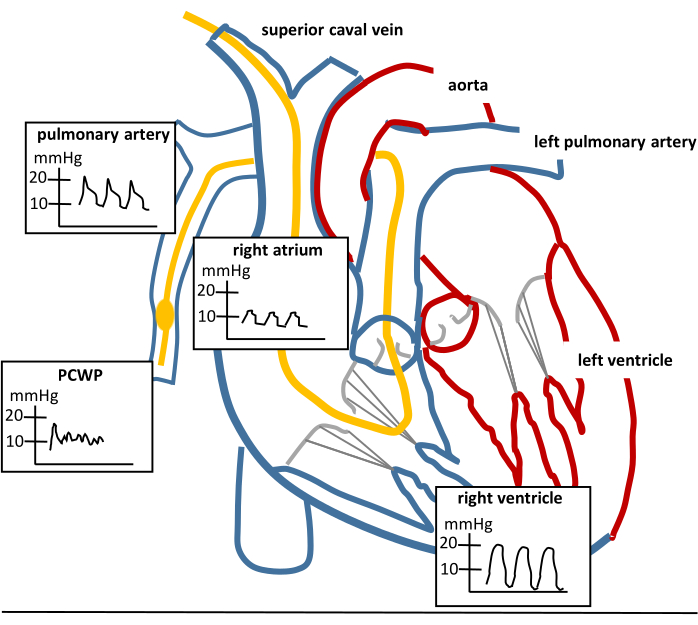
Figure 1: Pulmonary artery catheter placement. Sketch of the heart, a properly placed pulmonary artery catheter (PAC; yellow catheter) and the respective waveforms that can be seen while advancing a PAC. PCWP means pulmonary capillary wedge pressure. The PCWP waveform can only be seen in wedge position while the balloon is inflated. The PCWP curve should disappear and the pulmonary artery curve should be visible if the balloon is deflated and the PAC is placed properly. Please click here to view a larger version of this figure.
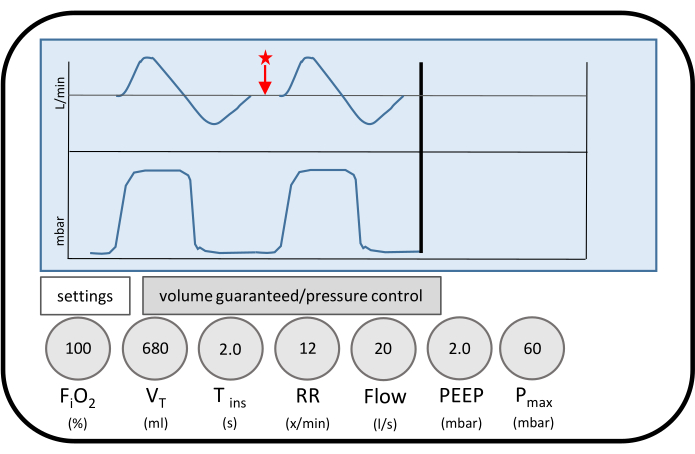
Figure 2: Ventilator settings of injurious ventilation. Displayed are the ventilator settings during ventilation to provoke ventilator-induced lung injury (VILI). The tidal volume corresponds to 17 mL/kg body weight in the respective animal. The flow pattern decreases to zero flow on expiration (red star). Zero flow is maintained for a relevant period of the respiratory cycle. Thus, almost complete inflation and deflation of the lungs is achieved to promote baro- and atelectrauma. Please click here to view a larger version of this figure.
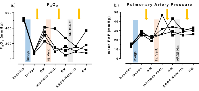
Figure 3: Systemic oxygenation and pulmonary artery pressure. (A) Individual results of the partial arterial pressure of oxygen. (B) Mean pulmonary arterial pressure of four animals are displayed as representative values for the induced lung injury. Test for statistical significance were not performed due to the small number of animals (n = 4). A recruitment maneuver was performed after each intervention (yellow arrows) to test for recruitability of the model. Note that PaO2 increases after lung lavage and after recruitment by at least 150 mmHg, but not after injurious ventilation. Please click here to view a larger version of this figure.
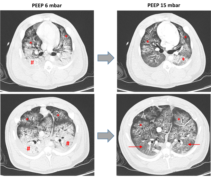
Figure 4: Computed tomography of the lungs. Representative computer tomographic scans (CT) of one animal after surfactant washout and mechanical ventilation with high tidal volumes and low PEEP to cause ventilator-induced lung injury (VILI). The scans were taken during ventilation with high positive end expiratory pressure of 15 mbar (PEEP 15 mbar) and low PEEP of 6 mbar (PEEP 6 mbar) with a tidal volume of 6 mL/kg body weight. The upper panels show the same apical region of the lungs. The lower panels show the same region of the lung at height of the heart. The # marks the dependent lung areas with basal atelectasis; the → marks the dependent lung areas/former atelectasis, which are recruited under ventilation with a PEEP of 15 mbar; the * marks extensive ground glass opacities with superimposed inter- and intralobular septeal thickening, which are not resolved during ventilation with a PEEP of 15 mbar, the + marks diffuse alveolar opacifications, which indicate alveolar hemorrhage and are not visible during ventilation with a PEEP of 6 mbar due to the extensive atelectasis. Please click here to view a larger version of this figure.
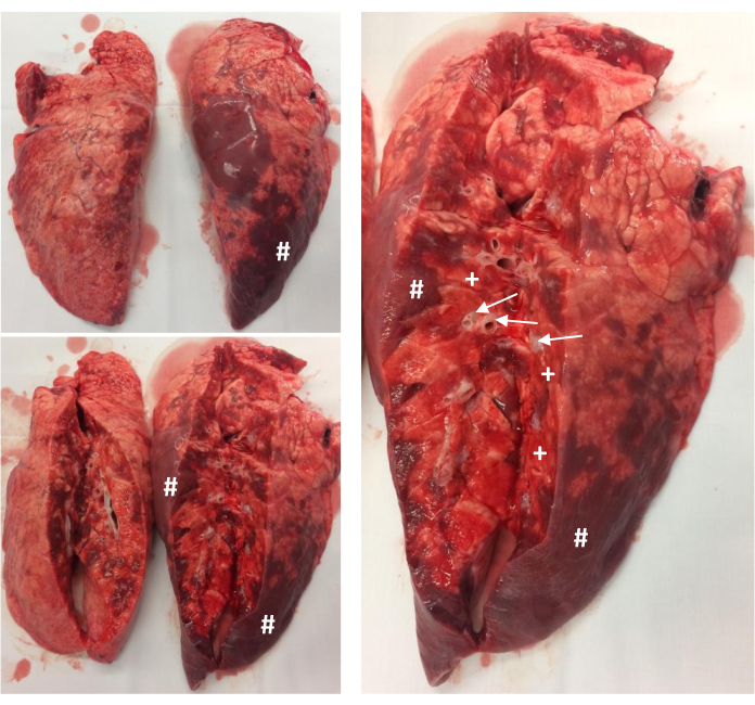
Figure 5: Postmortem examination of the lungs. Representative pathology of the unfixed lungs of one animal right after the experiment. The basal area of the lungs faces toward the reader. The # marks atelectasis; the + marks diffuse alveolar hemorrhage; the → marks distended, edematous peribronchial spaces. Please click here to view a larger version of this figure.
baseline | after lavage | RM | after injuriuos ventilation | RM | after ARDS-Net | RM | |
PaO2 (mmHg) |
514 ±13 |
87 ±12 |
324 ±78 |
197 ±134 |
147 ±95 |
128 ±37 |
185 ±129 |
PaCO2 (mmHg) |
48 ±6 |
86 ±10 |
82 ±12 |
66 ±5 |
96 ±4 |
92 ±5 |
123 ±10 |
pH | 7.39 ±0.09 |
7.14 ±0.05 |
7.17 ±0.08 |
7.26 ±0.06 |
7.11 ±0.04 |
7.14 ±0.04 |
7.04 ±0.03 |
lactate (mg/dL) |
4 ±3.9 |
6 ±5.0 |
6 ±5.9 |
4 ±3.6 |
4 ±3.5 |
4 ±3.6 |
6 ±5.3 |
heart rate (beats/min) |
86 ±8 |
90 ±11 |
92 ±12 |
104 ±18 |
129 ±30 |
147 ±13 |
149 ±5 |
CO (L/min) | 4 ±0.8 |
3.7 ±1.4 |
3.6 ±0.8 |
5.2 ±0.8 |
5.1 ±0.8 |
6.9 ±1.0 |
|
mAP (mmHg) |
93 ±4 |
101 ±21 |
108 ±31 |
78 ±8 |
96 ±31 |
65 ±12 |
72 ±9 |
SVR (dyn.sec.cm-5) |
1856 ±302 |
2552 ±777 |
1624 ±468 |
1179 ±237 |
903 ±292 |
711 ±166 |
|
mPAP (mmHg) |
14 ±1 |
27 ±2 |
22 ±2 |
33 ±10 |
33 ±8 |
29 ±3 |
30 ±3 |
PVR (dyn.sec.cm-5) |
106 ±170 |
267 ±442 |
170 ±258 |
92 ±126 |
108 ±160 |
66 ±88 |
|
PCWP | 6 ±2 |
10 ±2 |
8 ±2 |
9 ±1 |
10 ±4 |
11 ±5 |
|
Cdyn (mL/mbar) |
33 ±4 |
12 ±2 |
21 ±4 |
23 ±8 |
20 ±2 |
26 ±8 |
24 ±5 |
Table 1: Arterial blood gases, hemodynamic data and lung compliance. The table presents the respective arterial blood gases and hemodynamic data. RM: recruitment maneuver, PaO2: arterial partial pressure of oxygen, PaCO2: arterial partial pressure of carbon dioxide, CO: cardiac output, MAP: mean arterial pressure, SRV: systemic vascular resistance, mPAP: mean pulmonary arterial pressure, PVR: pulmonary vascular resistance, PCWP: pulmonary capillary wedge pressure. Data presented as mean ± SD.
baseline | after lavage | RM | ||||||
I | PaO2 (mmHg) | 540 | 81.3 | 270 | 21.9 | -the recruitment maneuver after surfactant depletion was preformed without prior injection of a muscle relaxant -the recruitment maneuver (RM) resulted in a tension pneumothorax with rapid cardiopulmonary deterioration (grey background) despite immediate chest drain insertion – following animals received a bolus injection of a muscle relaxant prior to a RM and the problem was not observed again |
||
PaCO2 (mmHg) | 42.6 | 69.4 | 84.9 | 93.9 | ||||
pH | 7.44 | 7.17 | 7.01 | 6.99 | ||||
Lactate (mmol/L) | 11 | 17 | 67 | 56 | ||||
heart rate (beats/min) | 138 | 155 | 141 | 221 | ||||
CO (L/min) | 7.7 | 3.6 | 1.6 | |||||
mAP (mmHg) | 82 | 60 | 143 | 53 | ||||
mPAP (mmHg) | 26 | 18 | 22 | 22 | ||||
PCWP (mmHg) | 10 | 12 | 12 | 17 | ||||
Cdyn (mbar/mL) | 35 | 11 | 19 | 13 | ||||
PCWP (mmHg) |
10 | 12 | 12 | 17 | ||||
Cdyn (mbar/mL) | 35 | 11 | 19 | 13 | ||||
baseline | after lavage | RM | after injurious ventilation | RM | ||||
II | PaO2 (mmHg) | 638 | 60 | 84 | 83.2 | 61.4 | 82.7 | -injurous ventilation was performed with tidal a volume of 17 ml/kg body weight for 3 hours -after injurious ventilation the animal deteriorated rapidly and could not be stabilized with e.g. bolus injections of epinephrine -the last blood gas analysis was obtained under ventilation with PEEP: 20 mbar. Ppeak: 35 mbar. resulting in a tidal volume of only 187 ml (4ml/kg body weight) – reduction of the injurious ventilation period was necessary in following experiments |
PaCO2 (mmHg) | 41 | 78 | 77 | 85.1 | 120 | 183 | ||
pH | 7.37 | 7.17 | 7.16 | 7.13 | 7.02 | 6.81 | ||
Lactate (mg/dL) | 16 | 18 | 20 | 17 | 30 | 65 | ||
heart rate (beats/min) | 86 | 64 | 109 | 133 | 150 | 185 | ||
CO (L/min) | 4.3 | 3.3 | 3.7 | 5.6 | 2.4 | |||
mAP (mmHg) | 77 | 82 | 61 | 53 | 77 | 40 | ||
mPAP (mmHg) | 15 | 30 | 24 | 35 | 35 | 32 | ||
PCWP (mmHg) | 7 | 8 | 9 | 8 | 9 | |||
Cdyn (mbar/mL) | 34 | 9 | 12 | 17 | 14 | 13 |
Table 2: Arterial blood gases and hemodynamic data during implementation of the protocol. The table presents the respective arterial blood gases and hemodynamic data of two animals, which died prematurely during the implementation of the protocol. Gray background highlights the last results before death. RM: recruitment maneuver, PaO2: arterial partial pressure of oxygen, PaCO2: arterial partial pressure of carbon dioxide, CO: cardiac output, mAP: mean arterial pressure, mPAP: mean pulmonary arterial pressure, PCWP: pulmonary capillary wedge pressure, PEEP: positive end-expiratory pressure, Ppeak: peak inspiratory pressure.
This article describes the induction of experimental ARDS in pigs combining surfactant depletion by repeated lung lavages and ventilation with high tidal volumes, low PEEP, and complete inflation/deflation of the lungs. This combination causes a reproducible and comparable deterioration in gas exchange and the resulting hemodynamic compromise but limits the recruitability of the lungs. Thus, this model mimics clinical ARDS with low recruitability and allows the investigation of new ventilation regimes.
There are a few limitations of the protocol. First, repeated lavages result in some of the histopathological properties of clinical (human) ARDS, including the formation of major atelectasis, perivascular edema formation, and an increase of the alveolar-capillary membrane thickness. High Tv/low PEEP ventilation adds some properties such as diffuse alveolar hemorrhage, which are not vulnerable to recruitment. Nevertheless, important features of human ARDS such as the formation of hyaline membranes cannot be induced within hours and are therefore missing in this model2,3. Second, the structural damage of the lungs is irreversible for hours or possibly days. But care must be taken to avoid an excessive baro-, volu-, and atelectrauma of the lungs, which would render the following experiment impossible. Using the ventilator settings described in the article, the protocol started with initially 3 h of VILI to test automated ventilation modes, which integrate recent clinical evidence regarding ventilation of ARDS patients. Unfortunately, some animals deteriorated during the course of the experiment and one case of a severe pneumothorax (Table 2) was observed. Reducing the VILI period to 2 h was suitable for the experimental design, but this time period may be adapted in other experimental settings. Third, lung lavages can result in abrupt right heart failure and death of the animal. About 10%-15% of the animals may die during the induction period. This number can be reduced following the recommendations published previously5. Finally, the study only presented the results of four animals and two further animals, which died prematurely during the implementation of the model. Strict local animal protection laws do not support experiments in further animals once the model is sufficiently implemented, but two-hit models consisting of surfactant depletion and injurious ventilation have been used by other research groups7.
Of importance, the aggravation of lung injury by ventilation with high Tv/low PEEP ventilation can result in uncontrollable structural damage of the lungs or hemodynamic decompensation. Hence, the tidal volumes have to be increased in steps over several minutes and an upper threshold for peak inspiratory pressure has to be set to avoid pneumothorax and hemodynamic instability. It was found that an upper threshold of 60 mbar was most suitable to cause VILI without losing animals prematurely.
The cyclic recruitment with high tidal volumes will result in sufficient oxygenation despite low PEEP. After the lung lavages, the PEEP was decreased to 2 mbar in a stepwise fashion parallel to increasing tidal volume in order to avoid intolerable hypoxemia.
Some investigators use higher respiratory rates to generate VILI7 due to a faster onset of VILI, but high respiratory rates can result in air trapping if the flow curve of the ventilator is not monitored closely. Air trapping may reduce VILI due to incomplete deflation of the lungs for one thing, while it also promotes hemodynamic instability caused by sustained high intrathoracic pressures. Thus, a slower respiratory rate was used with an ensured deflation of the lungs and longer VILI period in the described model.
Of note, markers of pulmonary inflammation such as interleukin 8 in the brochoalveolar fluid were not measured since prolonged ventilation in a reproducible model of low recruitability is the main application of the model. For research concerning specific inflammatory patterns (such as the hyper-inflammatory subphenotype of ARDS) a multiple hit model combining an inflammatory first hit such as i.v. lipopolysaccharide infusion with injurious ventilation could be favourable12.
The combination of surfactant washout and high Tv/low PEEP ventilation results in a time-efficient and reproducible model of human ARDS with respect to gas exchange and hemodynamic changes. The lung injury induced in this model presents low recruitability and permits the experimental investigation of therapeutic strategies, including mechanical ventilation.
The authors have nothing to disclose.
We gratefully acknowledge the excellent technical assistance of Birgit Brandt. This study was supported by a grant of the German Federal Ministry of Education and Research (FKZ 13GW0240A-D).
Evita Infinity V500 | Dräger | intensive care ventilator | |
Flow through chamber thermistor | Baxter | 93-505 | for measuring cardiac output |
Leader Cath Set | Vygon | 1,15,805 | arterial catheter |
Mallinckrodt Tracheal Tube Cuffed | Covidien | 107-80 | 8.0 mm ID |
MultiCath3 | Vygon | 1,57,300 | 3 lumen central venous catheter, 20 cm length |
Percutaneus Sheath Introducer Set | Arrow | SI-09600 | introducer sheath for pulmonary artery catheter of 4-6 Fr., 10 cm length |
Swan-Ganz True Size Thermodilution Catheter | Edwards | 132F5 | pulmonary artery catheter, 75 cm length |
urinary catheter | no specific model requiered | ||
Vasofix Braunüle 20G | B Braun | 4268113B | peripheral vein catheter |
Vigilance I | Edwards | monitor |