여기에서 우리는 출생 후 뇌 발달 과정에서 마우스 소뇌에서 과립 뉴런의 시냅스 형성을 시각화하는 방법을 설명합니다.이 세포는 시냅스 구조를 개선하고 시냅스를 형성하여 전체 뇌 회로에 통합됩니다.
이 콘텐츠를 보려면 JoVE 구독이 필요합니다. 로그인하거나 오늘 무료 평가판을 시작하세요.
이 논문에서
정오 공지
Important: There has been an erratum issued for this article. View Erratum Notice
요약
초록
뉴런은 뇌 발달 과정에서 구조와 기능에 역동적인 변화를 일으켜 다른 세포와 적절한 연결을 형성합니다. 설치류 소뇌는 시간이 지남에 따라 단일 세포 유형인 소뇌 과립 뉴런(CGN)의 발달과 형태 형성을 추적하는 이상적인 시스템입니다. 여기에서, 발달 중인 마우스 소뇌에서 과립 뉴런 전구체의 생체 내 전기천공을 사용하여 후속 형태학적 분석을 위해 세포를 드물게 라벨링했습니다. 이 기술의 효능은 CGN 성숙의 주요 발달 단계를 보여주는 능력에서 입증되며, 특히 이러한 세포가 시냅스 입력의 대부분을 받는 특수 구조인 수지상 발톱의 형성에 중점을 둡니다. 소뇌 발달 전반에 걸쳐 CGN 시냅스 구조의 스냅샷을 제공하는 것 외에도, 이 기술은 관심 유전자의 역할과 CGN 형태, 발톱 발달 및 시냅스 생성에 미치는 영향을 연구하기 위해 세포 자율적 방식으로 과립 뉴런을 유전적으로 조작하는 데 적용할 수 있습니다.
서론
뇌 발달은 배아 발생에서 출생 후의 삶으로 확장되는 장기간의 과정입니다. 이 기간 동안 뇌는 수상 돌기와 축삭 사이의 시냅스 배선을 조각하여 궁극적으로 행동을 유도하는 내적 자극과 외적 자극의 조합을 통합합니다. 설치류 소뇌는 단일 뉴런 유형인 소뇌 과립 뉴런(CGN)의 발달이 전구 세포에서 성숙한 뉴런으로 전환될 때 추적할 수 있기 때문에 시냅스가 어떻게 발달하는지 연구하는 이상적인 모델 시스템입니다. 이는 부분적으로는 소뇌 피질의 대부분이 출생 후 발달하여 출생 후 유전자 조작과 세포 표지가 용이하기 때문입니다1.
포유류에서 CGN 분화는 후뇌의 증식 세포 하위 집합이 마름모꼴 입술 위로 이동하여 소뇌 표면에 2차 발아 영역을 형성할 때 배아 발달이 끝날 때 시작됩니다 2,3,4. 과립 뉴런 전구체(GNP) 정체성에 완전히 전념하지만, 이 세포는 출생 후 14일(P14)까지 외부 과립층(EGL)의 바깥쪽 부분 내에서 계속 증식합니다. 이 층의 증식은 소뇌의 대규모 확장을 초래하는데, 이 세포들은 독점적으로 CGN을 생성하기 때문이다....
액세스가 제한되었습니다. 이 콘텐츠를 보려면 로그인하거나 체험판을 시작하세요.
프로토콜
참고: 모든 절차는 Duke University IACUC(Institutional Animal Care and Use Committee)에서 승인한 프로토콜에 따라 수행되었습니다.
1. 생체 내 전기천공법(in vivo electroporation) 또는 IVE를 위한 DNA 준비(수술 1일 전)
- 정제된 DNA(동물당 0.5-25μg), 3M 아세트산나트륨, 에탄올, 패스트 그린 염료, 초순수 증류수, 인산염 완충액(PBS)(재료 표 참조)을 수집합니다.
참고: DNA의 경우, 인간 유비퀴틴 프로모터 하에서 녹색 형광 단백질(GFP)을 발현하는 구축물을 Addgene(FUGW, https://www.addgene.org/14883/)으로부터 입수하였다. 유비쿼터스 프로모터의 제어 하에 GFP 또는 다른 형광 단백질을 발현하는 임의의 구축물은 작동해야 한다. 이 기술을 사용한 CGN 특정 라벨링은 구조에 의존하지 않고 전기천공에 의존합니다. - 원하는 양의 DNA, 10 부피 %의 3 M 아세트산 나트륨 및 250 부피 %의 100 % 얼음 차가운 에탄올을 혼합하여 전기 천공을 위해 DNA를 준비합니다. DNA는 용액에서 즉시 침전됩니다.
- DNA 혼합물을 -....
액세스가 제한되었습니다. 이 콘텐츠를 보려면 로그인하거나 체험판을 시작하세요.
결과

그림 4: 소뇌 발달 중 과립 뉴런 형태 분석. (A) 3-DPI에서 14-DPI(출생 후 연령 P10 내지 P21), 핵(파란색) 및 GFP(녹색)에서 전기천공된 CGN의 최대 투영 이미지; 화살촉은 개별 수상 돌기를 나타내며 스케일 바는 10 μm입니다. (B) 수상 돌기의 평균 수. (C) 소마의 기저부에서 수상돌기의 끝까지 측정한 평균 수상돌기 길이. (D) 발톱을 포함하는 수상 돌기의 분율; 1.00의 값은 100%이며, 즉, 모든 수상돌기에는 발톱이 있습니다. (E) 수지상 발톱의 전체 길이. 조건당 N > 30개의 세포, 조건당.......
액세스가 제한되었습니다. 이 콘텐츠를 보려면 로그인하거나 체험판을 시작하세요.
토론
소뇌 과립 뉴런은 포유류 뇌에서 가장 풍부한 뉴런으로 설치류 뇌의 전체 뉴런 인구의 거의 60-70 %를 차지합니다 1,14. 소뇌는 세포 증식, 이동, 수상돌기 형성 및 시냅스 발달의 메커니즘을 밝히기 위해 광범위하게 활용되었습니다 6,9,10,11,15,16,17,18,19,20
액세스가 제한되었습니다. 이 콘텐츠를 보려면 로그인하거나 체험판을 시작하세요.
공개 사항
저자는 이해 상충을 선언하지 않습니다.
감사의 글
이 연구는 NIH 보조금 R01NS098804 (AEW), F31NS113394 (UC) 및 Duke University의 Summer Neuroscience Program (DG)의 지원을 받았습니다.
....액세스가 제한되었습니다. 이 콘텐츠를 보려면 로그인하거나 체험판을 시작하세요.
재료
| 이름 | 회사 | 카탈로그 번호 | 댓글 |
|---|---|---|---|
| Betadine | Purdue Production | 67618-150-17 | |
| 시멘트 10 µ L 바늘 | Hamilton | 1701SN (80008) | 33 게이지, 1.27cm(0.5인치), 4점 스타일 |
| 치킨 안티-GFP | Millipore Sigma | AB16901 | 우리 실험실에서는 이 항체를 1:1000 농도로 사용합니다 |
| 튼 팁 애플리케이터 | |||
| 당나귀 안티 치킨 Cy2 | Jackson ImmunoResearch | 703-225-155 | 우리 실험실은 1:500 농도의 |
| 에탄올(200 증거) | Koptec | V1016 | |
| Electroporator ECM 830 | BTX Harvard Apparatus | 45-0052 | |
| Fast Green FCF | Sigma | F7252-5G | |
| FUGW plasmid | Addgene | 14883 | |
| Glass slides | VWR | 48311-703 | Superfrost plus |
| Glycerol | Sigma-Aldrich | G5516 | |
| 가열 패드 | Softheat | ||
| Hoescht 33342 형광 염료 | Invitrogen | 62249 | |
| Imaris | Bitplane | ||
| Isoflurane | Patterson Veterinary | 07-893-1389 | |
| 마이크로 커버 유리 | VWR | 48382-138 | |
| 매니큐어 | Sally Hansen | Color 109 | |
| 일반 염소 serum | Gibco | 16210064 | |
| O.C.T. 임베딩 컴파운드 | Tissue-Tek | 4583 | |
| Olympus MVX10 해부 스코프 | Olympus | MVX10 | |
| P200 피펫 리치 팁 | Fisherbrand | 02-707-138 | 니들 스페이서에 사용 |
| Parafilm | Bemis | PM-996 | |
| PBS pH 7.4 (10x) | Gibco | 70011-044 | |
| 단순 뉴라이트 트레이서 | FIJI | https://imagej.net/Simple_Neurite_Tracer:_Basic_ 지침 | |
| : 설탕 | 시그마 | S0389 | |
| 수술 도구 | RWD, 생명 과학 | 소형 가위 및 핀셋 | |
| Triton X-100 | Roche | 11332481001 | 비이온 세제 |
| 핀셋 | BTX, Harvard Apparatus | 45-0489 | 5mm, 백금 도금 핀셋 유형 전극, |
| 초순수 증류수 | Invitrogen | 10977-015 | |
| Vectashield 장착 매체 | Vectashield | H1000 | |
| Vetbond 조직 접착제 | 3M | 1469SB | |
| Zeiss 780 Upright Confocal | Zeiss | 780 |
참고문헌
- Altman, J., Bayer, S. A. Development of the cerebellar system : in relation to its evolution, structure, and functions. , CRC Press. (1997).
- Rahimi-Balaei, M., Bergen, H., Kong, J., Marzban, H. Neuronal migration during development of the cerebellum. Frontiers in....
액세스가 제한되었습니다. 이 콘텐츠를 보려면 로그인하거나 체험판을 시작하세요.
재인쇄 및 허가
정오
Formal Correction: Erratum: Utilizing In Vivo Postnatal Electroporation to Study Cerebellar Granule Neuron Morphology and Synapse Development
Posted by JoVE Editors on 4/06/2023. Citeable Link.
An erratum was issued for: Utilizing In Vivo Postnatal Electroporation to Study Cerebellar Granule Neuron Morphology and Synapse Development. A figure was updated.
Figure 2 was updated from:
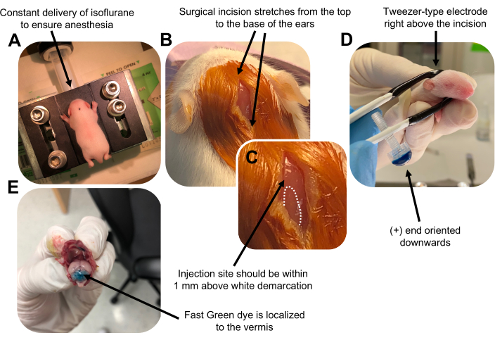
Figure 2: In vivo cerebellar electroporation of granule neuron progenitors in P7 wildtype mouse pups. (A) Pups are anesthetized with 4% isoflurane delivered at a rate of 0.8L/min to ensure anesthesia throughout the injection of the DNA solution. Isoflurane is delivered at a rate of 0.8 L/min. (B) After sterilizing the mouse 3 times with betadine and 70% ethanol, an incision is made that spans the distance of the ears, revealing the hindbrain. (C) A magnified image of a white demarcation on the cranium, a landmark for the injection site. DNA construct should be injected within 1 mm above the mark; dotted lines outline the demarcation, and black arrow denotes the injection site. The ridges of the cerebellar vermis may be visible and can be useful for finding the injection site. (D) Tweezer-type electrode orientation for efficient electroporation. Plus (+) end must be oriented downwards to pull negatively charged DNA into the cerebellar parenchyma prior to administration of electrical pulses. (E) Test injection of 1 µL of a 0.02% Fast Green dye shows injection is localized to the middle of the cerebellar vermis between lobules 5-7. Please click here to view a larger version of this figure.
to:
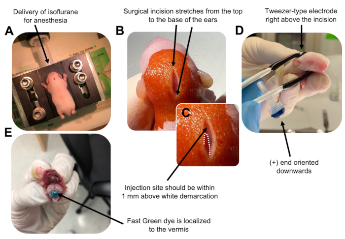
Figure 2: In vivo cerebellar electroporation of granule neuron progenitors in P7 wildtype mouse pups. (A) Pups are anesthetized with 4% isoflurane delivered at a rate of 0.8L/min to ensure anesthesia throughout the injection of the DNA solution. Isoflurane is delivered at a rate of 0.8 L/min. (B) After sterilizing the mouse 3 times with betadine and 70% ethanol, an incision is made that spans the distance of the ears, revealing the hindbrain. (C) A magnified image of a white demarcation on the cranium, a landmark for the injection site. DNA construct should be injected within 1 mm above the mark; dotted lines outline the demarcation, and black arrow denotes the injection site. The ridges of the cerebellar vermis may be visible and can be useful for finding the injection site. (D) Tweezer-type electrode orientation for efficient electroporation. Plus (+) end must be oriented downwards to pull negatively charged DNA into the cerebellar parenchyma prior to administration of electrical pulses. (E) Test injection of 1 µL of a 0.02% Fast Green dye shows injection is localized to the middle of the cerebellar vermis between lobules 5-7. Please click here to view a larger version of this figure.
