NOTE: See the Table of Materials for details related to all materials and equipment used in this protocol.
1. Culturing and cryopreservation of Wharton's jelly mesenchymal stem cells
- Remove the WJ-MSCs (from ATCC) from the -80 °C freezer. Seed the cells into a flask containing DMEM-F12 medium supplemented with 10% fetal bovine serum (FBS). Incubate them at 37 °C in an incubator containing 5% CO2.
- Change the culture medium every 2 days. Passage the cells when they reach 80% confluence. Wash the cells to be passaged with 5 mL of phosphate-buffered saline (PBS).
NOTE: Washing with PBS before adding trypsin ensures easy separation of cells from the flask surface. - Remove the PBS and add 5 mL of trypsin-EDTA solution to the flask. Incubate the flask for 5 min at 37 °C in an incubator containing 5% CO2.
- Collect the supernatant into the tube and centrifuge at 1,500 x g for 5 min. After centrifugation, remove the supernatant and add 1 mL of DMEM-F12 + 10% FBS to the tube by pipetting.
- Transfer the cells to a larger flask and continue the culture by adding DMEM-F12 + 10% FBS until sufficient exosomes are obtained35.
NOTE: Cultured cells are frozen at a density of 1 million/mL with 10% dimethyl sulfoxide (DMSO) and stored at -80 °C.
2. Production of exosomes from Wharton's Jelly mesenchymal stem cells
NOTE: Exosomes are isolated from cultured WJ-MSCs. Exosome isolation is performed when cells cover the flask surface and reach approximately 80% density.
- Remove the supernatant medium containing 10% FBS from the flask and wash the cells with 5 mL of PBS. Add serum-free DMEM-F12 medium to the cells after rinsing with PBS. Incubate flasks for 48 h at 37 °C in a 5% CO2 incubator. Following incubation, collect the serum-free medium in the tube and store at -20 °C.
NOTE: In step 2.1, 180 mL of serum-free medium is collected and stored at -20 °C. After thawing, a batch centrifugation process is started, as described below. - Isolation of exosomes
- Centrifuge the collected serum-free medium at 300 x g for 10 min at +4 °C. Carefully withdraw the supernatant and transfer it to another tube.
- Centrifuge the collected supernatant at 2,000 × g for 10 min at +4 °C. Carefully withdraw the supernatant and transfer it to another tube.
- Centrifuge the collected supernatant at 10,000 × g for 30 min at +4 °C. Carefully withdraw the supernatant and transfer it to another tube.
- Transfer the collected supernatant to ultracentrifuge tubes and ultracentrifuge at 110,000 × g for 130 min. Slowly remove the supernatant and add 1 mL of PBS to the pellet.
- Vortex the pellet and ultracentrifuge at 110,000 × g for 70 min at +4 °C36 (Figure 1). Slowly remove the supernatant and add 1 mL of PBS to the pellet.
NOTE: After the ultracentrifugation steps are completed, the exosomes obtained can be stored at -80 °C. The obtained pellet is now ready for characterization.
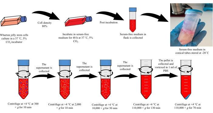
Figure 1: Exosome isolation. Please click here to view a larger version of this figure.
3. Characterization of exosomes
- Nanoparticle Tracking Analysis (NTA)
NOTE: Nanoparticle tracking analysis is performed to determine the size and concentration of the isolated exosomes. Tablets used for 1x PBS solution must be in the range of pH 7.3-7.5. 1x PBS Solution contains 10 mM phosphate buffer, 137 mM sodium chloride, and 2.7 mM potassium chloride. The solution is prepared by adding 1 PBS tablet in 100 mL of distilled water. This prepared solution is sterilized by autoclaving.- Dilute the exosomes 100-fold by adding 990 µL of PBS to 10 µL of exosomes. Take the diluted suspension into a disposable syringe.
- Turn on the NTA device and connect the computer. Open the software.
- Inject the sample in the syringe into the tubing in the cassette section of the device. Close the cassette cover of the device.
- In the software that opens, click on the Start Analysis button. Save the results obtained by pressing the Record button.
- Dynamic light scattering analysis
NOTE: Exosomes isolated for zeta potential and size measurement are also diluted in the same manner as for NTA analysis.- Add 980 µL of PBS to 20 µL of the exosomes.
- Open the zeta sizing device and the connected computer. Open the software.
- Add the suspension from step 3.2.1 to the disposable cuvette. Open the lid of the device, place the cuvette in the cassette, and close the lid.
- Select the cuvette type in the device's software. Click on the Start Analysis button for zeta potential and dimensional analysis.
NOTE: Analyses are performed separately for dimensional analysis and zeta potential.
4. Dopamine loading into exosomes
NOTE: After the characterization of WJ-MSCs exosomes is completed, dopamine-loaded exosomes were obtained as a drug delivery system. Drug loading into exosomes is performed using the incubation method.
- Add 3 mL of PBS to the exosome suspension from step 2.2.5. Sterilize the diluted suspension by filtration using 0.22 µm filters. Transfer 500 µL of the sterilized exosome suspension to another tube.
- Prepare dopamine solution (0.5 mg/mL) with distilled water. Add 500 µL of dopamine solution into the tubes containing the exosomes.
- Add saponin to the suspension in step 4.2. Incubate the prepared exosome-dopamine suspension for 24 h at 37 °C.
NOTE: Saponin concentration should not exceed 0.002% of the total solution. - Ultracentrifuge the suspension at 90,000 × g for 70 min to remove free dopamine and saponin from the medium. Store the isolated exosomes at -20 °C until use for further analysis37.
NOTE: Add 0.02% ascorbic acid for the stability of the prepared dopamine solution.
5. Characterization of dopamine loaded exosomes
- Nanoparticle tracking analysis (NTA)
NOTE: Nanoparticle tracking analysis is performed to determine the size and concentration of isolated exosomes.- Follow steps 3.1.1-3.1.4 to dilute the exosomes and perform NTA.
- Dynamic light scattering analysis
NOTE: Exosomes isolated for zeta potential and size measurement are also diluted similarly to NTA analysis.- Follow steps 3.2.1-3.2.4 to dilute the exosomes and perform dynamic light scattering analysis.
NOTE: Size and zeta potentials for each solution (saponin, dopamine, exosome-dopamine) are analyzed separately.
- Follow steps 3.2.1-3.2.4 to dilute the exosomes and perform dynamic light scattering analysis.
6. High-Performance Liquid Chromatography (HPLC)
NOTE: The amounts of dopamine loaded into exosomes are measured by the high-performance liquid chromatography (HPLC) method. To detect the presence of dopamine within the obtained formulation, exosomes are detonated by a special process.
- Put the formulation in a 75 °C heater to evaporate. Add acetonitrile (50:50 ratio) to the suspension and vortex. Sonicate the solution for 10 min.
- Centrifuge the solution for 10 min at 10,000 × g. Filter the supernatant through a 0.22 µm filter.
- Analyze all analytes using a C18 column at 30 °C at a flow rate of 1 mL/min (the mobile phase H2O/acetonitrile). Measure the absorbance at 230 nm38.
7. Drug loading capacity (DL) measurement and in vitro drug release kinetics
- Drug loading capacity (DL)
NOTE: The amount of dopamine loaded into exosomes is quantified using UV-Vis spectroscopy. The absorbance is read at 280 nm. The collected supernatants during synthesis are used to measure the amount of unloaded drug. The dopamine concentration in the supernatant is determined via a standard calibration curve for dopamine.- Prepare a stock solution of 1 mg/mL of dopamine. Prepare concentrations of 0.05, 0.1, 0.2, 0.3, 0.4, and 0.5 mg/mL from the stock solution using distilled water.
NOTE: Add 0.02% ascorbic acid for the stability of the prepared dopamine solution. - Generate a standard calibration curve by measuring the absorbance of each dilution of dopamine at 280 nm in a UV-Spectrophotometer.
- Measure the absorbance of the supernatant at 280 nm.
- Calculate the drug loading capacity for dopamine using Eq (1)39.
DL (%) =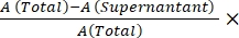 100 (1)
NOTE: The analysis is performed in triplicate.
- Prepare a stock solution of 1 mg/mL of dopamine. Prepare concentrations of 0.05, 0.1, 0.2, 0.3, 0.4, and 0.5 mg/mL from the stock solution using distilled water.
- In vitro drug release kinetics
NOTE: Drug release kinetics of dopamine-loaded exosomes are performed using a dialysis membrane. PBS, pH 7.4, is used as the release medium to simulate a physiological microenvironment.- Add 1 mL of dopamine-loaded exosomes to the dialysis membrane. Place the membrane in a beaker. Add 15 mL of PBS to the beaker.
- Sample 1 mL of release medium in a beaker at 0.5, 1, 2, 4, 6, and 8 h. At each time point, replace the volume of the sample with fresh PBS. Analyze the samples taken at specified intervals at 280 nm using a UV-Vis spectrometer.
- Calculate the results using Eq (2)40.
Release (%) = 100 (2)
NOTE: A calibration curve is prepared for the determination of in vitro drug release kinetics. Dopamine solution is prepared at 0.5, 1, 1.5, 2, 2.5, 3, 3.5, 4, 4.5, and 5.0 mg/mL concentrations. The UV absorbance value of each concentration is determined at 280 nm with a UV-Vis spectrophotometer.
8. In vitro cytotoxicity test
- Revitalization and culture of fibroblasts
- Remove the fibroblasts from the -80 °C freezer. Seed the cells into a flask containing DMEM-F12 + 10% FBS.
- Incubate the cells at 37 °C in an incubator containing 5% CO2. Change the culture medium every 2 days.
- Passage the cells until they reach 80% confluence. Follow steps 1.3-1.5. After centrifugation at 1,500 × g for 5 min, remove the supernatant, add 1 mL of DMEM-F12 + 10% FBS, and seed 10,000 cells per well in a 96 well plate.
- Add DMEM-F12 medium + 10% FBS and incubate the cells at 37 °C for 18 h in an incubator containing 5% CO2. Change the medium of the wells at the end of the incubation.
- Prepare the stock of dopamine-loaded exosome suspension. Dilute the dopamine-loaded exosome formulation with medium to obtain concentrations of 100 µL/mL, 250 µL/mL, 500 µL/mL, and 1,000 µL/mL.
- Add the prepared concentrations onto the cells within each well of the 96-well plate. Incubate the plates at 37 °C for 18 h in an incubator containing 5% CO241.
- MTT cell viability assay
NOTE: At the end of the incubation, the viability ratios of cells are determined by the MTT test.- Prepare the MTT solution (5 mg/mL) with PBS. Add 10 µL of MTT solution to each well. Incubate for 4 h at 37 °C.
- At the end of the incubation, add 100 µL of DMSO to each well. Incubate the plates for 30 min at room temperature in the dark. Measure the absorbance values in ELISA reader at 570 nm42,43.
NOTE: The MTT viability test is performed in triplicate.
Exosome isolation and characterization
Wharton jelly stem cells are cultured and incubated in a serum-free medium for 48 h when the culture reaches sufficient density. After the end of the incubation, the supernatant is stored at -20 °C. The collected supernatants are diluted with PBS and subjected to ultracentrifugation (Figure 1). The obtained solution is analyzed by NTA and DLS analyses. The exosomes are sterilized by passing through a 0.22 µm filter. The average size of the obtained exosomes was determined to be 98 nm (Video 1). Approximately 10 billion exosome particles were obtained at the end of centrifugation. The numbers of nanoparticles revealed by NTA and DLS analyses are consistent with each other. In addition, the zeta potential of exosomes originating from WJ-MSCs was measured as -15.7 mV (Figure 2 and Figure 3).
Video 1: Nanoparticle tracking analysis of Wharton Jelly-derived exosomes. Please click here to download this Video.
Loading of dopamine into exosomes
As calculated in NTA analysis, there were approximately 1.5 billion nanoparticles in 500 µL of the isolated samples from Wharton's jelly. Dopamine solution (5 mg/mL) was mixed with 500 µL of exosomes, and the suspension was incubated for 24 h at 37 °C. Following incubation, the obtained solution was characterized by NTA and DLS analyses. According to the NTA results, the average particle size of the solution was found to be 110 nm. A comparison of the particle sizes of exosomes and dopamine-loaded exosomes reveals that there was a significant increase in the dimensions of the dopamine-loaded exosomes. The numbers of nanoparticles revealed by NTA and DLS analyses were consistent with each other. In addition, the zeta potential of dopamine-loaded exosomes was measured as -17.6 Mv (Figure 2 and Figure 3).
Video 2: Nanoparticle tracking analysis of dopamine-loaded exosomes. Please click here to download this Video.
| Sample | Zeta potential |
| Exosome | -15.7 mV ± 1.1 |
| Saponin | -12 mV ± 0.7 |
| Dopamine | -14.4 mV ± 1.3 |
| Dopamine- loaded exosome | -17.6 mV ± 0.8 |
Table 1: Zeta potential of all solutions.
DSL analyzes, the Zeta sizer, and Zeta potential table of the dopamine, saponin, and dopamine-loaded exosome solution are shown in Table 1.
Figure 2: Nanoparticle tracking analysis of exosomes. (A) Exosomes; (B) dopamine-loaded exosomes. Please click here to view a larger version of this figure.
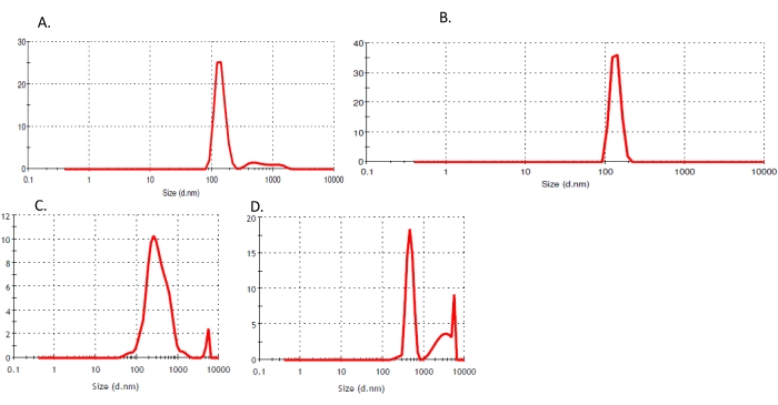
Figure 3: Zetasizer analysis. (A) Exosomes, (B) dopamine-loaded exosomes, (C) Saponin, (D) Dopamine. Abbreviation: d = size. Please click here to view a larger version of this figure.
The presence of dopamine in the solution was determined by HPLC analysis. Dopamine-loaded exosomes were ultracentrifuged and filtered to remove free dopamine. If dopamine loading into exosomes is successful at the end of incubation, it can be detected by direct measurement of dopamine following the rupture of the exosomes. For this purpose, the dopamine-loaded exosome suspension was heated with steam, acetonitrile added, and the suspension was sonicated. Absorbance was measured at 230 nm to assess the amounts of dopamine released from the disrupted exosomes.
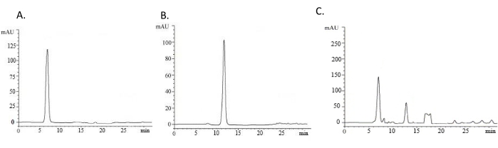
Figure 4: Presence of dopamine in the formulation determined by HPLC analysis. All analyses were performed using a C-18 column with a mobile phase of H2O/Acetonitrile at a flow rate of 1 mL/min at 30 °C. Absorbance is measured at 230 nm to monitor the elution of dopamine. (A) Dopamine, (B) Saponin, (C) Dopamine-loaded exosomes. Abbreviation: HPLC = high-performance liquid chromatography. Please click here to view a larger version of this figure.
As a result of HPLC analysis, the resulting peak for dopamine was observed at 6.45 min. The presence of saponin was also detected after exosome fragmentation (Figure 4).
Drug loading capacity and in vitro drug release kinetics
Dopamine loading capacity was calculated using UV spectrophotometry measurements and Eq (1) and Eq (2). Loading capacity percentage was found to be 10.89 ± 0.33. The cumulative drug release profile is shown in Figure 5. Drug release kinetics monitored for 8 h revealed that 74.8% of encapsulated dopamine was released from exosomes within the first 8 h (Figure 5).
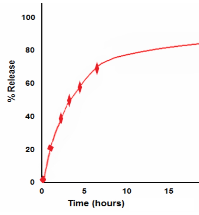
Figure 5: Cumulative drug release profile. Please click here to view a larger version of this figure.
In vitro cytotoxicity test
The cytotoxicity of dopamine-loaded and free exosomes were investigated on fibroblasts. Fibroblasts were exposed to different concentrations of formulations (100 µL/mL, 250 µL/mL, 500 µL/mL, and 1,000 µL/mL) and incubated for 18 h. Although dopamine-loaded exosomes did not demonstrate any remarkable cytotoxic effects, saponin added to the formulation to increase the encapsulation of dopamine decreased fibroblast viability (p < 0.05, Figure 6). There was an increase in cell viability when the fibroblasts were treated with dopamine-loaded exosomes up to 500 µL/mL concentration. At the end of the 18 h incubation, cell viability was determined by the MTT test (Figure 7). The statistical significance of the results was evaluated by performing one-way ANOVA.
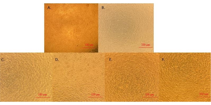
Figure 6: Microscope images (10x) after incubation of the formulation with the cells. (A) Dopamine, (B) Saponin, (C) Dopamine-loaded exosomes 100 µL/mL, (D) Dopamine-loaded exosomes 250 µL/mL, (E) Dopamine-loaded exosomes 500 µL/mL, (F) Dopamine-loaded exosomes 1,000 µL/mL. Scale bars = 100 µm. Please click here to view a larger version of this figure.
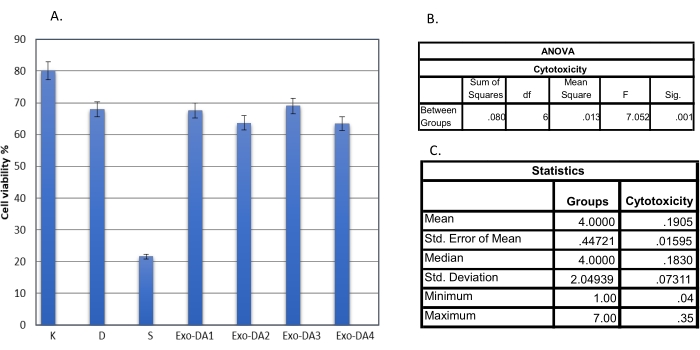
Figure 7: Statistical analysis of MTT viability test results in cells incubated for 18 h with different concentrations of dopamine, saponin, and dopamine-loaded exosomes. (A) Cytotoxicity results, (B,C) Statistical analyses. Abbreviations: D = Dopamine; S = Saponin; Exo-DA1 = Dopamine-loaded exosomes 100 µL/mL; Exo-DA2 = Dopamine-loaded exosomes 250 µL/mL; Exo-DA 3 Dopamine-loaded exosomes 500 µL/mL; Exo-DA 4 = Dopamine-loaded exosomes 1,000 µL/mL; MTT = 3-(4,5-dimethylthiazol-2-yl)-2,5-diphenyltetrazolium bromide. Please click here to view a larger version of this figure.
Additionally, the cytotoxic effects of free exosomes were also investigated on fibroblasts at similar concentrations (Figure 8). Before the experiment, exosomes were diluted with PBS to 60 million particles per mL. Then, 10 µL, 25 µL, 50 µL, and 100 µL of the exosome suspension was added to each well of a 96-well plate.
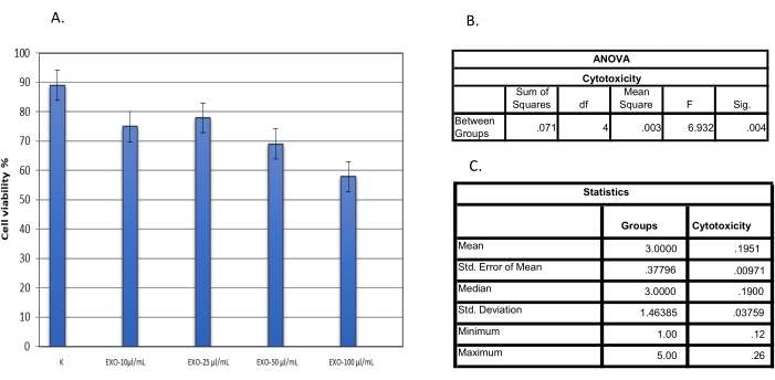
Figure 8: Statistical analysis of MTT viability test results in cells incubated for 18 h with different concentrations of exosomes. (A) Cytotoxicity results, (B,C) statistical analyses. Abbreviations: EXO = exosomes; MTT = 3-(4,5-dimethylthiazol-2-yl)-2,5-diphenyltetrazolium bromide. Please click here to view a larger version of this figure.
Fibroblast viability was found to be higher with 500 µL/mL dopamine-loaded exosomes and 25 µL/mL free exosomes compared to other concentrations. However, no significant cytotoxicity was observed at any concentration.
