This study provides a systematically optimized procedure of CRISPR/Cas9 ribonuclease-based construction of homozygous locust mutants as well as a detailed method for cryopreservation and resuscitation of the locust eggs.
The migratory locust, Locusta migratoria, is not only one of the worldwide plague locusts that caused huge economic losses to human beings but also an important research model for insect metamorphosis. The CRISPR/Cas9 system can accurately locate at a specific DNA locus and cleave within the target site, efficiently introducing double-strand breaks to induce target gene knockout or integrate new gene fragments into the specific locus. CRISPR/Cas9-mediated genome editing is a powerful tool for addressing questions encountered in locust research as well as a promising technology for locust control. This study provides a systematic protocol for CRISPR/Cas9-mediated gene knockout with the complex of Cas9 protein and single guide RNAs (sgRNAs) in migratory locusts. The selection of target sites and design of sgRNA are described in detail, followed by in vitro synthesis and verification of the sgRNAs. Subsequent procedures include egg raft collection and tanned-egg separation to achieve successful microinjection with low mortality rate, egg culture, preliminary estimation of the mutation rate, locust breeding as well as detection, preservation, and passage of the mutants to ensure population stability of the edited locusts. This method can be used as a reference for CRISPR/Cas9 based gene editing applications in migratory locusts as well as in other insects.
Gene editing technologies could be used to introduce insertions or deletions into a specific genome locus to artificially modify the target gene on purpose1. In the past years, CRISPR/Cas9 technology has developed rapidly and has a growing scope of applications in various fields of life sciences2,3,4,5,6. The CRISPR/Cas9 system was discovered back in 19877, and widely found in bacteria and archaea. Further research indicated that it was a prokaryotic adaptive immune system that depends on the RNA-guided nuclease Cas9 to fight against phages8. The artificially modified CRISPR/Cas9 system mainly consists of two components, a single guide RNA (sgRNA) and the Cas9 protein. The sgRNA is made up of a CRISPR RNA (crRNA) complementary to the target sequence and an auxiliary trans-activating crRNA (tracrRNA), which is relatively conserved. When the CRISPR/Cas9 system is activated, the sgRNA forms a ribonucleoprotein (RNP) with the Cas9 protein and guides Cas9 to its target site via the base pairing of RNA-DNA interactions. Then, the double-strand DNA can be cleaved by the Cas9 protein and as a result, the double-strand break (DSB) emerges near the protospacer adjacent motif (PAM) of the target site9,10,11,12. To mitigate the damage caused by the DSBs, cells would activate comprehensive DNA damage responses to efficiently detect the genomic damages and initiate the repair procedure. There are two distinct repair mechanisms in the cell: non-homologous end joining (NHEJ) and homology-directed repair (HDR). NHEJ is the most common repair pathway that can repair DNA double-strand breaks quickly and prevents cell apoptosis. However, it is error-prone because of leaving small fragments of insertions and deletions (indels) near the DSBs, which usually results in an open reading frame shift and thus can lead to gene knock-out. In contrast, homologous repairment is quite a rare event. On the condition that there is a repair template with sequences homologous to the context of the DSB, cells would occasionally repair the genomic break according to the nearby template. The result of HDR is that the DSB is precisely repaired. Especially, if there is an additional sequence between the homologous sequences in the template, they would be integrated into the genome through HDR, and in this way, the specific gene insertions could be realized13.
With the optimization and development of the sgRNA structures and Cas9 protein variants, the CRISPR/Cas9-based genetic editing system has also been successfully applied in research of insects, including but not limited to Drosophila melanogaster, Aedes aegypti, Bombyx mori, Helicoverpa Armigera, Plutella xylostella, and Locusta migratoria14,15,16,17,18,19. To the best of the authors' knowledge, although RNPs consisting of the Cas9 protein and in vitro transcribed sgRNA have been used for locust genome editing20,21,22, a systematic and detailed protocol for CRISPR/Cas9 ribonuclease mediated construction of homozygous mutants of the migratory locust is still lacking.
The migratory locust is an important agricultural pest that has a global distribution and poses substantial threats to food production, being especially harmful to gramineous plants, such as wheat, maize, rice, and millet23. Gene function analysis based on genome editing technologies can provide novel targets and new strategies for the control of migratory locusts. This study proposes a detailed method for knocking out migratory locust genes via the CRISPR/Cas9 system, including the selection of target sites and design of sgRNAs, in vitro synthesis and verification of the sgRNAs, microinjection and culture of eggs, estimation of mutation rate at the embryonic stage, detection of mutants as well as passage and preservation of the mutants. This protocol could be used as a basal reference for manipulation of the vast majority of locust genes and can provide valuable references for genome editing of other insects.
1. Target site selection and sgRNA design
2. Synthesis and verification of the sgRNA in vitro
3. Microinjection and culture of the eggs
4. Mutation rate estimation and the screening of mutants
5. Establishment and passaging of mutant lines
6. Egg cryopreservation and resuscitation
This protocol contains the detailed steps for generating homozygous mutants of the migratory locusts with the RNP consisting of Cas9 protein and in vitro synthesized sgRNA. The following are some representative results of CRISPR/Cas9-mediated target gene knockout in locusts, including target selection, sgRNA synthesis and verification (Figure 1A), egg collection and injection, mutant screening and passaging, cryopreservation, and resuscitation of the homozygous eggs.
In this study, the target site for CRISPR/Cas9 system is selected according to the results of three online programs (the E-CRISP, CRISPOR, and ZiFit) and located in the first exon (Figure 1B). According to the Cas9 cleavage assay in vitro (Table 1), the CRISPR/Cas9 RNP could digest the PCR fragment (containing the target site) with a cleavage rate of about 55% (Figure 1C). Then, this RNP was microinjected into 120 fertilized eggs at their single-cell stage using the standard microinjection system (Figure 2 and Figure 3). The sequencing results for embryonic stage mutation rate estimation suggested efficient genome editing at the target site (Figure 4A). Further, this study resulted in a 52.73% nymph hatching rate and 66.7% of the G0 adults were mosaic mutants (Table 4).
Further, the G0 chimeras were crossed with wild-type locusts to obtain heterozygous G1 individuals and the G1 heterozygotes with the same mutations (screened by TA-cloning) were in-crossed to generate the G2 animals. PCR-based phenotyping was used for detecting mutants and the obtained homozygotes were in-crossed to establish stable mutant lines (Figure 4B).
Meanwhile, the excess homozygous eggs were cryopreserved to improve the utilization rate of the homozygotes. Although the hatching rate of the cryopreserved eggs was reduced with the extension of cryopreservation time (Table 5), the remedial actions including applying filter paper fragments for keeping eggs wet and recovering these preserved eggs with a gradient of rising temperature were both helpful for the resuscitation (Figure 5 and Table 5). Finally, the homozygous population of mutants was successfully kept for subsequent research.
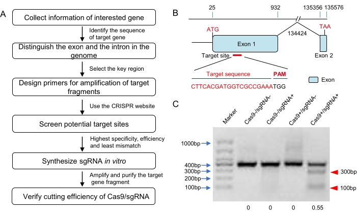
Figure 1: Design of the target and in vitro verification of the sgRNA. (A) The flow diagram for target selection, sgRNA synthesis, and bioactivity verification in vitro (including but not limited to locusts). (B) Schematic diagram of the gene structure and target site. The exons of this target gene are shown as blue domains and the target site was selected downstream of the start codon in exon 1. The target sequence is highlighted in red. (C) The agarose gel electrophoresis result of in vitro Cas9 cleavage assay. A DNA fragment harboring the target sequence (about 400 bp in length) was amplified and used as the substrate for Cas9 digestion. The expected small bands (about 100 bp and 300 bp here; marked with red triangles) suggested that the synthesized sgRNA could induce effective Cas9 cleavage. The cleavage rate was about 55% according to the grayscale analysis. Please click here to view a larger version of this figure.
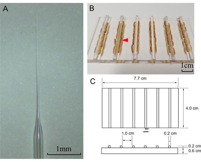
Figure 2: Needle preparation and the microinjection pad for locust eggs. (A) Tip of a prepared needle for microinjection of locust eggs. Scale bar: 1 mm. (B) Picture of a microinjection pad with eggs (indicated with a red triangle) arranged on it. The scale bar represents 1 cm. (C) The size of the microinjection pad. Please click here to view a larger version of this figure.
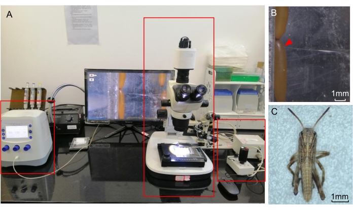
Figure 3: Egg microinjections. (A) A representative microinjection system labeled with red boxes indicating a microscope (middle) and a micromanipulator (right) connected to a microinjector (left). The computer is used for data storage and for providing auxiliary observation. (B) Injection of the locust egg. The injection site is marked with a red triangle. (C) A hatched nymph under the microscope. The scale bars represent 1 mm. Please click here to view a larger version of this figure.
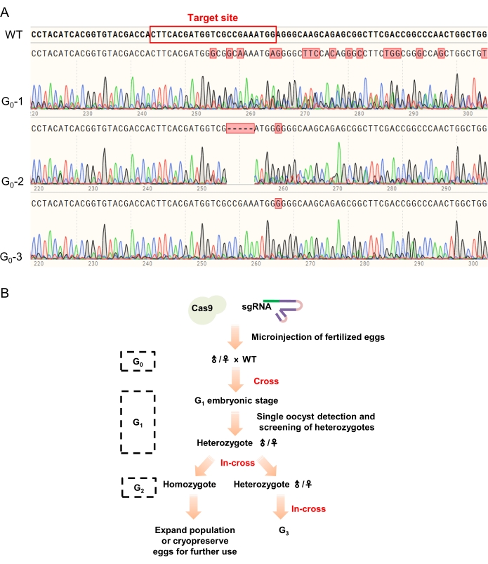
Figure 4: The sequencing results of G0 animals and passaging strategy of the mutant lines. (A) The typical sequencing results in the G0 screening. Multiple peaks near the target site (marked with a red rectangle in the wild-type sequence) indicated that the tested egg/locust was successfully edited by CRISPR/Cas9 system (as shown in G0-1 and G0-2), while the individual without any change in the sequencing result was considered as not edited and abandoned (e.g., G0-3). (B) The passaging strategy of the mutant lines. The G0 animals were firstly screened by PCR-mediated genotyping and those with multiple peaks in their sequencing results were crossed with wild-type locusts to generate the G1 animals. PCR-mediated genotyping and TA cloning were used to identify the G1 heterozygotes. Then, heterozygotes with the same mutations were in-crossed to generate the G2 animals (homozygotes and heterozygotes) for further research and passaging. Please click here to view a larger version of this figure.
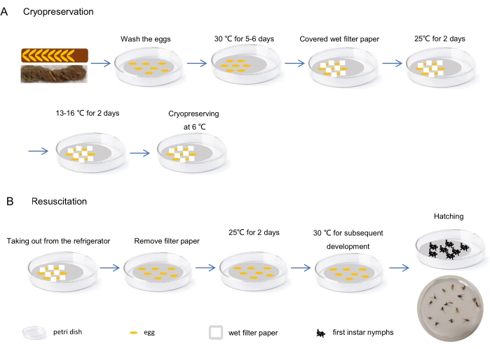
Figure 5: Egg cryopreservation and resuscitation. (A) The procedure of cryopreservation: Isolate the eggs from egg pods and incubate them at 30 °C for 5-6 days after washing with sterile water. Then, gather the developed eggs together in the Petri dish and cover them with small scraps of moist filter paper. Wrap the entire Petri dish with a paraffin film and keep it at 25 °C for 2 days, followed by another 2 days at a relatively lower temperature (e.g., 13-16 °C). Finally, refrigerate it at 6 °C. (B) The procedure of resuscitation: Take the Petri dishes with cryopreserved eggs out from the refrigerator and keep them at 25 °C for 2 days after removing the filter paper scraps. Then, the eggs can be cultured for subsequent development at 30 °C until the nymphs hatch. Please click here to view a larger version of this figure.
Reagent | Volume (μL) |
Cas9 protein (300 ng/μL) | 1 |
sgRNA (300 ng/μL) | 1 |
PCR product (200 ng/μL) | 1 |
10×NEBuffer r3.1 | 1 |
Nuclease-free water | 6 |
Table 1: In vitro digestion system. 200 ng of purified target fragment (PCR product) was mixed with the CRISPR/Cas9 system (300 ng of each component) for testing the bio-activity of the synthesized sgRNAs. A commercial buffer was used and nuclease-free water was added to make up the total volume to 10 µL.
Reagent | Volume (μL) |
2xEs Taq MasterMix | 12.5 |
Forward primer | 0.5 |
Reverse primer | 0.5 |
Template (lysis product) | 1 |
Nuclease-free water | 10.5 |
Table 2: PCR system for target gene fragment amplification. To amplify the genomic fragment of the target gene, a commercial Taq mix was used and primers were added according to the manufacturer's instructions. 1 µL of the lysis product was used as the template and nuclease-free water was used to make the total volume to 25 µL.
Temperature (°C) | Time | Cycles |
95 | 5 min | 1 |
95 | 30 s | 35 |
55 | 30 s | |
72 | 40 s | |
72 | 10 min | 1 |
Table 3: PCR program for target gene amplification. The PCR program for target gene amplification was: 95 °C for 5 min; 35 cycles of 95 °C for 30 s, 55 °C for 30 s, 72 °C for 40 s; 72 °C for 10 min.
Items | Data |
No. of injected eggs | 120 |
Tested embryos | 10 |
No. of hatched nymphs | 58 |
Hatching rate | 52.73% |
No. of G0 adult | 12 |
No. of G0 mutants | 8 |
Mutation efficiency in G0 adults | 66.67% |
Table 4: Summary of editing efficiency. 120 eggs were injected to knock out the target gene and 10 eggs were taken for testing during the embryo stage. 58 nymphs hatched from the rest embryos (the hatching rate was 52.73%). Finally, 12 G0 locusts developed to the adult stage and 8 of them were successfully edited at the target site. The mutation rate in G0 adults was 66.67%.
Control group | Group 1 | Group 2 | Group 3 | Group 4 | |
No. of eggs | 120 | 120 | 120 | 120 | 120 |
Temperature treatment for storage | 30 °C | 30°C (5-6 d)→6 °C | 30°C (5-6 d)→25°C (2 d)→13-16°C (2 d)→6 °C | 30°C (5-6 d)→25°C (2 d)→13-16°C (2 d)→6 °C | 30°C (5-6 d)→25°C (2 d)→13-16°C (2 d)→6 °C |
Storage time | 14 days | 14 days | 1 month | 3 months | 5 months |
Temperature treatment for resuscitation | 30 °C | 6°C→30 °C | 6°C→25°C (2 d)→30 °C | 6°C→25°C (2 d)→30 °C | 6°C→25°C (2 d)→30 °C |
No. of hatched locusts | 108 | 0 | 96 | 86 | 78 |
Hatching rate | 90.00% | 0 | 80.00% | 71.67% | 65.00% |
No. of adult locusts | 81 | 0 | 60 | 46 | 31 |
Eclosion rate | 75.00% | 0 | 62.50% | 53.49% | 39.74% |
Table 5: Summary of the cryopreservation and resuscitation of eggs. 600 eggs were divided into five groups for cryopreservation and resuscitation study. The first group (control) of 120 eggs was always incubated at 30 °C and stored for 14 days. The second group (Group 1) of 120 eggs was transferred to a refrigerator after incubation at 30 °C for 5-6 days and stored at 6 °C for 14 days. Then, these eggs were transferred to 30 °C for resuscitation. The other groups (group 2, group 3, and group 4, each group contained 120 eggs) experienced a gradient cooling and recovering temperature treatment as described in the protocol (steps 6.1-6.3) with a different storage time (1 month for group 2, 3 months for group 3, and 5 months for group 4). At last, 108 nymphs hatched from the control group (at a hatching rate of 90%) and 81 of them developed to the adult stage (75% of the eclosion rate). No locusts hatched in the second group (Group 1). The hatching rate and eclosion rate of the other groups were lower compared to the control group and declined with the storage time. 96 nymphs hatched in group 2 and 60 of them developed to the adult stage. 86 nymphs hatched in group 3 and 46 of them developed to the adult stage. In group 4, 78 nymphs hatched and 31 of them developed to the adult stage.
Locusts have been among the most devastating pests to agriculture since the civilization of human beings23. CRISPR/Cas9-based genome editing technology is a powerful tool for providing better knowledge of the biological mechanisms in locusts as well as a promising pest control strategy. Thus, it is of great benefit to develop an efficient and easy-to-use method of CRISPR/Cas9-mediated construction of homozygous locust mutants. Although some great works have been reported and provided some basic workflow for genome editing in locusts18,20,21,22, a systematic optimization of the whole procedure is still lacking. In general, the homozygous locust mutant generated by embryonic injection of CRISPR/Cas9 system can be divided into three major steps: a) sgRNA design, synthesis, and screening in vitro, b) egg collection, preparation, and injection; and c) mutant screening and homozygote maintaining. This protocol provides an optimized workflow and the steps of target site selection, in vitro sgRNA screening, microinjection of the eggs as well as detecting mutants are especially critical for effectively generating homozygous mutant locusts with the CRISPR/Cas9 system.
Firstly, the target site, the form of corresponding sgRNA, and the concentration of sgRNA have been suggested as the critical factors of genome editing efficiency in insects18,27,28. To solve this problem, it is recommended to first analyze the gene structure and design more than one target site on exon 1 or near the start codon using the online resources. Then, synthesize sgRNA in vitro and mix it with Cas9 protein at different concentrations to optimize the cutting efficiency of RNP. This procedure will help to identify a sgRNA with high bioactivity and optimize the composition of the RNP complex for the candidate target gene.
Secondly, collecting as many fresh eggs as possible in a limited time and improving the hatching rate as well as mutation rate are also big challenges for genome editing operations. The gregarious adult locusts reared at the condition of 16 (light):8 (dark) photoperiod are used for collecting enough eggs in a short period during 9:00-11:00 am. It is important to gently apply downward pressure between the eggs using a paintbrush or tweezers to carefully isolate the eggs from the pod. With enough practice, multiple users can reliably separate individual eggs from the pod. After each egg is isolated and washed with sterile water, the eggs are aligned on the designed injection pads (Figure 2B,C) to stabilize the eggs during microinjection. The egg is injected with the optimized RNP near the micropyles. To limit the mechanical damage and minimize the pollution of eggs, fresh eggs are allowed to stay in the pods for about 30 min to make sure that the eggshell tanning is ideal for microinjection21, owing to its condensed structure and high defense ability on invasion after enough tanning29,30. Meanwhile, a published report indicated that the syncytial division31 of locust eggs occurs at some point between 0 h and 4 h after laying. With the current standard microinjection protocol, injection of about 100 eggs can be accomplished within 40 min. Taken together, the procedure of tanning and microinjection of 100 fertilized eggs can be completed within 2 h. Thus, there is at least 2 h left for CRISRP/Cas9-mediated cleavage and the repair of target genes to achieve efficient mutation in the injected eggs and future adult locusts.
Lastly, effective strategies for passaging and mutant identification are important for generating homozygotes in most mutant animals. In locusts, for genes that are nonlethal after editing, the cross strategy suggested here is that G0 mutants cross with wide-type locusts and further in-crosses can be performed using the G1 heterozygotes with the same mutations to generate homozygous mutants in the following generation. To verify the mutation in each locust during the mutant inheriting process, it is recommended to use the alkaline lysis method to digest a short segment of antennae as the template for PCR-based genotyping. Finally, use the obtained homozygous adults for further in-cross to expand the population. Limited by the population size and development stage differences between homozygous locusts, the excess eggs are recommended for cryopreservation at 6 °C and resuscitation by gradient temperature rise (Figure 5), which is based on the principle of low-temperature diapause and high-temperature diapause release of migratory locust eggs32,33.
In short, the CRISPR/Cas9 system has been proven to be a reliable tool for facilitating the study of functional genomics of migratory locusts. This detailed protocol can be used as a reference for CRISPR/Cas9 based gene editing applications in migratory locusts as well as in other insects.
The authors have nothing to disclose.
This work was supported by the National Natural Science Foundation of China (32070502, 31601697, 32072419 and the China Postdoctoral Science Foundation (2020M672205).
10×NEBuffer r3.1 | New England Biolabs | B7030S | The buffer of in vitro Cas9 cleavage assays |
2xEs Taq MasterMix (Dye) | Cwbio | CW0690 | For gene amplification |
2xPfu MasterMix (Dye) | Cwbio | CW0686 | For gene amplification |
CHOPCHOP | Online website for designing sgRNAs, http://chopchop.cbu.uib.no/. | ||
CRISPOR | Online website for designing sgRNAs, http://crispor.org. | ||
CRISPRdirect | Online website for designing sgRNAs, http://crispr.dbcls.jp/. | ||
Electrophoresis power supply | LIUYI BIOLOGY | DYY-6D | Separation of nucleic acid molecules of different sizes |
Eppendorf Tube | Eppendorf | 30125177 | For sample collection, etc. |
Fine brushes | Annigoni | 1235 | For cleaning and isolating eggs. Purchased online. |
Flaming/brown micropipette puller | Sutter Instrument | P97 | For making the microinjection needles |
Gel Extraction Kit | Cwbio | CW2302 | DNA recovery and purification |
Gel Imaging Analysis System | OLYMPUS | Gel Doc XR | Observe the electrophoresis results |
GeneTouch Plus | Bioer | B-48DA | For gene amplification |
Glass electrode capillary | Gairdner | GD-102 | For making injection needles with a micropipette Puller |
Incubator | MEMMERT | INplus55 | For migratory locust embryo culture |
Metal bath | TIANGEN | AJ-800 | For heating the sample |
Micro autoinjector | Eppendorf | 5253000068 | Microinjection of embryos early in development |
Micro centrifuge | Allsheng | MTV-1 | Used for mixing reagents |
Microgrinder | NARISHIGE | EG-401 | To ground the tip of injection needle |
Microloader | Eppendorf | 5242956003 | For loading solutions into the injection needles. |
Micromanipulation system | Eppendorf | TransferMan 4r | An altinative manipulation system for microinjection |
Microscope | cnoptec | SZ780 | For microinjection |
Motor-drive Manipulator | NARISHIGE | MM-94 | For controling the position of the micropipette during the microinjection precedure |
Multi-Sample Tissue Grinder | jingxin | Tissuelyser-64 | Grind and homogenize the eggs |
ovipisition pot | ChangShengYuanYi | CS-11 | Filled with wet sterile sand for locust ovipositing in it. The oocysts are collected from this container. Purchased online. |
Parafilm | ParafilmM | PM996 | For wrapping the petri dishes. |
pEASY-T3 Cloning Kit | TransGen Biotech | CT301-01 | For TA cloning |
Petri dish | NEST | 752001 | For culture and preservation of the eggs. |
Pipettor | Eppendorf | Research®plus | For sample loading |
plastic culture cup | For rearing locusts seperately and any plastic cup big enough (not less than 1000 mL in volume) will do. Purchased online. | ||
Precision gRNA Synthesis Kit | Thermo | A29377 | For sgRNA synthesis |
Primer Premier | PREMIER Biosoft | Primer Premier 5.00 | For primer design |
SnapGene | Insightful Science | SnapGene®4.2.4 | For analyzing sequences |
Steel balls | HuaXinGangQiu | HXGQ60 | For sample grinding.Purchased online. |
Tips | bioleaf | D781349 | For sample loading |
Trans DNA Marker II | TransGen Biotech | BM411-01 | Used to determine gene size |
TrueCut Cas9 Protein v2 | Thermo | A36496 | Cas9 protein |
UniversalGen DNA Kit | Cwbio | CWY004 | For genomic DNA extraction |
VANNAS Scissors | Electron Microscopy Sciences | 72932-01 | For cutting off the antennae |
Wheat | To generate wheat seedlings as the food for locusts. Bought from local farmers. | ||
ZiFiT | Online website for designing sgRNAs, http://zifit.partners.org/ZiFiT/ChoiceMenu.aspx. |