Here, we present a protocol to detect bacterial motility based on a color reaction. Key advantages of this method are that it is easy to evaluate and more accurate, and does not require specialized equipment.
Bacterial motility is crucial for bacterial pathogenicity, biofilm formation, and drug resistance. Bacterial motility is crucial for the invasion and/or dissemination of many pathogenic species. Therefore, it is important to detect bacterial motility. Bacterial growth conditions, such as oxygen, pH, and temperature, can affect bacterial growth and the expression of bacterial flagella. This can lead to reduced motility or even loss of motility, resulting in the inaccurate evaluation of bacterial motility. Based on the color reaction of 2,3,5-triphenyl tetrazolium chloride (TTC) by intracellular dehydrogenases of living bacteria, TTC was added to traditional semisolid agar for bacterial motility detection. The results showed that this TTC semisolid agar method for the detection of bacterial motility is simple, easy to operate, and does not involve large and expensive instruments. The results also showed that the highest motility was observed in semisolid medium prepared with 0.3% agar. Compared with the traditional semisolid medium, the results are easier to evaluate and more accurate.
Bacterial motility plays a critical role in bacterial pathogenicity, biofilm formation, and drug resistance1. Bacterial motility is closely associated with pathogenicity and is necessary for bacterial colonization during early infection of host cells2. Biofilm formation is closely related to bacterial motility, where bacteria adhere to the surface of solid media through motility. Bacterial motility has long been considered to be positively correlated with biofilm formation. A high degree of bacterial drug resistance due to biofilm can lead to persistent infections that are a threat to human health3,4,5. Therefore, it is important to detect bacterial motility. The bacterial motility test is mainly used to examine the motility of different forms of bacteria in the living state, which can indirectly determine the presence or absence of flagella and, thus, has an important role in the identification of bacteria.
There are direct and indirect methods to detect bacterial motility6. As bacteria with flagella show motility, it is possible to detect whether bacteria are motile indirectly by detecting the presence or absence of flagella. For example, it is possible to detect motility indirectly by electron microscopy and flagellar staining to indicate that bacteria are motile. It is also possible to detect by direct methods, such as suspension drop and semisolid puncture methods.
The semisolid puncture method commonly used in undergraduate microbiology laboratories to detect bacterial motility inoculates the bacteria into the puncture in the semisolid agar medium containing 0.4-0.8% agar, according to the direction of bacterial growth. If the bacteria grow along the puncture line to spread around, cloud-like (brush-like) growth traces appear, indicating the presence of flagella and, therefore, motility. If there are no puncture-line growth traces, the bacterium is neither flagellated nor motile.
However, this method has its drawbacks: the bacteria are colorless and transparent, the flagellar activity is affected by the physiological characteristics of the living bacteria and other factors, and the concentration of agar and the small diameter of the test tube. Moreover, aerobic bacteria are only suitable for growth on the agar surface, affecting the observation of bacterial motility. Hence, to improve this experiment, 2,3,5-triphenyltetrazolium chloride (TTC) (colorless) was added to the medium to establish a more reliable and intuitive method of determining bacterial motility than the current direct-puncture method using intracellular dehydrogenases to catalyze the formation of a red product of TTC7,8,9,10.
1. Preparation of semisolid medium
2. Bacterial strains
NOTE: Eighty strains were isolated from the aquatic environment and identified using an automated bacteria identification instrument (see the Table of Materials), including Escherichia coli, Pseudomonas aeruginosa, Salmonella spp., Vibrio spp., Klebsiella pneumoniae, and Aeromonas hydrophila (Table 1). Staphylococcus aureus (see the Table of Materials) was used as a negative nonmotile control; Escherichia coli, Pseudomonas aeruginosa, and Salmonella typhimurium (see the Table of Materials) were used as positive control strains.
3. TTC-enhanced bacterial motility observation
4. Effect of different agar concentrations on bacterial motility
Both standard strains and isolated strains were compared for motility detection, and the results are shown in Table 1. Due to the absence of flagella, Staphylococcus aureus and Klebsiella pneumoniae only grew along the inoculated line on both traditional and TTC semisolid media. In contrast, Pseudomonas aeruginosa, Escherichia coli, and Salmonella typhimurium showed growth in all directions around the inoculated line after culturing for 24 h on TTC semisolid medium. This was even more obvious after 48 h culture (Figure 1). Although the bacteria grew in all directions in the traditional semisolid medium, it was much more difficult to visualize than in TTC medium due to the small number of bacteria at the outer side of the inoculated line.
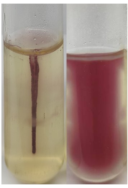
Figure 1: Motility test results using TTC semisolid medium. Staphylococcus aureus on the left, Escherichia coli on the right. Please click here to view a larger version of this figure.
Strain | Semisolid medium with 0.4% agar | Semisolid medium with 0.4% agar and 0.005% TTC | |||
24 h | 48 h | 24 h | 48 h | ||
P. aeruginosa ATCC27853 | + | + | + | + | |
E. coli ATCC25922 | + | + | + | + | |
S. typhimurium ATCC14028 | + | + | + | + | |
S. aureus ATCC25923 | – | – | – | – | |
E. coli (15) | 12 | 14 | 13 | 14 | |
Salmonella spp. (8) | 7 | 8 | 8 | 8 | |
A. hydrophila (20) | 18 | 20 | 20 | 20 | |
Vibrio spp. (8) | 7 | 8 | 8 | 8 | |
P. aeruginosa (24) | 18 | 20 | 22 | 23 | |
K. pneumoniae (5) | -5 | -5 | -5 | -5 | |
Numbers indicate the numbers of positive strains (+) and negative stains (-). |
Table 1: Comparison of bacterial motility.
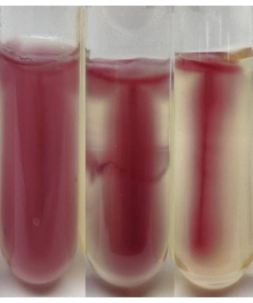
Figure 2: Observation of Escherichia coli motility activity at different agar concentrations. Left to right: 0.3%, 0.5%, 0.8% agar. Please click here to view a larger version of this figure.
The influence of agar concentration on bacterial motility is shown in Figure 2. We found that the highest motility was observed in semisolid medium prepared with 0.3% agar. The medium color in the tube turned almost entirely red. In contrast, the area of red diffusion decreased, and the diffusion was prolonged with increasing agar concentration.
The detection of bacterial motility by the semisolid medium method is affected by many factors13,14. Bacterial growth conditions, such as oxygen (aerobic on agar surface, nonaerobic at the bottom of the tube with the semisolid medium), pH, and temperature, can affect the viability of bacterial flagella, which can lead to reduced motility or even loss of motility15. In addition, some mucus-type bacteria as their motility can be affected by the production of podoconjugates.
The addition of TTC to the semisolid medium helps the observation of motility. The fermentative bacteria with motive power can grow in all directions along the puncture line after incubation. Hence, the medium around the puncture line becomes red. The nonfermentative bacteria with motive power have low oxygen content in the lower part of the medium. Hence, these bacteria grow poorly, so that only the upper layer of the medium is red. Bacteria without motive power can only grow on the inoculation line, and only the puncture line appears red.
When detecting bacterial motility with TTC-semisolid medium, the longer the culture time, the more obvious are the results, especially with lower agar concentrations. If the result is difficult to interpret, the culture time should be extended appropriately. This may be related to the agar concentration of the semisolid medium, the number of bacteria, and their motility16. In addition, this method revealed that some strains of E. coli and P. aeruginosa could not grow around on conventional semisolid agar medium and showed only a faint red color on the surface of the medium on TTC semisolid agar medium. This may be due to the production of capsules by such strains, which affects their motility17. This phenomenon also occurs in Neisseria meningitidis18 because of its capsule. In conclusion, the detection of bacterial motility using a chromogenic semisolid medium containing TTC reduces the influence of bacterial factors on the test results and makes the results easier to observe with the naked eye. The advantage of a high detection rate makes this an effective method that can replace the traditional semisolid medium for detecting bacterial motility.
The authors have nothing to disclose.
This study was supported by the Priority Academic Program Development of Jiangsu Higher Education Institutions (PAPD) and Teaching Reform Research Project of China Pharmaceutical University (2019XJYB18).
Bacto Agar | Difco | ||
Escherichia coli | ATCC | ATCC25922 | Positive control |
Pseudomonas aeruginosa | ATCC | ATCC27853 | Positive control |
Salmonella typhimurium | ATCC | ATCC14028 | Positive control |
Staphylococcus aureus | ATCC | ATCC25923 | Negative nonmotile control |
Tryptose | OXOID | ||
TTC | Sigma | 298-96-4 | |
VITEK 2 automated microbial identification system | Bio Mérieux |