Summary

تقنيات الفحص المجهري لتفسير الاستعمار الفطري في أنسجة النباتات المتغايرة والإنبات التكافلي للبذور

Published: May 17, 2022
doi:

Summary

يهدف هذا البروتوكول إلى توفير إجراءات مفصلة لجمع وتثبيت وصيانة عينات النباتات المتغايرة ، وتطبيق تقنيات الفحص المجهري المختلفة مثل المسح المجهري الإلكتروني ونقله ، والمجهر الضوئي ، والكونفوكال ، والمجهر الفلوري لدراسة الاستعمار الفطري في الأنسجة النباتية والبذور المنبتة بالفطريات الفطرية.

Abstract

علم النبات الهيكلي هو منظور لا غنى عنه لفهم كامل للبيئة وعلم وظائف الأعضاء والتنمية وتطور النباتات. عند البحث في النباتات المتغايرة (أي النباتات التي تحصل على الكربون من الفطريات) ، يمكن للجوانب الرائعة من تكيفاتها الهيكلية ، وأنماط استعمار الأنسجة بواسطة الفطريات ، والتشريح المورفوغرافي للأعضاء الجوفية أن تنير استراتيجياتها التنموية وعلاقاتها مع hyphae ، مصدر العناصر الغذائية. دور مهم آخر للفطريات التكافلية يرتبط بإنبات بذور السحلية. جميع أنواع Orchidaceae هي mycoheterotrophic أثناء مرحلة الإنبات والشتلات (المتفطرة الأولية) ، حتى تلك التي تقوم بعملية التمثيل الضوئي في مراحل البالغين. بسبب نقص الاحتياطيات الغذائية في بذور السحلية ، فإن التكافل الفطري ضروري لتوفير الركائز وتمكين الإنبات. يمكن أن يجيب تحليل مراحل الإنبات من خلال وجهات النظر الهيكلية أيضا على أسئلة مهمة تتعلق بتفاعل الفطريات مع البذور. يمكن تطبيق تقنيات تصوير مختلفة للكشف عن النباتات الداخلية للفطريات في الأنسجة النباتية ، كما هو مقترح في هذه المقالة. يمكن تلطيخ الأجزاء اليدوية والرقيقة من الأعضاء النباتية ثم ملاحظتها باستخدام المجهر الخفيف. يمكن تطبيق الفلوروكروم المترافق مع أجغلوتينين جنين القمح على الفطريات واحتضانه مع Calcofluor White لتسليط الضوء على جدران الخلايا النباتية في المجهر البؤري. بالإضافة إلى ذلك ، يتم تفصيل منهجيات المسح المجهري الإلكتروني ونقله لبساتين الفاكهة المتغايرة ، ويتم استكشاف إمكانيات تطبيق هذه البروتوكولات في النباتات ذات الصلة. يتم وصف الإنبات التكافلي لبذور السحلية (أي في وجود الفطريات الفطرية) في البروتوكول بالتفصيل ، إلى جانب إمكانيات إعداد الهياكل التي تم الحصول عليها من مراحل مختلفة من الإنبات للتحليل باستخدام المجهر الضوئي والمشبك والإلكتروني.

Introduction

البحوث الهيكلية في علم النبات ، والتي تغطي مورفولوجيا النبات والتشريح ، أساسية في فهم الكائن الحي بأكمله1،2 ، وتوفر وجهات نظر لا غنى عنها لدمج والمساهمة في المعرفة المتعلقة بالبيئة وعلم وظائف الأعضاء والتنمية وتطور النباتات3. تشمل الطرق في مورفولوجيا النبات والتشريح حاليا البروتوكولات والمعدات والمعرفة التي تم تطويرها مؤخرا وكذلك منذ أكثر من قرن من الزمان2. إن التنفيذ المستمر والتكيف مع الأساليب الكلاسيكية (مثل المجهر الضوئي) جنبا إلى جنب مع التقنيات الحديثة (مثل المجهر البؤري ، التصوير المقطعي المصغر بالأشعة السينية) لها نفس الأساس الأساسي: المعرفة النظرية التي تمكن من تطوير منهجية.

الأداة الرئيسية في تشريح النبات والمورفولوجيا هي الصورة. على الرغم من الاعتقاد الخاطئ بأن هذه التحليلات هي ملاحظات بسيطة ، مما يعطي مساحة للتفسيرات الذاتية2 ، فإن تحليل وفهم الصور في هذا المجال يتطلب معرفة الأساليب المطبقة (المعدات ، ونوع التحليل ، والإجراءات المنهجية) ، ومكونات الخلية ، والكيمياء النسيجية ، وجسم النبات (تنظيم الأنسجة ووظيفتها ، وعلم الوراثة ، والتكيفات المورفولوجية). يمكن أن يؤدي تفسير الصور التي تم الحصول عليها عبر مجموعة متنوعة من الطرق إلى ربط الشكل والوظيفة ، وفك رموز التركيب الكيميائي للبنية ، والتأييد في وصف الأصناف ، وفهم العدوى بمسببات الأمراض النباتية ، وغيرها من التقييمات المماثلة.

عند البحث في النباتات المتغايرة (MH) (أي النباتات غير الضوئية التي تحصل على الكربون من الفطريات الفطرية 4,5) ، يمكن للجوانب الرائعة من تكيفاتها الهيكلية ، وأنماط استعمار الأنسجة بواسطة الفطريات ، والتشريح المورفوغرافي للأعضاء الجوفية أن تنير استراتيجيات تنميتها وعلاقاتها مع hyphae ، والتي هي مصدر العناصر الغذائية. عادة ما تظهر الأعضاء الجوفية لنباتات MH تكيفات مهمة تتعلق بارتباطها بفطريات التربة ، وبالتالي من الضروري إجراء هذه التحقيقات التشريحية والمورفولوجية6. لا ينبغي تجاهل الأعضاء الهوائية لأنواع MH ، حيث يمكن أن تكون النباتات الداخلية موجودة أيضا في هذه الأنسجة ، حتى لو لم تكن فطريات فطرية (ملاحظات شخصية ، لم تنشر بعد).

إلى جانب الجوهرية الراسخة لارتباط الفطريات الفطرية بأنواع MH خلال دورة حياتها بأكملها7 ، فإن كل أنواع السحلية ، حتى الأنواع ذاتية التغذية ، لها مرحلة أولية ملزمة من التغذية الفطرية في البيئات الطبيعية. يحدث ذلك لأن جنين بساتين الفاكهة غير متمايز ويفتقر إلى السويداء أو النبتات ، وبالتالي فهو غير قادر على التطور وتأسيس نفسه في البيئات الطبيعية دون الدعم الغذائي من الشركاء الفطريين 4,8. وبالنظر إلى أنه يمكن تطبيق بروتوكولات الإنبات التكافلي ليس فقط على أنواع MH ولكن أيضا على بساتين الفاكهة الضوئية ، بهدف التحقيق في خصوصية فطريات السحلية في الإنبات وتطوير البروتوكروم ، وهي منهجية مطبقة على نطاق واسع في مبادرات الحفاظ على الأنواع المهددة بالانقراض9،10،11.

في تجميع الطرق هذا ، نصف الخطوات المهمة التي ينطوي عليها جمع وتثبيت وتخزين عينات نبات MH للدراسات التشريحية (القسم 1) ، وتحليل السطح واختيار العينات (القسم 2) ، وطرق التقسيم (اليدوي: القسم 3 ، الميكروتوميا: القسم 4 ، الاستئصال المجهري بالتبريد: القسم 5) ، التلطيخ والتركيب (القسم 6) ، التألق والمجهر البؤري للنباتات الداخلية الفطرية (القسم 7) ، المجهر الإلكتروني الماسح (القسم 8) ، والمجهر الإلكتروني الناقل (القسم 9). بالإضافة إلى ذلك ، نصف طريقة الإنبات التكافلي لبذور السحلية (MH و autotrophic ، القسم 10) ، حيث يمكن تطبيق طرق التصوير المذكورة سابقا بنجاح لتحليل الاستعمار الفطري للبذور والبروتوكورمات والشتلات في عملية الإنبات.

Figure 1
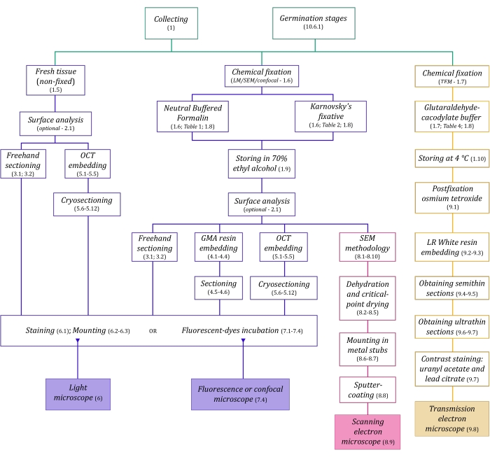
الشكل 1: التلخيص التخطيطي لطرق التصوير. توفر المخططات مؤشرات على خطوات البروتوكول التي يتم تفصيلها فيها. الاختصارات: GMA = ميثاكريلات غليكول ، OCT = مركب درجة حرارة القطع الأمثل ، SEM = المجهر الإلكتروني الماسح. يرجى النقر هنا لعرض نسخة أكبر من هذا الرقم.

يسبق تقنيات الفحص المجهري الموصوفة هنا بالتفصيل (الشكل 1) الخطوات الأساسية التالية: جمع العينات وإصلاحها وتجفيفها وتضمينها وتقسيمها. نظرا لأن الخطوات متغيرة (الشكل 1) اعتمادا على التقنية (التقنيات) المختارة ، فمن المهم التفكير في المستقبل ، مع مراعاة المثبتات التي سيتم إعدادها ونقلها إلى موقع التجميع ، وكيفية إعداد العينات قبل التثبيت ، وعمليات الجفاف التي سيتم استخدامها (القسم 1) ، وإمكانيات التضمين المختلفة وطرق التقسيم (الأقسام 4 ، 5 ، و 9). يلخص الشكل 1 بالتتابع جميع الخطوات المطلوبة لكل تقنية من تقنيات الفحص المجهري الموضحة بدقة أدناه.

Protocol

1. جمع العينات وتثبيتها وصيانتها ملاحظة: يمكن العثور على نباتات MH بالكامل عادة في الغابة المظلمة تحت الأرض 12,13 ، خاصة في المناطق الرطبة والوفيرة بالقمامة ، في حين يمكن العثور على نباتات MH جزئيا في الغابات الأكثر انفتاحا 12,13.<sup class…

Representative Results

بعد المراحل الأساسية لتثبيت الأنسجة النباتية ينتج هياكل خلوية مشابهة قدر الإمكان للحالة الحية ، مع الأخذ في الاعتبار مورفولوجيا المكونات والأنسجة الخلوية وحجمها وتنظيمها المكاني16. لاحظ هذه السمات في العينات بعد التثبيت الكيميائي (الشكل 4). يمثل ا?…

Discussion

تتمتع تحليلات الصور في تشريح النبات ومورفولوجيا بإمكانات مهمة لتحقيق الأهداف والمساعدة في فهم العلاقات بين النباتات المتغايرة والنباتات الداخلية الفطرية التي لا غنى عنها ، كما يتضح من دراسات الأعضاء الجوفية 6,40 ، والتحليلات الهيكلية للإنبات التكافلي للبذور3…

Disclosures

The authors have nothing to disclose.

Acknowledgements

يشكر المؤلفون التمويل المقدم من FAEPEX و FAPESP (2015/26479-6). يشكر MPP Capes على منحة درجة الماجستير (العملية 88887.600591/2021-00) و CNPq. JLSM يشكر CNPq على منح الإنتاجية (303664/2020-7). ويشكر المؤلفون أيضا الوصول إلى المعدات والمساعدة التي يقدمها مختبر المجهر الإلكتروني – IB/Unicamp)، والمعهد الوطني للعلوم والتكنولوجيا في مجال الضوئيات المطبقة على بيولوجيا الخلية – Unicamp)، ومختبر بيولوجيا الأوعية الدموية – DBEF/IB/Unicamp)؛ LAMEB (UFSC) وإليانا دي ميديروس أوليفيرا (UFSC) للمساهمات في بروتوكول الحماية من التبريد ؛ LME للمساهمات في بروتوكول TEM.

Materials

Acetone Sigma-Aldrich 179124 (for SEM stubs mounting)
Agar-agar (AA) Sigma-Aldrich A1296 (for seeds germination tests)
Calcofluor White Stain Sigma-Aldrich 18909 fluorescent dye (detects cellulose)
Citrate Buffer Solution, 0.09M pH 4.8 Sigma-Aldrich C2488 (for toluidine blue O staining)
Conductive Double-Sided Carbon Tape Fisher Scientific 50-285-81 (for SEM)
Confocal Microscope Zeiss (any model)
Copper Grids Sigma-Aldrich G4776 (for TEM)
Critical-point dryer Balzers (any model)
Cryostat Leica Biosystems (any model)
Dissecting microscope Leica Biosystems (= stereomicroscope, any model)
Entellan Sigma-Aldrich 107960 rapid mounting medium for microscopy
Ethyl alcohol, pure (≥99.5%) Sigma-Aldrich 459836 (= ethanol, for dehydration processes)
Formaldehyde solution, 37% Sigma-Aldrich 252549 (for NBF solution preparation)
Formalin solution, neutral buffered, 10% Sigma-Aldrich HT501128 histological tissue fixative
Gelatin capsules for TEM Fisher Scientific 50-248-71 (for resin polymerisation in TEM)
Gelatin solution, 2% in H2O Sigma-Aldrich G1393 (dilute for slides preparation – OCT adherence)
Glutaraldehyde solution, 25% Sigma-Aldrich G6257 (for Karnovsky’s solution preparation)
HistoResin Leica Biosystems 14702231731 glycol methacrylate (GMA) embedding kit
Iodine Sigma-Aldrich 207772 (for Lugol solution preparation)
Lead(II) nitrate Sigma-Aldrich 228621 Pb(NO3)2 (for TEM contrast staining)
Light Microscope Olympus (any model)
LR White acrylic resin Sigma-Aldrich L9774 hydrophilic acrylic resin for TEM
Lugol solution Sigma-Aldrich 62650 (for staining)
Metal stubs for specimen mounts Rave Scientific (for SEM, different models)
Microtome Leica Biosystems manual rotary microtome or other model
Oatmeal agar (OMA) Millipore O3506 (for seeds germination tests)
OCT Compound, Tissue-Tek Sakura Finetek USA 4583 embedding medium for frozen tissues
Osmium tetroxide Sigma-Aldrich 201030 OsO4 (for TEM postfixation)
Parafilm M Sigma-Aldrich P7793 sealing thermoplastic film
Paraformaldehyde Sigma-Aldrich 158127 (for Karnovsky’s solution preparation)
Poly-L-lysine solution, 0.1% in H2O Sigma-Aldrich P8920 (for slides preparation – OCT adherence)
Poly-Prep Slides Sigma-Aldrich P0425 poly-L-lysine coated glass slides
Polyethylene Molding Cup Trays Polysciences 17177A-3 (6x8x5 mm, for embbeding samples in GMA resin)
Polyethylene Molding Cup Trays Polysciences 17177C-3 (13x19x5 mm, for embbeding samples in GMA resin)
Potassium iodide Sigma-Aldrich 221945 (for Lugol solution preparation)
Potato Dextrose Agar (PDA) Millipore 70139 (for seeds germination tests)
Scanning Electron Microscope Jeol (any model)
Silane [(3-Aminopropyl)triethoxysilane] Sigma-Aldrich A3648 (for slides preparation – OCT adherence)
Silane-Prep Slides Sigma-Aldrich S4651 glass slides coated with silane
Silica gel orange, granular Supelco 10087 (for dessicating processes)
Sodium cacodylate trihydrate Sigma-Aldrich C0250 (for glutaraldehyde-sodium cacodylate buffer)
Sodium hydroxide Sigma-Aldrich S5881 NaOH (for Karnovsky’s solution preparation and TEM contrast staining)
Sodium hypochlorite solution Sigma-Aldrich 425044 NaClO (for seeds surface disinfection)
Sodium phosphate dibasic, anhydrous Sigma-Aldrich 71640 Na2HPO4 (for NBF solution and PB preparation)
Sodium phosphate monobasic monohydrate Sigma-Aldrich S9638 NaH2PO4·H2O (for NBF and PB)
Sputter coater Balzers (any model)
Sucrose Sigma-Aldrich S0389 C12H22O11 (for cryoprotection and germination test)
Sudan III Sigma-Aldrich S4131 (for staining)
Sudan IV Sigma-Aldrich 198102 (for staining)
Sudan Black B Sigma-Aldrich 199664 (for staining)
Syringe (3 mL, any brand, for TEM contrast staining)
Syringe Filter Unit, Millex-GV 0.22 µm Millipore SLGV033R PVDF, 33 mm, gamma sterilized (for TEM contrast staining)
Tek Bond Super Glue 793 Tek Bond Saint-Gobain 78072720018 liquid cyanoacrylate adhesive, medium viscosity
Toluidine Blue O Sigma-Aldrich T3260 (for staining)
Transmission Electron Microscope Jeol (any model)
Triphenyltetrazolium chloride Sigma-Aldrich T8877 (for the tetrazolium test in seeds germination)
Trisodium citrate dihydrate Sigma-Aldrich S1804 Na3(C6H5O7)·2H2O (for TEM contrast staining)
Ultramicrotome Leica Biosystems (any model)
Uranyl acetate Fisher Scientific 18-607-645 UO2(CH3COO)2 (for TEM contrast staining)
Vacuum pump (any model)
Wheat Germ Agglutinin, Alexa Fluor 488 Conjugate TermoFisher Scientific W11261 fluorescent dye-conjugated lectin (detects sialic acid and N-acetylglucosaminyl residues)

References

  1. Evert, R. F. . Esau’s Plant Anatomy: Meristems, Cells, and Tissues of the Plant Body: Their Structure, Function, and Development. , (2006).
  2. Yeung, E. C. T., Stasolla, C., Sumner, M. J., Huang, B. Q. . Plant Microtechniques and Protocols. , (2015).
  3. Sokoloff, D. D., Jura-Morawiec, J., Zoric, L., Fay, M. F. Plant anatomy: at the heart of modern botany. Botanical Journal of the Linnean Society. 195 (3), 249-253 (2021).
  4. Leake, J. R. The biology of myco-heterotrophic (‘saprophytic’) plants. New Phytologist. 127 (2), 171-216 (1994).
  5. Bidartondo, M. I. The evolutionary ecology of myco-heterotrophy. New Phytologist. 167 (2), 335-352 (2005).
  6. Imhof, S., Massicotte, H. B., Melville, L. H., Peterson, R. L. Subterranean morphology and mycorrhizal structures. Mycoheterotrophy. , 157-214 (2013).
  7. Rasmussen, H. N., Rasmussen, F. N. Orchid mycorrhiza: implications of a mycophagous life style. Oikos. 118 (3), 334-345 (2009).
  8. Rasmussen, H. N., Dixon, K. W., Jersáková, J., Těšitelová, T. Germination and seedling establishment in orchids: a complex of requirements. Annals of Botany. 116 (3), 391-402 (2015).
  9. Zettler, L. W. Terrestrial orchid conservation by symbiotic seed germination: techniques and perspectives. Selbyana. 18 (2), 188-194 (1997).
  10. Stewart, S. L., Kane, M. E. Symbiotic seed germination and evidence for in vitro mycobiont specificity in Spiranthes brevilabris (Orchidaceae) and its implications for species-level conservation. In Vitro Cellular & Developmental Biology – Plant. 43 (3), 178-186 (2007).
  11. Zhao, D. -. K., et al. Orchid reintroduction based on seed germination-promoting mycorrhizal fungi derived from protocorms or seedlings. Frontiers in Plant Science. 12, 701152 (2021).
  12. Selosse, M. A., Roy, M. Green plants that feed on fungi: facts and questions about mixotrophy. Trends in Plant Science. 14 (2), 64-70 (2009).
  13. Merckx, V. S. F. T., Mennes, C. B., Peay, K. G., Geml, J. Evolution and diversification. Mycoheterotrophy: The Biology of Plants Living on Fungi. , 215-244 (2013).
  14. Boon, M. E., Drijver, J. Routine Cytological Staining Techniques: Theoretical Background and Practice. Macmillan International Higher Education. , (1986).
  15. Karnovsky, M. A formaldehyde-glutaraldehyde fixative of high osmolality for use in electron microscopy. Journal of Cell Biology. 27 (2), 137-138 (1964).
  16. Hayat, M. . Fixation for Electron Microscopy. , (1981).
  17. Roland, J. C., Vian, B. General preparation and staining of thin sections. Electron Microscopy of Plant Cells. 1, 675 (1991).
  18. Gerrits, P. O., Horobin, R. W. Glycol methacrylate embedding for light microscopy: basic principles and trouble-shooting. Journal of Histotechnology. 19 (4), 297-311 (1996).
  19. Zhang, Z., Niu, L., Chen, X., Xu, X., Ru, Z. Improvement of plant cryosection. Frontiers in Biology. 7 (4), 374-377 (2012).
  20. BeneŠ, K. On the media improving freeze-sectioning of plant material. Biologia Plantarum. 15 (1), 50-56 (1973).
  21. Fischer, A. H., Jacobson, K. A., Rose, J., Zeller, R. Preparation of slides and coverslips for microscopy. Cold Spring Harbor Protocols. 2008 (5), (2008).
  22. Sakai, W. S. Simple method for differential staining of paraffin embedded plant material using toluidine blue O. Stain Technology. 48 (5), 247-249 (1973).
  23. O’Brien, T., Feder, N., McCully, M. E. Polychromatic staining of plant cell walls by toluidine blue O. Protoplasma. 59 (2), 368-373 (1964).
  24. Ventrella, M. C., Almeida, A. L., Nery, L. A., Coelho, V. P. d. e. M. Métodos Histoquímicos Aplicados às Sementes. Universidade Federal de Viçosa. , (2013).
  25. Pearse, A. G. E. . Histochemistry, Theoretical and Applied. , (1960).
  26. Andrade-Linares, D. R., Franken, P. Fungal endophytes in plant roots: taxonomy, colonization patterns, and functions. Symbiotic Endophytes. , 311-334 (2013).
  27. Wymer, C. L., Beven, A. F., Boudonck, K., Lloyd, C. W. Confocal microscopy of plant cells. Confocal Microscopy Methods and Protocols. , 103-130 (1999).
  28. Marques, J. P. R., Soares, M. K. M. Manual de Técnicas Aplicadas à Histopatologia Vegetal. FEALQ. , (2021).
  29. Navarro, B. L., Marques, J. P. R., Appezzato-da-Glória, B., Spósito, M. B. Histopathology of Phakopsora euvitis on Vitis vinifera. European Journal of Plant Pathology. 154 (4), 1185-1193 (2019).
  30. Marques, J. P. R., et al. Sugarcane cell wall-associated defense responses to infection by Sporisorium scitamineum. Frontiers in Plant Science. 9, 698 (2018).
  31. Jeffree, C. E., Read, N. D. Ambient-and low-temperature scanning electron microscopy. Electron Microscopy of Plant Cells. , 313-413 (1991).
  32. Bozzola, J. J., Russell, L. D. . Electron Microscopy: Principles and Techniques for Biologists. , (1999).
  33. Murray, S. Basic transmission and scanning electron microscopy. Introduction to electron Microscopy for Biologists. , 3-18 (2008).
  34. . Glossary of TEM terms Available from: https://www.jeol.co.jp/en/words/emterms/ (2021)
  35. Seaton, P. T., et al. Orchid seed and pollen: a toolkit for long-term storage, viability assessment and conservation. Orchid Propagation: From Laboratories to Greenhouses—Methods and Protocols. , 71-98 (2018).
  36. Otero, J. T., Ackerman, J. D., Bayman, P. Differences in mycorrhizal preferences between two tropical orchids. Molecular Ecology. 13 (8), 2393-2404 (2004).
  37. Koch, R. A., et al. Marasmioid rhizomorphs in bird nests: Species diversity, functional specificity, and new species from the tropics. Mycologia. 112 (6), 1086-1103 (2020).
  38. Webster, J., Weber, R. . Introduction to Fungi. , (2007).
  39. Sisti, L. S., et al. The role of non-mycorrhizal fungi in germination of the mycoheterotrophic orchid Pogoniopsis schenckii Cogn. Frontiers in Plant Science. 10, 1589 (2019).
  40. Martos, F., et al. Independent recruitment of saprotrophic fungi as mycorrhizal partners by tropical achlorophyllous orchids. New Phytologist. 184 (3), 668-681 (2009).
  41. Alves, M. F., et al. Reproductive development and genetic structure of the mycoheterotrophic orchid Pogoniopsis schenckii Cogn. BMC Plant Biology. 21 (1), 332 (2021).
  42. Merckx, V. S. F. T. Mycoheterotrophy: an introduction. Mycoheterotrophy: The Biology of Plants Living on Fungi. , 1-17 (2013).
  43. Hall, J. L., Hawes, C. . Electron Microscopy of Plant Cells. , (1991).
check_url/kr/63777?article_type=t

Play Video

Cite This Article
Pena-Passos, M., Sisti, L. S., Mayer, J. L. S. Microscopy Techniques for Interpreting Fungal Colonization in Mycoheterotrophic Plants Tissues and Symbiotic Germination of Seeds. J. Vis. Exp. (183), e63777, doi:10.3791/63777 (2022).

View Video