The article describes the method for isolating conditionally immortalized glomerular endothelial cells from the kidneys of transgenic mice expressing the thermolabile simian virus 40 and photo-activatable mitochondria, PhAMexcised. We describe the procedure for glomeruli isolation from whole kidneys using beads, digestion steps, seeding, and culturing of GECs-CD31 positive.
Glomerular endothelial cell (GEC) dysfunction can initiate and contribute to glomerular filtration barrier breakdown. Increased mitochondrial oxidative stress has been suggested as a mechanism resulting in GEC dysfunction in the pathogenesis of some glomerular diseases. Historically the isolation of GECs from in vivo models has been notoriously challenging due to difficulties in isolating pure cultures from glomeruli. GECs have complex growth requirements in vitro and a very limited lifespan. Here, we describe the procedure for isolating and culturing conditionally immortalized GECs with fluorescent mitochondria, enabling the tracking of mitochondrial fission and fusion events. GECs were isolated from the kidneys of a double transgenic mouse expressing the thermolabile SV40 TAg (from the Immortomouse), conditionally promoting proliferation and suppressing cell differentiation, and a photo-convertible fluorescent protein (Dendra2) in all mitochondria (from the photo-activatable mitochondria [PhAMexcised] mouse). The stable cell line generated allows for cell differentiation after inactivation of the immortalizing SV40 TAg gene and photo-activation of a subset of mitochondria causing a switch in fluorescence from green to red. The use of mitoDendra2-GECs allows for live imaging of fluorescent mitochondria's distribution, fusion, and fission events without staining the cells.
The glomerulus is critical for blood filtration by restricting the passage of large molecules through the glomerular filtration barrier1,2. The glomerulus contains four cell types: parietal epithelial cells, podocytes (visceral epithelial cells), glomerular endothelial cells (GEC), and mesangial cells3. The glomerular endothelium is characterized by a unique vascular structure, as per the presence of fenestrae required for large filtration volumes4. The apical surface of the glomerular endothelium is covered with a negatively charged glycocalyx layer and a coat called the endothelial surface layer that creates a space between the endothelium and blood. This structure provides high charge selectivity restricting the passage of negatively charged molecules such as albumin and preventing leukocyte and platelet adhesion5.
GECs are very sensitive to metabolic changes, such as the hyperglycemia associated with the diabetic milieu. Indeed, diabetes leads to increased circulation of noxious substances, the saturation of glucose metabolism pathways, and disturbed cellular redox balance3,6. Moreover, the increase in reactive oxygen species induces mitochondrial dysfunction, which affects endothelial function7.
The overall goal of the current protocol is to isolate immortalized glomerular endothelial cells with fluorescent mitochondrial features. Indeed, the cell culture of primary GECs has a limited proliferative cycle and early senescence8. In addition, the presence of fluorescent mitochondria helps examine fission and fusion events in response to hyperglycemia or any other treatment. As an alternative method, other labs used h-TERT to immortalize cells in vitro9.
The method described here allows for the isolation of conditionally immortalized mitoDendra2 glomerular endothelial cells from 4-6-week-old animals (Figure 1). This detailed protocol describes the use of transgenic mice (H-2Kb-tsA58) harboring the simian virus 40 large tumor antigen (SV40 TAg) gene10,11 for generating thermolabile conditionally immortalized cells. The tsA58 TAg gene product is functional at the permissive temperature of 33 °C under the control of the inducible 5' flanking promoter of the mouse H-2Kb gene, which is increased above basal levels upon exposure to interferon gamma (IFNγ), therefore maintaining the conditional proliferation phenotype12. H-2Kb is rapidly degraded at the non-permissive temperature of 37 °C in the absence of IFNγ, removing the immortalizing function of the tsA58Tag in cells and allowing the cells to develop a more differentiated phenotype13,14,15. Optional crossing of H-2Kb-tsA58 transgenic mice with PhAM mice, which express a mitochondria-specific (subunit VIII of cytochrome c oxidase) Dendra2-green, allows the live detection of fluorescent mitochondria16. Dendra2 green fluorescence switches to red fluorescence after exposure to a 405 nm laser16. When mitochondria fuse after photo-switching, they form elongated shapes that appear yellow from the exchange of green and yellow material or appear red when they undergo fission7,17. The mitoDendra2-GECs are a great tool for studying the cellular responses of GEC mitochondria to different stimuli.
All animal procedures described here were approved by the IACUC at Icahn School of Medicine at Mount Sinai. We used three male mice (H-2Kb-tsA58 transgenic mice with photo-activatable mitochondria [PhAM]) purchased from Jackson lab and kept on a normal chow diet.
1. Working conditions and preparations
2. Magnetic particle concentrator
3. Isolation of mouse kidney glomerular cells
4. Preparation of beads for endothelial cell isolation
5. Isolating the glomerular endothelial cells with CD31-coated beads
6. Cultivating the mitoDendra2-GECs
7. Passaging of mitoDendra2-GECs
8. Characterization of mitoDendra2-GECs
9. Live cell imaging of mitochondria structural changes in response to oxidative stress or high glucose
In this article, a detailed protocol for the isolation of conditionally immortalized glomerular endothelial cells with stable fluorescent mitochondria (mitoDendra2-GECs) is described (Figure 1). The use of young 6-10-week-old mice is essential to obtain a substantial number of healthy cells. After 3 days of culture, cells start growing slowly from the isolated glomeruli, as shown in Figure 3G. After 7 days, cells are heterogeneous, showing other glomerular cell types, such as podocytes, parietal epithelial cells, and mesangial cells (Figure 4A). After reaching 70% to 80% confluency, other glomerular cells were removed using a positive selection of endothelial cells with magnetic CD31 labeled beads (Figure 4B–C). The mitoDendra2-GECs expressed CD31 (Figure 4D) and were negative for podocyte marker as shown by synaptopodin negative staining (Figure 4E-F).
The fluorescent mitochondrial feature in the mitoDendra2-GECs allows for studying mitochondria morphology under various conditions in vitro. Here, we tested the effect of high glucose (25mM) on the mitochondria structure of mitoDendra2-GECs (Figure 5D-F). Compared to the elongated mitochondria visible in cells under normal glucose (5mM; Figure 5A-C), high glucose-induced fragmentation or fission of mitochondria as observed by prominent spheroid-shaped mitochondria (Figure 5E). Furthermore, we used a 405 nm laser to photo-convert a selected subpopulation of mitochondria in a single live mitoDendra2-GEC (Figure 5A,D). In Figure 5B and Figure 5E, successful photo-switching of mitochondria from green (488nm) to red (561 nm) in the selected area is presented. Importantly, it allowed for witnessing the fusion events in mitoDendra2-GECs grown under normal glucose, as can be observed by the yellow merged green and red fluorescence as a result of mitochondria matrix fusion (Figure 5C). In contrast, in the high glucose-treated mitoDendra2-GEC, the mitochondria were fragmented and were mainly red (Figure 5F), suggesting that, in the time frame tested, the fission/fusion events were delayed or inhibited due to the damage caused by the high glucose treatment, and this is consistent with previous reports18,19.
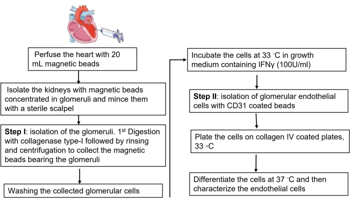
Figure 1: Representative description of murine GEC isolation. The diagram represents the significant steps of GEC isolation, starting from perfusing mice heart with magnetic beads to isolate the kidney glomeruli, the digestion of collagenase, the culture of glomerular cells, and finally, the purification of GEC using CD31-coated beads. Please click here to view a larger version of this figure.
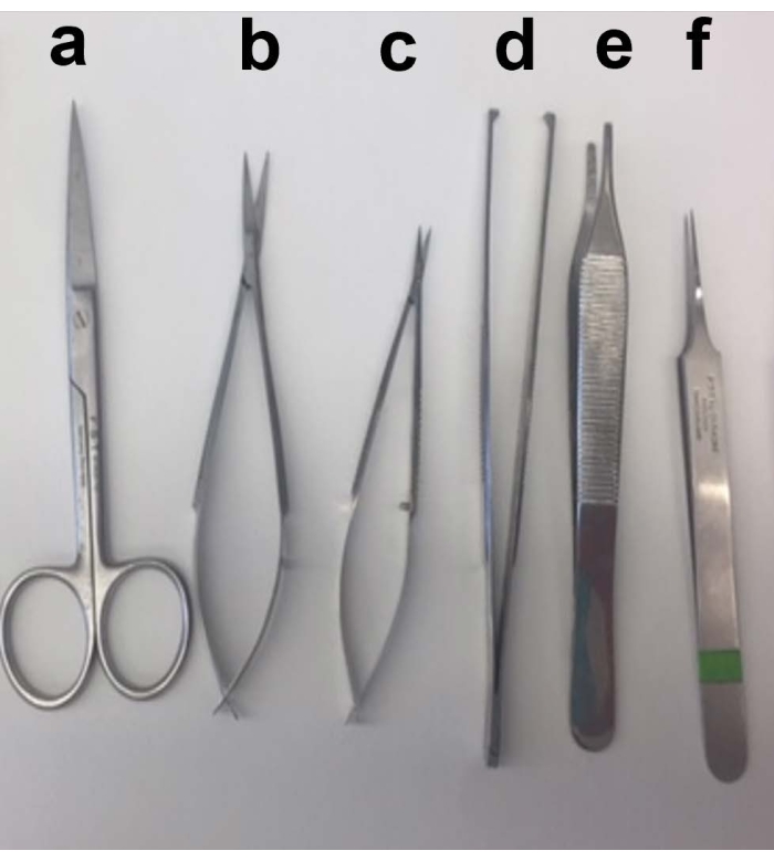
Figure 2: Description of kidney dissection. Representative image of all the sterile surgical instruments needed for the dissection. Scissors (a) are needed to open the skin of the mouse, and a separate pair of scissors (b,c) are needed to isolate the kidney. Tweezers (d) and (e) are needed to hold the skin. Tweezer (f) is needed to collect the dissected kidney. Please click here to view a larger version of this figure.
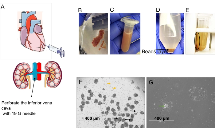
Figure 3: Description of glomeruli harvesting. (A) Perfuse the left ventricle with the HBSS-beads solution using a 20 mL syringe. Perforate the inferior vena cava with a 19 G needle. (B) After perfusion, remove the fat from the kidney and cut it into 1 mm pieces. (C) Digest the tissue with collagenase type II. (D) After digestion and centrifugation, three layers are formed; the beads bearing the glomeruli are in the middle layer, as indicated by the black arrow. (E) Separate the isolated beads bearing the glomeruli using the magnetic concentrator. (F) Representative image of freshly isolated glomeruli. (G) Growing cells from isolated glomeruli after 3 days. Please click here to view a larger version of this figure.
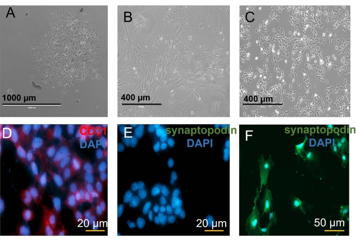
Figure 4: Mitochondrial assay in vitro. (A) Glomerular cells after 3 days of culture. (B) GEC after 7 days. (D) Purification of GEC with CD31-labeled beads. Microscopic imaging of CD31 staining. (E) GECs are negative for synaptopodin. (F) Podocytes stained with synaptopodin (488 nm, green fluorescence) as a negative control. Please click here to view a larger version of this figure.
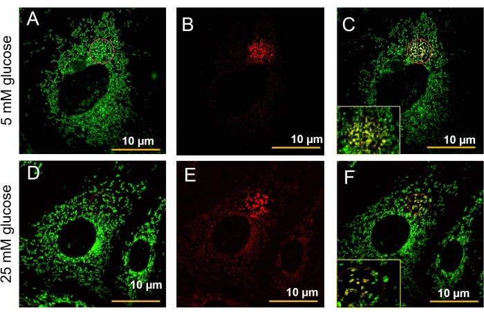
Figure 5: Microscopic imaging of fluorescent mitochondrial fusion in vitro. Images were acquired with a confocal microscope using a 40x water objective. (A–B) Selected area (red circle) was illuminated with a 405 nm line (4% laser power) for 300 bleaching iterations at a speed of 6.3-12.61/pixel, as previously described16. (A–C) Cells in growth media containing 5 mM glucose; green fluorescence (488 nm) shows healthy mitochondria. (B,E) Mitochondria were photo-switched into red (567 nm). (C) Yellow fluorescence detects an active fusion event of GEC treated with 5mM glucose. (D) Cells were treated with high glucose 25 mM for 30 min. Green fluorescence shows fragmented mitochondria and (E) a portion that was photo-switched into red. (F) The figure shows a decreased fusion event as shown by reduced yellow fluorescence. Please click here to view a larger version of this figure.
Mitochondria are critical for cellular metabolism, homeostasis, and stress responses, and their dysfunction is linked to many diseases, including kidney disease. Mitochondria have a role in the pathologic generation of excessive reactive oxygen species (ROS), the regulation of intracellular calcium levels, cell death pathways, and cytoskeletal dynamics21,22,23.
The isolation of murine GECs is challenging because of their small number, size, matrix factors, and interdependent nature with other glomerular cells that are essential for their survival. The current article presents a detailed protocol for mouse GEC isolation. Specifically, we used transgenic mice as a tool to obtain conditionally immortalized cell lines with high proliferation potential. The transgenic mice, expressing simian virus 40 (SV40) large T antigen tsA58, enable the activation of continuous proliferation under permissive conditions at 33 °C and were here crossed with the PHAMexcised mice expressing mitochondrial-specific fluorescence. The double transgenic mice offered an excellent tool for studying mitochondria structure. Indeed, PHAMexcised mice bear mitochondrial-specific (subunit VIII of cytochrome c oxidase) Dendra2 green, allowing the live detection of fluorescent mitochondria and the observation of fission and fusion events through green-red photo-switching16.
The protocol consists of three significant steps: extracting glomeruli using beads, the proliferation of glomerular cells, and the subsequent purification of GEC using CD31 beads once the cells are in culture. The use of only one digestion with collagenase is sufficient to isolate glomerular cells. Other protocols make use of a second digestion with DNAse and proteinase24. However, we have noticed better cell yields with only one digestion step. The DNAse may remain active and slow down the proliferation of the cells. Furthermore, GEC could be isolated using differential sieving as previously described20, which is not always easy when using mice kidneys.
It is crucial to autoclave all the surgical tools before starting the isolation of the kidneys and the glomeruli to avoid contamination. Moreover, it is important to filter and supplement the solutions with antibiotics to eliminate any source of contaminants. Indeed, mycoplasma infections may cause cytopathology that consequently interferes with every parameter measured in cell culture25.
The isolation of glomeruli relies on the intra-cardiac perfusion of beads. Certainly, slow perfusion with 20 mL of 1x HBSS-containing beads is essential to harvest a high number of glomeruli. The beads will be trapped in the glomeruli, which eases their purification using a magnetic concentrator. The beads might be prepared and stored at 4 °C for up to 15 days before using them to ease the experimental procedure.
Furthermore, digesting glomeruli with continuous shaking is crucial for dissociating a good number of glomeruli from the kidney. On the other hand, the isolation of murine GECs is challenging because of their small size and the presence of other glomerular cells. Therefore, the stepwise isolation of glomeruli, the growth of all glomerular cells in culture, and the subsequent use of CD31-coated beads to purify endothelial cells are vital, specifically for obtaining more significant numbers of viable and conditionally proliferative GECs. Nevertheless, validating the purity of cells with immunostaining and flow cytometry is needed.
In addition, the protocol describes the characterization of the GEC phenotype and the mitochondria fusion/fission assay in vitro. The mitoDendra2-GECs are an excellent tool for studying the cellular responses of GEC mitochondria to different stimuli in vitro without the need for transfection or staining.
The authors have nothing to disclose.
The authors thank Professor Cijiang He and Dr. Fu Jia for their insights in mice endothelial cell isolation and thank Professor Mone Zaidi for providing the PhAMexcised mice and valuable discussions. The authors would also like to acknowledge the Microscopy CORE at the Icahn School of Medicine at Mount Sinai and staff for the guidance we received. This work was supported by grants from National Institutes of Health grant R01DK097253 and Department of Defense CDMRP grant E01 W81XWH2010836 to I.S.D.
100 µm cell strainer | Fisher | 22-363-549 | |
1ml Insulin Syringes | BD | 329424 | |
25G butterfly | BD | 367298 | |
3 mm cutting edge scissors | F.S.T | 15000-00 | |
30ml syringe | BD Biooscience | 309650 | |
40 µm cell strainer | Fisher | 22-363-547 | |
40 µm nylon mesh | |||
Bonn Scissors | F.S.T | 14184-09 | |
Bovine serum albumin | Fisher | BP1600-100 | |
CD31 | abcam | ab7388 | |
Collagenase type I | Corning | 354236 | |
Collagenase type II | SIGMA | C6885 | 125CDU/mg |
Collagene type IV | SIGMA | C5533-5M | |
Dnase-I | Qiagen | 79254 | |
Dynabeads 450 | Thermofisher Scientific | 14013 | |
endothelial cells growth medium | Lonza | cc-3156 | |
Extra fine graefe forceps | F.S.T | 11150-10 | |
FBS | Gemini | 100-106 | Heat inactivated |
Fibronectin | Thermofisher | 33016015 | |
Fine forceps | F.S.T Dumont | E6511 | |
HBSS | GIBCO | 14065-056 | |
IFNg | Cell Science | CRI001B | |
Immortomouse | Jackson laboratory | 32619 | Tg(H2-K1-tsA58)6Kio/LicrmJ |
L-Glutamine 100x | Thermofisher Scientific | 25030081 | |
Magnetic particle concentrator | Thermofisher Scientific | 12320D | |
mitotracker | Thermofisher Scientific | M7512 | |
PBS 1X | Corning | 46-013-CM | |
penecillin streptomycin 100x | Thermofisher Scientific | 10378016 | |
PhaM mice | Jackson laboratory | 18397 | B6;129S-Gt(ROSA)26Sortm1.1(CAG-COX8A/Dendra2)Dcc/J |
Protease (10 mg/ml) | SIGMA | P6911 | |
RPMI | GIBCO | 3945 | |
Sodium Pyruvate 100mM | Thermofisher Scientific | 11360070 | |
Standard pattern forceps | F.S.T | 11000-12 | |
Surgical Scissors – Sharp-Blunt | F.S.T | 14008-14 | |
synaptopodin | Santa Cruz | sc-515842 | |
Trypsin 0.05% | Thermofisher Scientific | 25300054 |